622 11/11 GLA pt 2 Skin Infections
 Call Kai
 Learn
 Practice Test
 Spaced Repetition
 Match
 Flashcards
 Knowt Play
1/80

There's no tags or description
Looks like no tags are added yet.
81 Terms
Deep Tissue Bacterial Infections
ecthyma
necrotizing fasciitis
gas gangrene
Epidermis to Bone
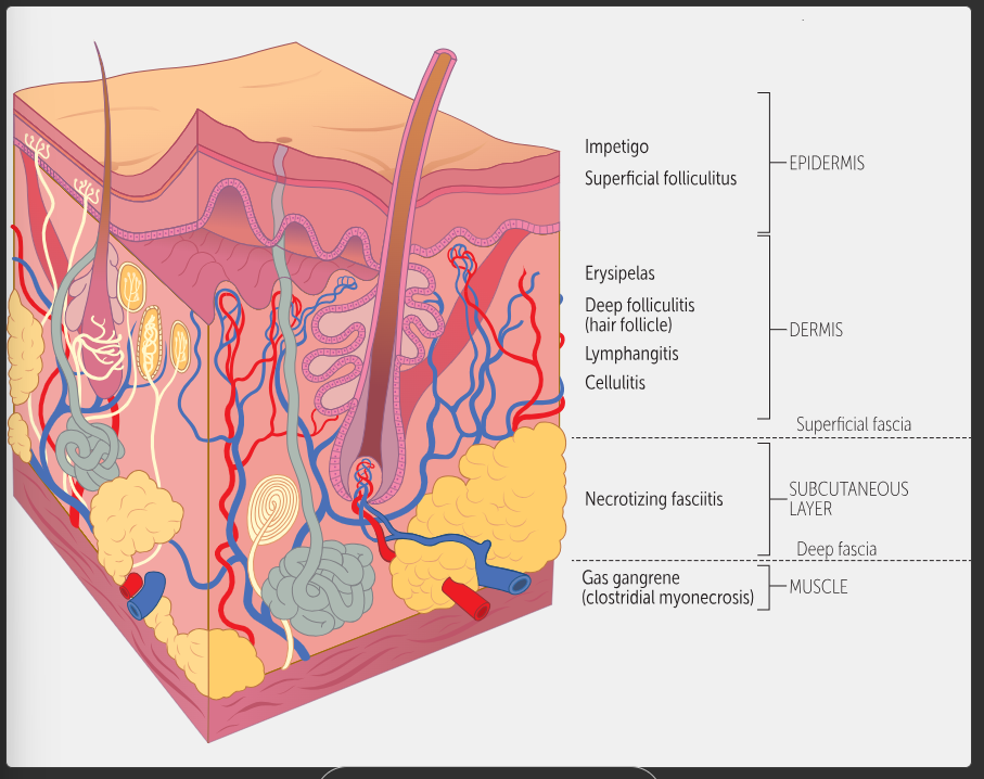
epidermis → papillary dermis → reticular dermis → subcutaneous fat → fascia → muscle → bone
Skin Layers and Infections
Ecthyma (Simple)
bacterial skin infection that begins superficially and extends into the dermis
Ecthyma Gangrenosum (Simple)
skin infection of dermal and subcutaneous layer, most commonly associated with pseudomonas, but other orgs have been implemented (s aureus, s pyogenes)
Necrotizing Fasciitis (Simple)
deep soft tissue infection that spreads along muscle fascia, destroying it and its overlaying subcutaneous fat
Gas Gangrene (Simple)
serious muscle infection that occurs either traumatically (clostridium perfringens) or spontaneously (clostridium septicum)
Ecthyma
dermal infection with ulcerations and crust
can result from chronic, untreated impetigo
Ecthyma Bacteria
s pyogenes
s auerus
Ecthyma Risk Factors
children, elderly, immunocompromized (HIV, diabetes)
poor hygiene and crowded living conditions, high temp and humidity in tropical places, minor injuries or other skin conditions, untreated impetigo (esp in pt with poor hygiene)
Ecthyma Clinical Presentation
butt, thighs, legs, ankles, feet
local lymph nodes may become swollen and painful
begin as vesicles or pustule on inflamed skin that burst, hard crust forms, and an ulcer forms underneath
Ecthyma Ulcers
red, swollen, ooze pus
may stay same size an resolve on own, or grow to create a sore 0.5-3 cm in diameter
can persist for weeks or months and leave a scar

Ecthyma Diagnosis
apperance, clinical signs, history
regional lymphadenopathy
can do culture or gram stain, skin biopsy shows dermal necrosis and inflammation with granulomatous perivascular infiltrate
Ecthyma Differential
non-bullous impetigo
Methicillin-resistant Staphylococcus aureus (MRSA) infections
Pyoderma grangrenosum
Insect bit reaction
Sporotrichosis
At-Home Ecthyma Management
soak crusted areas in mixture of half cup of white vinegar in liter of warm water, apply compress to area for 10 mins 5x a day
improved hygiene (daily bathing, frequent laundering of clothes and linens, using separate towels and washcloths, use of screens/nets to prevent insect bites)
mortality is 38-77% in pts with EG and septicemia caused by pseudomonas, but only abt 15% it pts without sepsis
Medical Treatment of Ecthyma
topical antiseptics such as povidone iodine, Manuka honey, or hydrogen peroxide cream may be applied to the are three times per day--after removing crusts
topical antibiotic ointment, mupirocin, can be applied three times per day
oral antibiotics are prescribed if the infection is extensive or not responding adequately to topical treatment, penicillin is the antibiotic of choice, treatment varies but 2 weeks to several weeks may be necessary depending on response
Bacteria of Ecthyma Gangrenosum
pseudomonas
Ecthyma Gangrenosum Risk Factors
critically ill and immunocompromised
leukemia, lymphoma, severe burns, malnutrition, organ transplant, immunosupressive therapy, diabetes, recent viral illness, catheters, IV placements, tracheostomies
Ecthyma Gangrenosum Clinical Presentation
initially painless, round, red patches that rapidly become pustular with surrounding inflammation
blister then forms, spreads, and evolves into a gangrenous ulcer with a black/gray scab (eschar) surrounded by a red to purple halo
transformation can be as little as 12 hours, on axillae and perineal area
fever, unstable vital signs, altered level of consciousness

Possble Causes of Ecthyma Gangrenosum

Ecthyma Gangrenosum Differential
Pyoderma gangrenosum
Cutaneous anthrax
Antiphospholipid antibody syndrome
Acute meningococcemia (Neisseria meningitidis in the bloodstream)
Disseminated candidiasis
Necrotizing fasciitis
Necrotizing vasculitis
Ecthyma Gangrenosum Lab Diagnosis
gram stain of fluid if present, swab of underside of scab if not
two blood cultures should be drawn from different locations and during a temp spike prior to initiating antibiotic therapy
two skin biopsies can be taken to rule out other organisms
Biopsy of Ecthyma Gangrenosum
vascular necorsis with few inflammatory cells and many surrounding bacteria
second biopsy sent for tissue culture for bacteria, fungi, yeasts, and mycobacteria
Gram Stain of Ecthyma Gangrenosum
numerous gram - rods surrounding necrotic vessels
Ecthyma Gangrenosum Antibiotics
piperacillin and aminoglycoside
2 Types of Necrotizing Fasciitis
polymicrobial (type 1)
monomicrobial (type 2)
Polymicrobial (Type 1) Necrotizing Fasciitis
aerobic and anaerobic bacteria
commonly at least one anaerobic spp (bacteriodes, clostridium, peptostreptococcus) is isolated in combination with enterobacteriaceae
Monomicrobial (Type 2) Necrotizing Fasciitis
caused by b-hemolytic streptococci
Necrotizing Fasciitis Risk Factors
skin damage and immunosupression, major penetrating trauma, recent surgery, malignancy
diabetes in lower extremity, perineum, head and neck
Why is muscle typically spared during the early stages of necrotizing fasciitis?
a more robust blood supply provides a better immune response and protects the muscle
Necrotizing Fasciitis Clinical Presentation
acute and rapid leading to systemic toxicity, limb loss, and death
ill-defined erythema, edema, severe pain, fever, crepitus, skin bullae, necrosis, ecchymosis

When should you suspect Necrotizing Fasciitis?
pt with soft tissue infection and signs of systemic illness such as fever and hemodynamic instability
early recognition is critical as rapid progression and extensive destruction can occur
Necrotizing Fasciitis Diagnosis
surgical exploration to reveal extend of involvement and allow for next stage of treatment
radiographic (gas in soft tissue) and lab studies can aid diagnosis, but surgery should be prioritized

Necrotizing Fasciitis Treatment
early and aggressive surgical exploration and debridement in addition to broad-spectrum empiric antibiotic therapy and hemodynamic support
Gas Gangrene (Clostridial myonecrosis)
comes from clostridium species that are common in nature and form enodspores, muscle is savagely destroyed
Main Bacteria of Gas Gangrene
clostidium perfringens
Gas Gangrene Risk Factors
traumatic wound that compromises blood supply
deep penetrating injuries such as knife wounds, gunshot wounds, and crush injuries
Clostridial a Toxin
major cause of damage to surrounding tissues in gas gangrene, blunts immune response to exacerbate the infection
Gas Gangrene Clinical Presentation
sudden and rapid after surgery, septic abortion, or trauma
fever, severe muscle pain at site of infection, crepitus
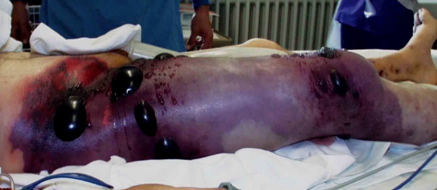
tissue may turn dark purple and necrotic, and large bullae or blisters may form
Gas Gangrene Image
Gas Gangrene Diagnosis
pain at site of injury coupled with symptoms of systemic toxicity (tachycardia, fever, shock) and gas in soft tissue
radiographic images can be used but should not delay treatment
Gas Gangrene Gram Stain and Tissue Cultures
clostridial, no granulocytes (neutrophils, basophils, eosinophils)
a toxin is responsible for inability of granulocytes to respond appropriately during gas gangrene by upregulating adhesive factors on granulocytes
this increase in adhesive factors make it impossible for granulocytes to leave the blood vessels and fight the infection
Gas Gangrene Management
surgical debridement and antibitotic therapy, might have to do multiple operations
survival is 80% when treated promptly
clindamycin
Clindamycin
used to treat gas gangrene, helps neutralize the a toxin produced by the clostridia that is responsible for the bulk of the damage because ti has anti-protein synthesis effects
Dysbiosis
normal microbiota is out of balance
Bacteria that Cause Skin Infections
staphylococcus aureus
streptococcus pyogenes
pseudomonas aeruginosa
vibrio
vuinificus
aeromonas hydrophila
clostridium perfringens
pasteurella multicida
staphylococcus epidermidis
bacillus anthracis
cutibacterium (Propionibatcterium) ances
bartonella henselae
erysipeiothrix rhusiopathiae
mycobacterium spp
non-spore forming anaerobic bacteriaClostridium

Non-Spore Forming Anaerobic Bacteria that Cause Skin Infections
actinomyces spp
peptostreptcoccus anaerobius
fusobacterium nucleatum
porphyromonas gingivalis
prevotella melaninogenica
bacteriodes fragillis
Bacterial Causes of Cellulitis

Necrotizing Fasciitis Terminology

Staphylococcus aureus Overview
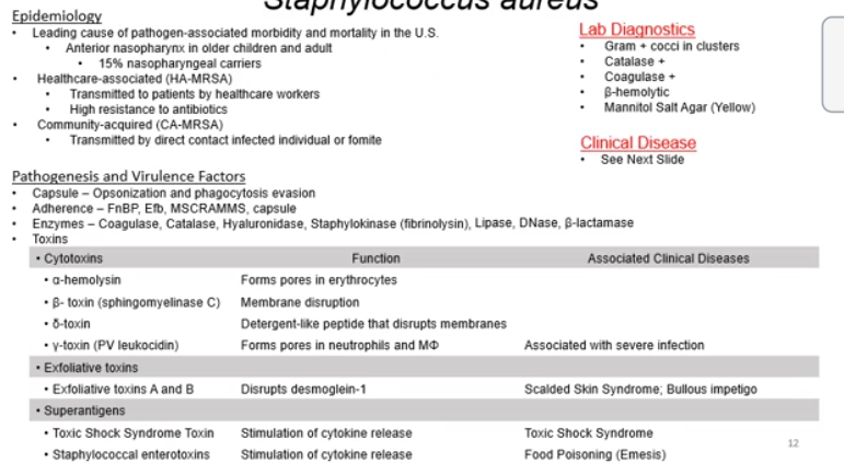
S aureus Clinical Disease
SS
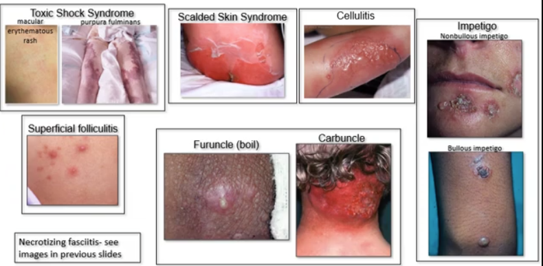
Streptococcus pyogenes Overview
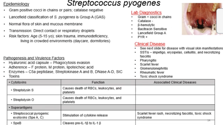
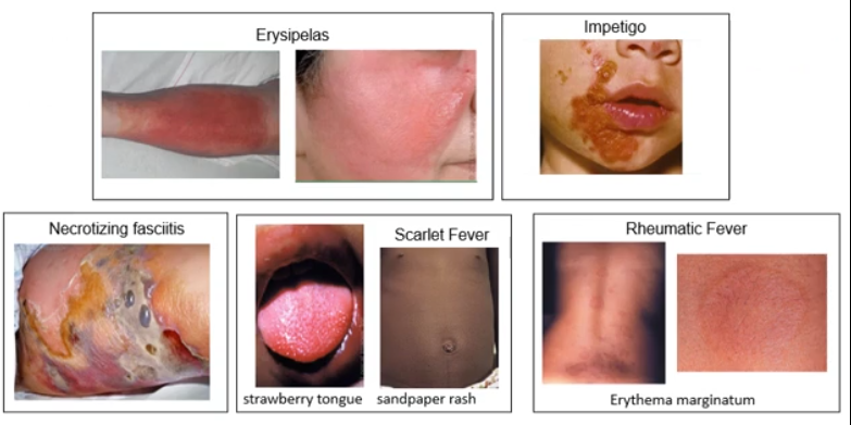
S pyogenes Clincal Disease
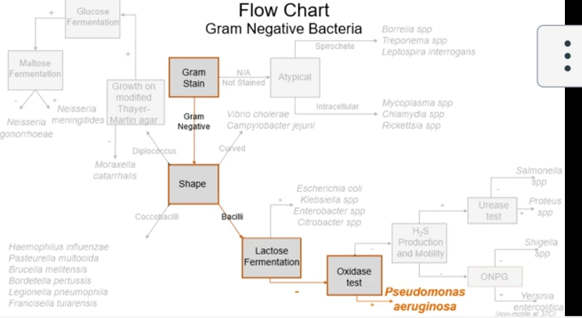
Pseudomas aeruginosa Flow Chart
Pseudomas aeruginosa General Information

Pseudomas aeruginosa Epidemiology
hospital sites (improper hygiene, medical equipment, ICU)
hot tubs/swimming pools (folliculitis, external otitis)
improper eye care (contact lens-associated keratitis)
Pseudomas aeruginosa Clinical Disease
skin and soft tissue infections
pulmonary infections
osteochondritis, osteomyelitis
ear and eye infections
UTI, endocarditi, bactereia, meningitis
Pseudomas aeruginosa Skin and Soft Tissue Infections
colonization of burn wounds (vascular damage, tissue necrosis, bacteremia)
folliculitis from immersion in contaminated water
hot tubs, whirlpools, swimming pools
ecthyma gangrenosum
necrotizing fasciitis
Pseudomas aeruginosa: Folliculitis
exposure to contained hot tub and swimming pool water
prutric follicular, macuoplular, vesicular, or pustule lesions
Pseudomas aeruginosa: Necrotizing Fascitis
rare
Pseudomas aeruginosa: Green Nail Syndrome
infection of paronychia

Pseudomas aeruginosa: Burn Wound Infections
localized immunosupression at burn sites, neutrophil recruitment limited, often colonizes after gram + cocci
symptoms: blue-green exudate, extending cellulitis, fever in some
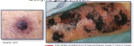
Pseudomas aeruginosa: Ecthyma Gangrenosum
skin lesions due to bacteremia, indicates sepsis
symptoms are hemorrhagic vesicles/pustules that become necrotic ulcers with erythematous border
Pseudomas aeruginosa: Pulmonary Infections
chronic can be susceptible to cystic fibrosis
acute can be ventilator-associated pneumonias
Pseudomas aeruginosa Virulence Factors Diagram

Pseudomas aeruginosa Secretion Systems
has multiple, type 2-4
type 3 is very important virulence factor
Exotoxin A
major virulence factor of Pseudomas aeruginosa, secreted through type 2
ribosylates the diphthamide amino acid residue on the eukaryotic elongation factor (EF2), resulting in disruption of protein synthesis and cytotoxicity
kills cells it gets into
Exotoxin A Pathogenesis (P aeruginosa)
most likely contributes to the dermatonecrosis (black) that occurs in burn wounds
Elastase
virulence factor of Pseudomas aeruginosa, secreted through type 2
degrades elastin in host cell that damages elastic-containing tissues
Elastase Pathogenesis (P aeruginosa)
causes hemorrhagic lesions (ecthyma gangrenosum)
Phospholipase C
virulence factor of Pseudomas aeruginosa, hemolosyn that breaks down phospholipids
Pseudomas aeruginosa Virulence Factors
secretion systems: 3 secretion systems, exotoxin A, elastase, phospholipase, ExoS & ExoU, pyocyanin, pyoverdine
adherence/motility: type iv pili, flagella for motility, OmpF adhesin, aliginate
Pseudomas aeruginosa Secretion Virulence Factors
3 secretion systems, exotoxin A, elastase, phospholipase, ExoS & ExoU, pyocyanin, pyoverdine
Pseudomas aeruginosa Adherence/Motility Virulence Factors
type iv pili, alginate, flagella for motility, OmpF adhesin
Alginate
major surface adhesion virulence factor of Pseudomas aeruginosa, exopolysaccharide helps with attachment and biofilm development
Alginate Pathogenesis (P aeruginosa)
important virulence factor in patients with cystic fibrosis
helps bacteria stick to host and to each other to form biofilm
Quorum Sensing
the ability of p aeruginosa cells to communicate with each other (group communication) because of the alginate virulence factor
Type VI Pilli
major surface adhesion virulence factor of Pseudomas aeruginosa, helps it stick to the host cell
ExoS & ExoU
virulence factor of Pseudomas aeruginosa, from type iii secretion systems
S disrupts host cell signaling, resulting in rapid cellular death
U causes lysis of host membrane
Pyocyanin
virulence factor of Pseudomas aeruginosa, gives it blue-green pigment
causes some of the immune system to activate and trigger apoptosis
Pyoverdine
virulence factor of Pseudomas aeruginosa, yellow-green fluorescent pigment
used for identification in culture