Human Anatomy Vocab Quiz 3 (27-44)
 Call Kai
 Learn
Learn Practice Test
 Spaced Repetition
 Match
Match Flashcards
 Knowt Play
1/17

There's no tags or description
Looks like no tags are added yet.
Name | Mastery | Learn | Test | Matching | Spaced | Call with Kai | Chat |
|---|
No analytics yet
Send a link to your students to track their progress
18 Terms
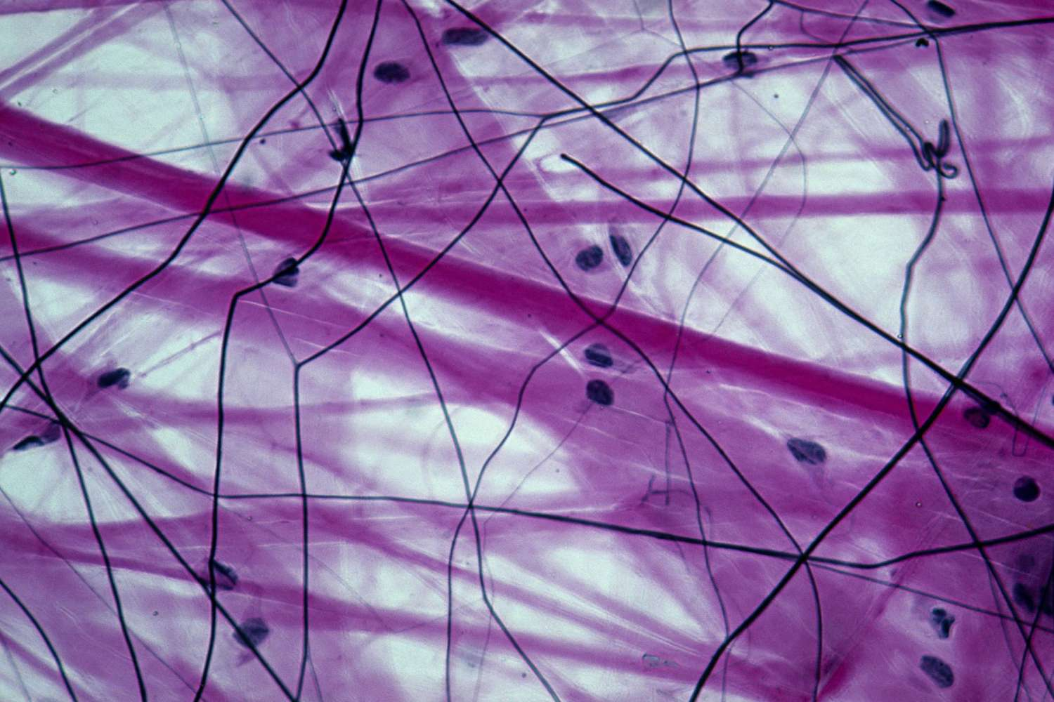
Connective Tissue
Most abundant, widely distributed tissue; few cells in lots of extracellular matrix
Functions: binding and support, protection, insulation, transport of substances
Loose – adipose
Dense – tendons and ligaments
Supporting – bone and cartilage
Fluid – blood.

Collagen Fibers
Fibers in the matrix that provide strength
One of 3 fiber types in the extracellular matrix (collagen = strength, elastic = stretch, reticular = fine network).
Collagen is the main fiber in dense connective tissue, forms a network in hyaline cartilage, thick fibers in fibrocartilage, and many are found in bone matrix
Made by fibroblasts.

Bone
Osseous tissue: hard, calcified matrix with blood vessels
Supporting connective tissue.
Functions: support and protect (by enclosing), stores calcium/minerals/fat, marrow is the site of blood cell formation (hematopoiesis).
Cells: osteoblasts, osteocytes (lie in lacunae).
Location: bones.

Osteocytes
Mature bone cells that lie in lacunae
Osteocytes lie in small spaces called lacunae, arranged in rings between the lamellae around the central (Haversian) canal.

Osteon
Haversian system: the structural unit of compact bone
Osteon is a central (Haversian) canal, which carries blood vessels, surrounded by concentric rings of matrix called lamellae.
Osteocytes sit in lacunae between the lamellae

Cartilage
Flexible supporting tissue with no nerves or blood
Functions: support, resists compression.
Cells: chondroblasts (produce the matrix) and chondrocytes (mature cells that lie in lacunae).
Locations: larynx, joints, tip of nose, ear, intervertebral discs, rib-breastbone, knee joint.
Subclasses: hyaline, elastic, fibrocartilage.

Hyaline Cartilage
Amorphous but firm matrix; collagen fibers form an imperceptible network
Function: supports and reinforces, has resilient cushioning properties, resists compressive stress.
Locations: forms most of the embryonic skeleton; covers ends of long bones in joint cavities; costal cartilages of the ribs; cartilages of the nose, trachea, and larynx.

Elastic Cartilage
Similar to hyaline cartilage, but with more elastic fibers in the matrix
Function: maintains the shape of a structure while allowing great flexibility.
Location: supports the external ear (pinna); epiglottis.

Fibrocartilage
Matrix similar to but less firm than hyaline; thick collagen fibers predominate
Function: tensile strength with the ability to absorb compressive shock.
Locations: intervertebral discs, pubic symphysis, discs of the knee joint.
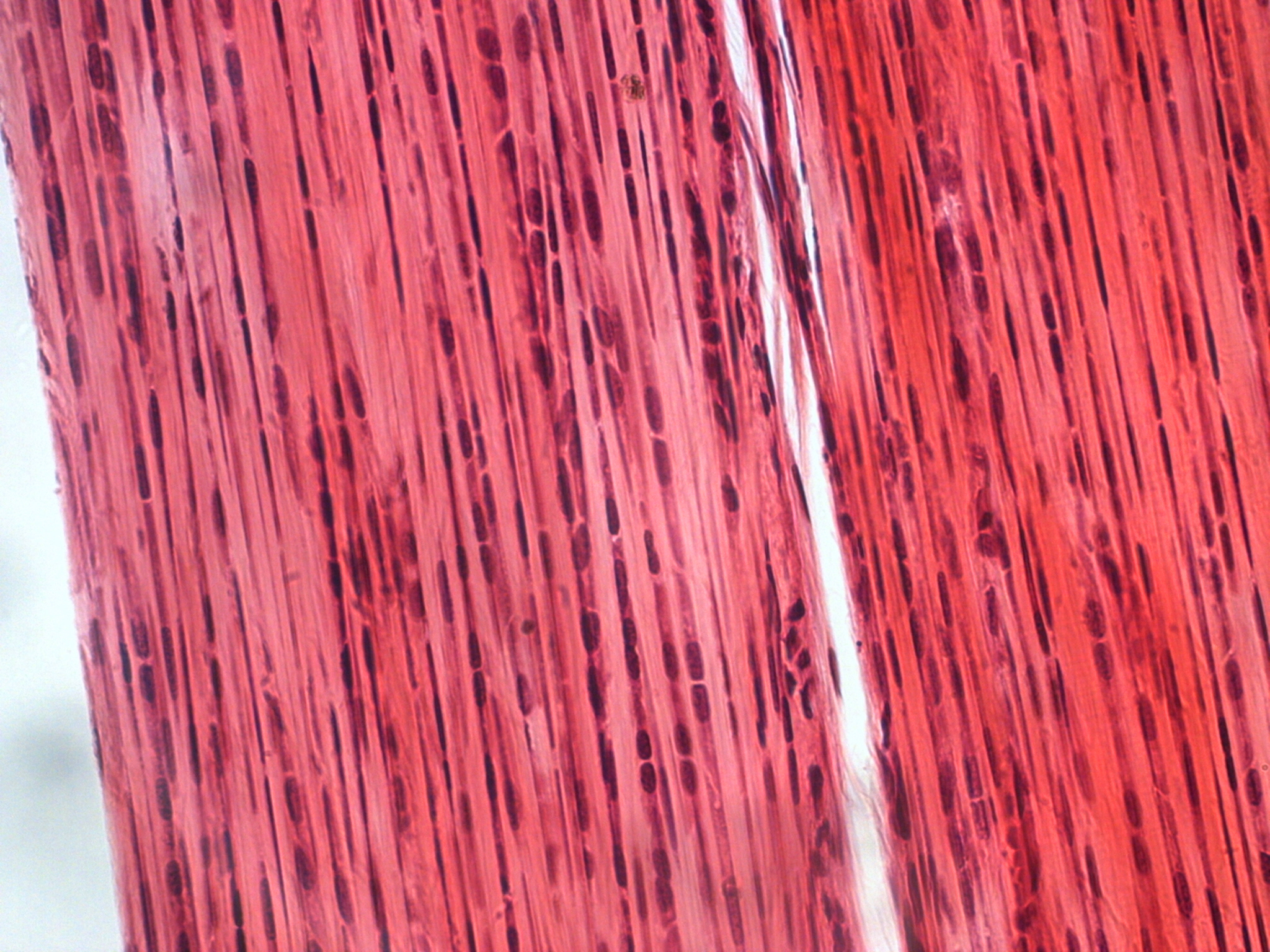
Dense Connective Tissue
Connective tissue made mainly of collagen fibers that resists tension
Subclass: dense regular (tendons and ligaments).
Structure: primarily parallel collagen fibers, a few elastic fibers.
Functions: elastic, resists tension, withstands great tensile stress when pulling force is applied in one direction.
Cell: fibroblasts.
Locations: tendons (muscle-bone), ligaments (bone), aponeuroses, lower layers of skin.
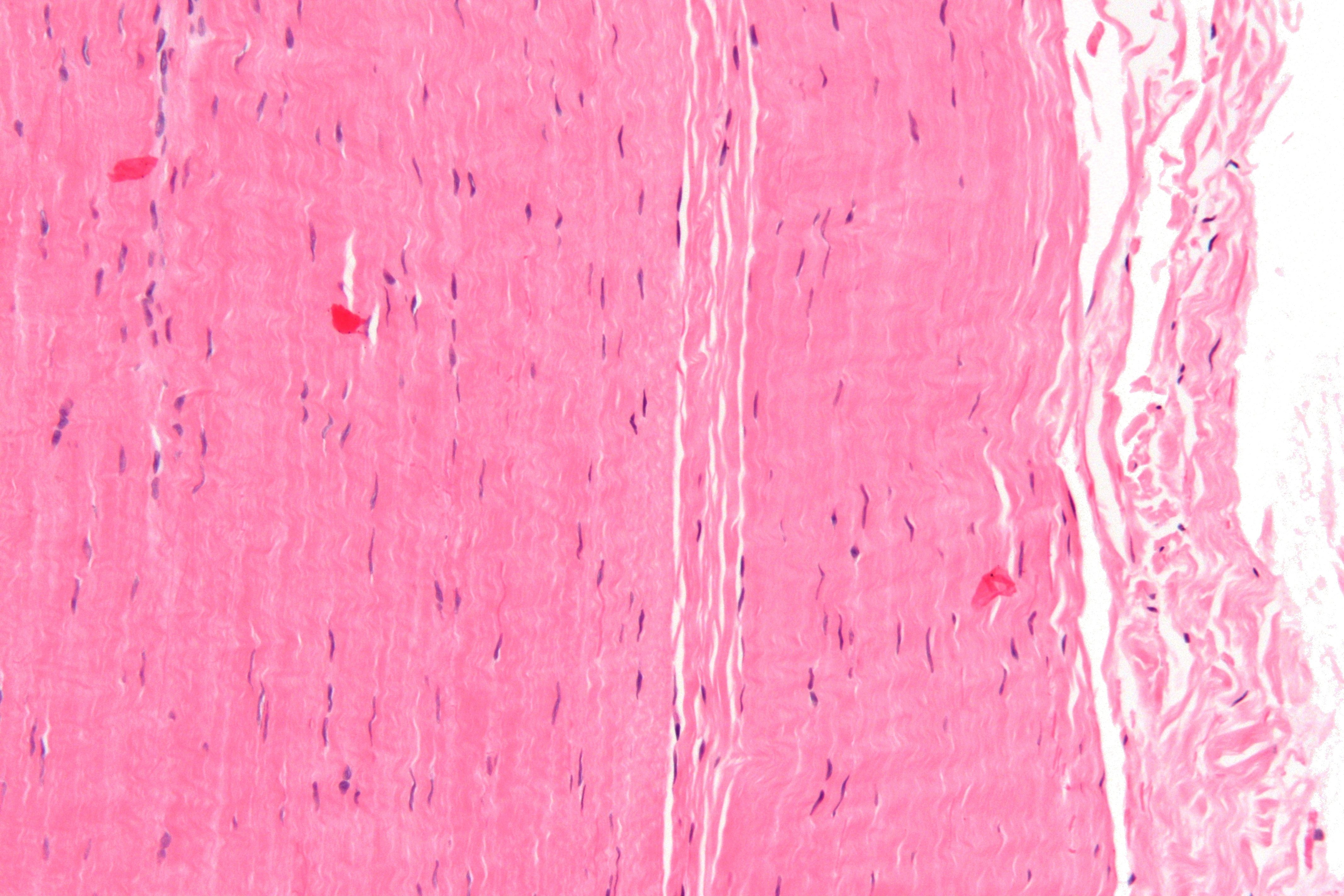
Tendons
Dense regular connective tissue attaching muscle to bone
Type of dense regular connective tissue.
Mainly parallel collagen fibers with fibroblasts in between.
Attach muscles to bones (or to muscles) and withstand great tensile stress when pulling force is applied in one direction.

Ligaments
Dense regular connective tissue attaching bone to bone
Type of dense regular connective tissue (most ligaments).
Mainly collagen fibers, with fibroblasts as the major cell.
Resist tension and hold bones together at joints, for example the shoulder joint.

Loose Connective Tissue
'Universal packing material': softer, fewer fibers, gel-like matrix
Subclasses: areolar, adipose, reticular.
Functions: cushion and protect organs (areolar, fat), store nutrients (fat), internal framework (reticular), fight infection (areolar).
Cells: fibroblasts, adipocytes.
Locations: under skin, lymph nodes, hips, behind eyeballs.
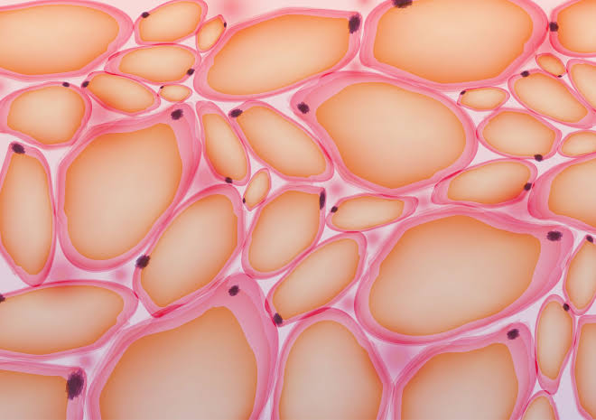
Adipose Tissue
Loose connective tissue of closely packed fat cells (adipocytes)
Adipocytes have the nucleus pushed to the side by a large fat droplet (vacuole).
Function: provides reserve food fuel; insulates against heat loss; supports and protects organs.
Locations: under skin in the hypodermis; around kidneys and eyeballs; within abdomen; in breasts.

Blood
Fluid connective tissue within blood vessels with no fibers
Subclasses: blood cells and plasma.
Red and white blood cells in a fluid matrix (plasma).
Function: transports respiratory gases, nutrients, wastes, hormones, and other substances.
Cells: white blood cells (leukocytes), red blood cells (erythrocytes), platelets.
Location: contained within blood vessels.

Erythrocytes
Red blood cells
They are the most numerous cells in a blood smear and surround the white blood cells.
Function: transport respiratory gases, they carry oxygen using hemoglobin
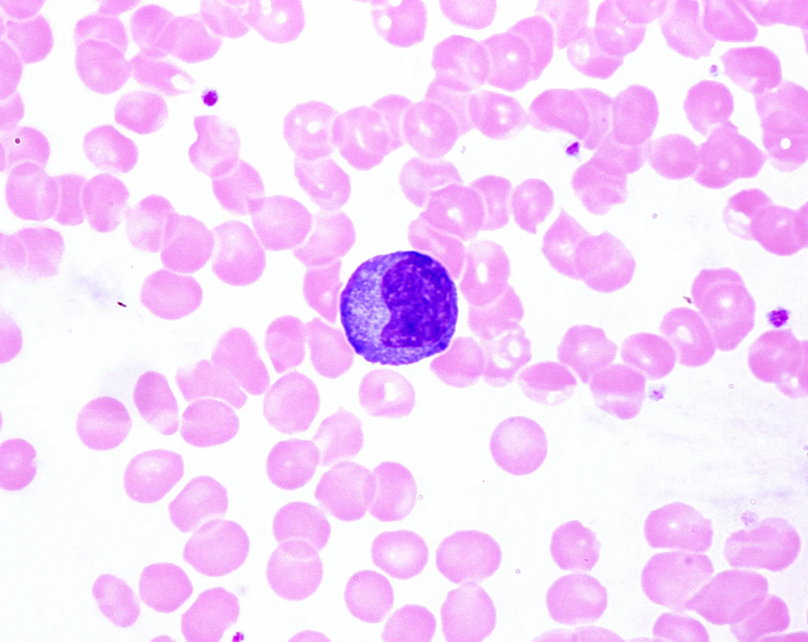
Leukocytes
White blood cells
Five types: neutrophil, eosinophil, basophil, lymphocyte, monocyte.
In the blood smear, a neutrophil and a lymphocyte are surrounded by red blood cells.
They defend the body against infection.

Plasma
The fluid matrix of blood in which cells are suspended
A subclass of blood (blood cells + plasma).
Blood is a fluid matrix with no fibers.
Transports nutrients, wastes, gases, and hormones.