Structure, Function of the Salivary Glands: Lesson 55
 Call Kai
 Learn
Learn Practice Test
 Spaced Repetition
 Match
Match Flashcards
 Knowt Play
1/54

There's no tags or description
Looks like no tags are added yet.
Name | Mastery | Learn | Test | Matching | Spaced | Call with Kai | Chat |
|---|
No analytics yet
Send a link to your students to track their progress
55 Terms
Function of salivary glands
Saliva has digestive, lubricating and protective functions; as well as important buffering functions
Salivary glands
Exocrine glands in the mouth which produce saliva
Three pairs of major salivary glands and several minor salivary located throughout
the oral mucosa
Major salivary glands in the mouth
parotid, mandibular, and sublingual glands

Minor salivary glands
named according to their location → labial, lingual buccal, palatine, molar and zygomatic
Parotid Salivary Gland
Structurally similar in all species and composed of numerous lobules separated by a thin CT septa
Roughly triangular, relatively thin, and molded around the ventral portion of the auricular cartilage
Occupies a depression between the masseter, the wing of the atlas, and the auricular cartilage.
Parotid salivary gland in the horse
partially overlays the lateral wall of the guttural pouch, and should be considered when using an external surgical approach to the guttural pouch
Image of the parotid gland

Parotid Duct: trajectory and termination varies amongst species
Close proximity to the external carotid artery, maxillary vein and branches of the facial and trigeminal nn. L
The parotid duct terminates on a papilla, located opposite an upper third to fourth cheek tooth
Parotid duct in carnivores and sheep
In carnivores and sheep, it crosses the lateral surface of the masseter muscle
The parotid duct terminates on a papilla, located opposite an upper third to fourth cheek tooth
❑ Dog- opposite premolar 4
Parotid duct in horse, ox, goat and pig
it courses medial to the angle of the jaw and travels along the ventral surface of the masseter muscle
The parotid duct terminates on a papilla, located opposite an upper third to fourth cheek tooth
❑ Horse, small ruminant and pig- 3rd or 4th superior cheek teeth
❑ Ox- opposite the 5th superior cheek tooth
Species difference in the parotid duct

Mandibular Salivary Gland
Located close to the angle of the jaw and partially covered by the parotid salivary gland.
largely ovoid gland in carnivores and situated subcutaneously and caudal to the monostomatic salivary gland between the linguofacial(a) and maxillary vein(b).
Contained within a strong fibrous capsule which continues rostrally into the monostomatic (compact) part of the sublingual gland.
mandibular salivary gland relations
rostrally, close to the angle of the jaw with the mandibular lymph nodes, sublingual gland, masseter and digastricus muscles
medially with the digastricus, medial retropharyngeal lymph node and external carotid artery.
caudally the muscles of the neck
Image of the canine mandibular salivary gland

Conditions with the mandibular salivary gland
In the dog the mandibular and monostomatic salivary glands are of practical importance since they undergo cystic changes (ranula) which require removal
May Result in the disruption of mastication swallowing, prehension and can cause excess salivation (ptyalism)
Mandibular duct
drained by a single duct that travels ventral to the mucous membrane floor of the mouth, close to the frenulum, and opens on the sublingual caruncle

Sublingual Salivary Gland: Monostomatic (compact)
Situated more caudally
Drains by a single duct
Shares a common opening with the mandibular salivary duct on top of the sublingual caruncle
Sublingual salivary gland: polystomatic(diffuse)
Situated more rostrally, Drained by several ducts
openings are located on a longitudinal fold in the lateral sublingual recesses
In cattle: on top the conical papillae located on the fold
The only part present in the horse
Salivary glands
Comprise a series of secretory units(glandular epithelium) that originate from the oral ectoderm and grow into the underlying mesoderm as large aggregates of compound glands
The secretory component of each gland is the parenchyma, and the supporting connective tissue is the stroma.
Types of secretion by salivary glands
serous, seromucous, or mucous, depending on its mucin content
Each salivary secretory unit is composed of?
three epithelial cell types
Serous cells, Mucous cells, Myoepithelial cells
Serous cells
Pyramidal shape protein(enzyme) secreting cells
Contain spherical nucleus, numerous RER and apical secretory granules
Joined apically by tight junctions and adherent junctions creating a spherical unit called an acinus
Mucous cells
Columnar shaped cells with more compressed basal nuclei.
Contain apical granules with hydrophilic mucins that provide lubricating properties in saliva
Can be organized in acini but often are found in cylindrical tubules
Histology of mucous cells

Composition of the parotid salivary gland
Compound acinar gland composed of numerous lobules separated by thin connective tissue septa
Each lobule consists of acini formed by pyramid-shaped cells with basal nuclei
Myoepithelial cells are located between the secretory cells and the basement membrane
Parotid salivary gland duct system

Description of the parotid salivary gland duct system
The lumen of the acinus opens into a short intercalated duct, simple cuboidal epithelium → joins a larger striated duct which extend to the edge of the lobule
At the edge of the lobule, they join interlobular ducts located in the CT septa between the lobules.
The interlobular ducts then converge to form the parotid duct
Interlobular ducts within the duct system
Interlobular ducts are lined by simple columnar epithelium which changes to stratified columnar epithelium as the ducts become larger and fuse with similar ducts draining other lobules.
Mandibular Salivary Gland produces?
two-thirds of all saliva.
Seromucous (mixed) compound tubuloacinar gland
Mandibular salivary gland: mixed salivary gland
In this type of gland, the serous and mucous units may be separate, or the serous cells may form a halfmoon shaped cap on one side of the mucous unit called a serous demilune.
Myoepithelial cells are present around the secretory units
Sublingual salivary gland composition
Seromucous (mixed) compound tubuloacinar gland(Similar to the mandibular) Smallest of the major glands
In addition to the typical mucous acini and demilunes, the glands of dogs and cats contain clusters of serous acini.
The mucous cells form tubular secretory units that connect the serous acini with the intercalated ducts
Striated and intercalated ducts are present but not prominent
Sublingual glands in large animals
Sublingual glands of cattle, sheep and pigs are almost entirely mucous, with relatively few serous demilunes
Minor salivary glands in the cat

Lingual glands
located in the submucosa between the intrinsic muscles of the tongue
Gustatory glands
associated with the vallate and foliate papillae and are entirely serous
Labial, palatine, buccal and pharyngeal glands
also contribute mucous and serous secretory products to the saliva
Zygomatic salivary gland
is only present in carnivores
Molar salivary gland is similar to the zygomatic salivary gland
Saliva has numerous functions relating to metabolic and nonmetabolic activities
including
Providing a medium for dissolved and suspended food materials that chemically stimulate taste buds.
Buffering the content of the oral cavity, because of its high concentration of bicarbonate ions
Digesting carbohydrates with the digestive enzyme alpha amylase, which breaks down one to four glycosidic bonds and continues to act in the esophagus and stomach
Parasympathetic innervation of the salivary glands
Parasympathetic innervation → cranial nerves VII and IX
PS originate from two salivatory nuclei(rostral salivatory nucleus and caudal salivatory nucleus located) in the brainstem
The mandibular and sublingual glands receive parasympathetic innervation via
cranial nerves VII
The parotid gland receives parasympathetic innervation via
cranial nerves IX
Sympathetic innervation of the salivary glands
originate from the caudal thoracic segment of the spinal cord from synapses in the cranial cervical ganglion and reaches the salivary glands in the tunica adventitia of the arteries
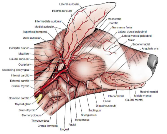
Arterial supply to the head
leaves the aorta through the brachiocephalic and then the left and right common
carotid arteries.
The common carotid arteries terminate near the body of the hyoid bone by dividing into the internal and external carotid arteries
The internal carotid artery is smaller and runs through the deep structures of the head and brain.
The external carotid is the main continuation of the common carotid and supply either half of the head:
Its branches consists of; occipital, cranial laryngeal, ascending pharyngeal, lingual, facial, caudal auricular, parotid, superficial temporal and maxillary arteries.
The two terminating branches of the external carotid: a. Superficial temporal artery (smaller)
the branches of the artery are; the messeteric, transverse facial, rostral auricular, temporal, superior and inferior palpebral arteries.
The two terminating branches of the external carotid: b. Maxillary artery (larger)
gives branches that supply the deep structures of the head lying outside of the brain. It is considered the main continuation of the external carotid.
Can be divided intro three portions:; mandibular:, pterygoid: and Pterygopalatine:
Anatomy of the arterial supply to the head
Anatomy of the venous drainage of the head

Arterial and venous drainage of the parotid salivary gland
The main blood supply to the gland is via the parotid artery (blue) a branch of the external carotid.
Minor branches are sent to the gland by the caudal auricular, masseter, transverse facial and rostral auricular arteries.
The primary veins draining the gland are radicles of the superficial temporal (green) and great auricular veins
Anatomy of Arterial and venous drainage of the parotid salivary gland

Arterial and Venous Drainage of the Mandibular Salivary Gland
The largest artery supplying the gland is the facial artery (red) and smaller branches from the caudal auricular artery (green)
The chief vein draining the gland terminates in the lingual vein
(yellow) which then drains into the external maxillary vein. A smaller vein also drains the gland an terminates either in the facial, maxillary or lingual vein.
Anatomy of Arterial and Venous Drainage of the Mandibular Salivary Gland

Arterial and Venous Drainage of the Sublingual Salivary Gland
The main supply to the monostatic potion is via the glandular branch of the facial artery and the polystomatic portion the sublingual artery
(grey) a branch of the lingual artery
The satellite veins of the arteries drain the gland
Anatomy of Arterial and Venous Drainage of the Sublingual Salivary Gland

Rate of Saliva Secretion
horse produce ~50L of saliva daily.
The pig produces ~15 L/day
The cow produces ~ 100-200 L/day: flow rate increases during rumination and helps to buffer the fatty acids that are produced