MCB 170 Midterm 2
 Call Kai
 Learn
 Practice Test
 Spaced Repetition
 Match
 Flashcards
 Knowt Play
1/53

There's no tags or description
Looks like no tags are added yet.
54 Terms
electrophysiology
study of electrical activity in neurons
use electrodes
by nearby neurons or inside them
extracellular/intracellular
voltage clamp
Cole, Hodgkin, Huxley - studied w/ squids because you can see their axon
cells held at certain membrane potential and measure current through voltage-gated ion channels
study how action potential is impacted by ion flow/currents and if currents depend on voltage/membrane potential
on segment of axon
reference electrode outside
recording electrode inside
current-electrode sets membrane potential
Steps of voltage clamp
depolarize axon to set membrane potential
voltage-gated sodium channels open —> Na+ entering cell
equal but opposite charge (aka - ) added to keep at 0mV
voltage-gated Na channels inactivate as sodium ones open —> K+ entering cell
equal but opposite charge (aka + ) added to keep at 0 mV
electroencephalogram (EEG)
measure electrodes on surface of cell to pick on activity of multiple neurons
less invasice
study sleep
patch clamping
measure electrical current by ion channels by clamping onto the membrane/channels
local field potential (LFP)
measure electrodes directly in brain & tissue to pick of activity of multiple neurons
more invasive
can pick up dendritic activity
staining & microscopy
preserve and cut tissue
use stain to visualize cells
formalin (FFPE): used to cut tissue
golgi stain
staining tech. using Ag
detailed, but only look at a small subset of neurons
nissl stain
staining technique using cresyl violet
binds to nucleic aids and ribsomal DNA to examine the cell body
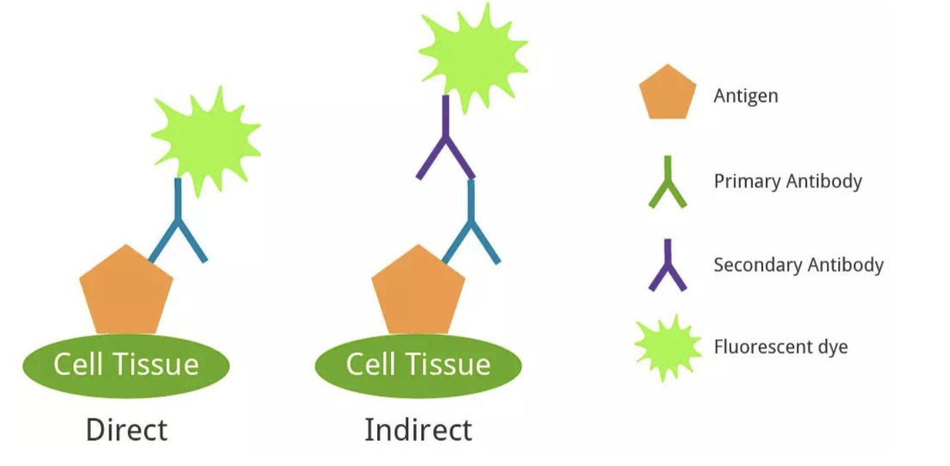
immunohistochemistry
primary antibody directed onto antigen on cell of interest
secondary binds to primary
tagged w/ fluorescent dye for visualization
strengthens signal
electronmicroscopy (EM)
view thin sections of tissue - ultra-structure
view w/ fine detail & resolution
view molecular structure (ex. myelinating axons)
connectomics
determine structure of NS by mapping neural connections
neuroatomical track-tracing techniques
antereograde: cell body —> axon
retrograde: axon —> cell body
fluorescent + immunohistochemistry + EM
see what neurons lead to which one
harder to see circuits

viruses and tracing techniques
infect neurons and spread to other neurons
question of toxicity of viruses (need to find safe ones)
use antero and retro directionality
more specific than conventional tracing (can cause false positives)
viral vectors + immuno & fluorescent proteins to help visualize
model organisms
compare effects w/o worrying about ethics
ex. mice
model organisms and studying neurons/glia
use fluorescent proteins (light shined will have light shine back)
genetic engineering in cells of interest
GCaMP
engineered Ca2+ indicator
fluorescent light glows when Ca2+ binds, showing neuronal activity
in vivo
testing within live organsim
in vitro
in glass/dish cell culture allows for study cellular environment outside of the organism
can manipulate one cell type and see how it responds to drugs, chem. cues, etc.
can harvest DNA, RNA. proteins
see change in gene expression/proteins
see genes involved in growth & injury
glia also isolated (microglia/astrocytes)
embryonic origin of NS
formed by ectodermal layer germ layer
formation of CNS from ectodermal layer
folding of neural tube —> vesicles forming regions of the brain
neural plate neuralizes first
know order

formation of PNS from ectodermal layer
neural crest folds to from the neural plate
neural crest cells are derived from neural fold
migrate to periphery to create other cells (peripheral neurons & Schwann cells)

radial glial cells
parents cells (progenitors) for other cell types (neurons & glia)
neurons 1st, then glia
neuronal migration
raidal glial cells provide scaffold of newborns to migrate
neural precursors (cells that become neurons) migrate along radial glial cells
start at ventricular zone and migrate up
process, trailing, migrating, leading formation
symmetrical cell division
one parent radial glial cell = two daughter radial glial cells
asymmetrical cell division
one parent radial glial cell = one neural precursor daughter cell (which will become
neuron or glia) + one daughter radial glial cell
techniques to study radial glial cells
fluorescent protein (GFP)
viruses
video microscopy to study w/ timelapsing
order layers of neurons in the cerebral cortex form
neurons migrate from deepest layer —> top (inside —> out)
way neurons know where to go
axons sense neurotrophic factors: guidance cues and growth factors
bind to specific receptors on growth cone: growing tip of axon/dendrite
no receptor = grows towards nowhere
cause cytoskeleton to remodel to grow towards/away (attracted/repulsed)
goal: to go to muscle, organ, skin, other neurons
neurite: growing extension of neuron that becomes dendrite/axon
use Ca2+ indicator dye to show how calcium is important for signaling movement of growth cone and axon
motor neurons traveling long distance to find target
cross over midline from one side of cell body to another at the pyramidal decussation in corticospinal track
require series of repulsive/attractive gradient cues to guide it
target secretes factors promoting growth of axons towards them
take foreverrrrrr tho ;-;
polarity by guidance cues
semaphorin 3A is an example of guidance cue protein
determines polarity
basil end (low S-3A; repulsed); apical end (high S-3A; attracted)
how do synapses form
when the axon reaches the target!
formation of motor neuron synpases @ neuromuscular junctions
motor neuron axon secretes factors (Agrin?) to receive ACh
sets up post-synaptic side
action potential —> synapse —> ACh release
muscle fibers open so Na+ enters muscle fiber
EPSP so muscle contracts
pruning
form of neuroplasicity occurring when too many synapses made in development
made redundantly for functionality (in case something goes wrong)
synapse elimination at neuromusclar junction (PNS)
before: muscle fibers receive input from multiple motor neurons
after:
AChR blockage in small area (toxin) causes synapse to be eliminated, so neuron retracks
one neuron per fiber (one neuron can connect to multiple fibers though)
determined by activity: stronger input causes specific neuron to be kept
loss of all input = death
synapse elimination in CNS
neuron-neuron connection
fliopodium - portion of neuron - contracts axon
vesicle recruits on pre-synaptic side
postsynaptic NT receptors form clusters
microglia do pruning by engulfing the synapse
trophic support
provide nourishment to target tissue by secretion by other tissues
lowered strength connections means lowered trophic support so the cell dies
“support” —> tells cell to keep going <3
limited supply means ones more likely to survive get it
what do memory & learning impact?
#plasicity
Aplysia study
studied because of fewer nerves & simple circuits
study gill withdrawal reflect
stimulating siphon —> sensory neuron releases glutamate —> stimulated motor neuron —> withdrawal
habituation: constant weak signals result in no response (no withdrawal and lowered glutamate release)
shocking tail —>sensory neuron serotonin level increase —> glutamate released from 1st circuit —> stimulated motor neuron —> withdrawal
strengthen withdrawal response
sensitization of tail if touched
olidendrocytes and response to injury (CNS)
demyelination of myelin changes action potential propagation
can lead to Multiple Sclerosis (MS)
olidendrocyte precursor cells (OPCs): stimulated to promote remyelination following injury
make into olidendrocytes
MS - inflammatory environment makes inhibits OPC from becoming olidendrocytes
astrocytes and response to injury (CNS)
become reactive following injury
GFAP gene expression increases
astroglyosis: proliferate/divide and create scars
inc. immune cell infiltration & BBB maintenance/repair
coral peripheral immune cells/amplify inflammatory response
microglia and response to injury (CNS)
typical function: reg. neuronal activity, synaptic plasticity,, BBB maintenance
after injury:
large-scale cytokine prod. (tell cells to move/act + coord. immune response)
recruit peripheral immune cells
pathogen destruction
debris clearence
tissue repair
microglia changes shape
neurovascular unit and response to injury (CNS)
after traumatic brain injury (can lead to neurodegen. disorders if constant)
reactive astroglosis
neuronal death
olidendrocyte dysfunction
microglia activates
pro-inflammatory cytokines
damage can be caused by peripheral vascular problems (can lead to dementia)
hypotension, high BP as cell walls lose plasicity
BBB breakdown w/ natural aging
PNS nerve regeneration
by Schwann cells, immune fibroblasts (mesenchymal) ,macrophages
Schwann cells migrate along vasculature, taking axon to target as it grows along track made by it
repair Schwann cells help axon regrow to debris
everything past injury is GONE
Bands of Bünger: forms new bridge for axon to grow w/ factors to help w/ regrowth
CNS nerve regeneration
inhibitory to axon regeneration
CNS molecules inhibiting its growth
ex. astrocytes secreting extracellular matrices telling it not to grow
no macrophages to clean debris