3.1.1. exchange surfaces
 Call Kai
 Learn
 Practice Test
 Spaced Repetition
 Match
 Flashcards
 Knowt Play
1/41

There's no tags or description
Looks like no tags are added yet.
42 Terms
what is the difference between sa; v ratio and metabolism in small and big animals
Small and single–celled organisms have:
High surface area to volume (SA:V) ratio – can rely on diffusion to obtain oxygen and nutrients and remove wastes.
Low metabolic activity – requirements for oxygen and nutrients are lower and production of wasteis slower.
Large organisms have:
Low SA:V ratios – diffusion cannot provide the organism with enough oxygen and nutrients.
Higher metabolic activity – higher requirements for oxygen and nutrients and faster production of toxic waste.
Larger organisms require specialised exchange surfaces to meet their oxygen, nutrient and wasteremoval needs.
Efficient exchange surfaces have the following features to increase the rate of diffusion:
high surface area
thin layers
maintaining a diffusion gradient
selectively permeable.
The nasal cavity carries out the following functions:
Warms the air. A good blood supply and large surface area, ensure the air entering the lungs is a similar temperature to the air already present.
The nasal cavity moistens inhaled air to prevent the respiratory system from drying out and losing too much water.
Traps dust and pathogens. The lining of the nasal cavity is hairy and secretes mucus, which prevents irritation and infection in the respiratory system.

how is the trachea adapted to its function
he trachea (windpipe) connects the mouth to the bronchi.
The trachea is lined with incomplete rings of strong, flexible cartilage which maintains its shape and prevents collapse.
The trachea has elasticfibres, which allow it to stretch and recoil during ventilation.
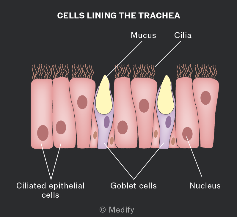
The cells lining the trachea are mostly ciliated epithelial cells, with goblet cells in between.
Goblet cells produce mucus, which trap dirt and microorganisms.
The ciliated epithelial cells waft and move mucus away from the lungs and towards the throat, where it is swallowed.
what ar e some adaptations of the bronchi
The trachea branches into two smaller bronchi, which lead to each lung. The bronchi have a similarfunction to the trachea and also contain supporting rings of cartilage. The bronchi then branch into bronchioles.
The smaller bronchioles do not have cartilage rings and contain smooth muscle.
The smooth musclecan contract and relax, controlling the air reaching the lungs. Bronchioles are lined with a thin layer of flattened epithelial cells. Due to the thin layer of cells, a small amount of gas exchange can happen in the bronchioles.
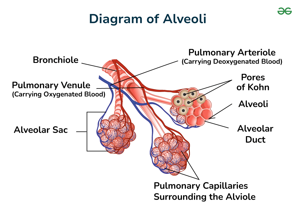
The branches of the small bronchioles end at the alveoli. The alveoli are tiny air sacs in the lungs where the majority of gas exchange takes place.

The alveoli have many adaptations:
Lined with elastic tissue, allowing them to stretch and recoil during ventilation.
Large surface area for increased rate of diffusion.
Good ventilation and blood supply maintain a steep concentration gradient, which increases the rate of diffusion.
Surfactant makes the surface of the alveoli moist, which aids diffusion.
Each alveoli is lined with flattened epithelial cells,(are cells that cover the surfaces of the body and line internal organs and cavities.)
and the capillaries are lined with a thin layer of endothelial cells( thin, flat cells that line the inside of blood vessels.) . This makes the distance required for diffusion short, increasing the rate of gas exchange.
The factors that affect the rate of diffusion can be combined into an equation called Fick’s law to determine:
rate of diffusion = surface area x difference in conc
/ thickness of exchange surface
what are the components of the respiratory system, also the function
The respiratory system carries out the process of ventilation:
Ribs: bones which surround and protect the lungs and heart and move during ventilation.
Diaphragm: a dome-shaped sheet of muscle which contracts and relaxes to facilitate breathing.
External intercostal muscles: contract during inhalation and move the ribs up and outwards.
Internal intercostal muscles: contract only during forced expiration.
Pleural membranes: surround the lungs and make pleural fluid.
Pleural fluid: reduces friction during ventilation when the pressure in the lungs changes.
what happens during inspiration
During inspiration:
The diaphragm contracts and flattens.
The external intercostal muscles contract and pull the rib cage up and out.
The volume in the thorax increases, and the pressure decreases.
Air moves into the lungs.
what happens during expiration
During expiration:
The diaphragm relaxes and moves upward into relaxed dome shape
The external intercostal muscles relax and the rib cage moves down and in.
The volume in the thorax decreases, and the pressure increases.
Air moves out of the lungs.
During forced expiration, the internal intercostal muscles will also contract, forcing air out of the lungsquickly.
what is a spirometry used to investigate, why’s this usefu, how does it work
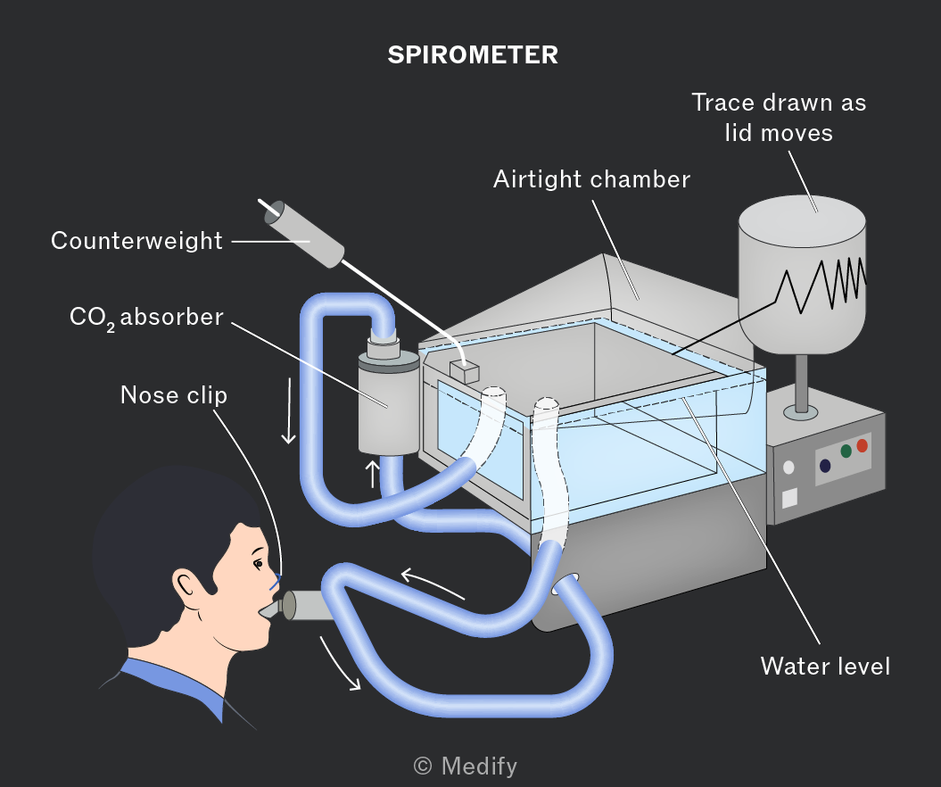
Spirometry is used to investigate breathing patterns and lung volume. This can be used to help diagnose lung diseases such as COPD and asthma.
A spirometer measures the changes in the volume of air in the lungs during inhalation and expirationand produces a trace.
The soda lime absorbs carbon dioxide to prevent toxic buildup.
The nose clip prevents a person from breathing out of their nose, as this would make the trace inaccurate.
what does tidal volume show
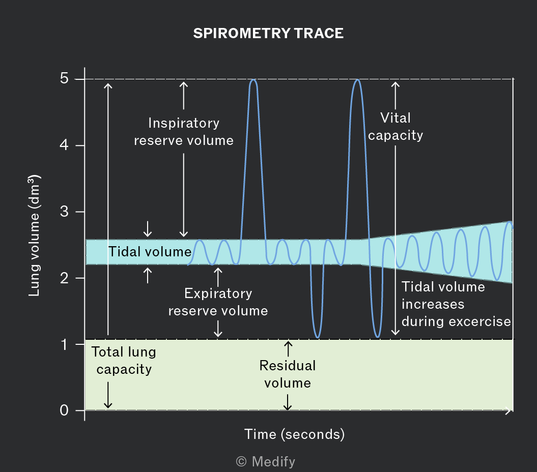
Tidal volume: volume of air which moves in and out of the lungs during ventilation at rest. Tidal volume is about 0.5 for most adults.

what is the inspiratory reserve volume
Inspiratory reserve volume: the volume of air that can be inhaled during forced inhalation. This is the volume above the normal inhalation.
what does the expiratory reserve volume show
Expiratory reserve volume: the volume of air that can be exhaled during forced expiration. This is the volume above the normal expiration.
what is the vital capacity
Vital capacity: is the maximum volume of air a person can breathe out after taking the deepest breath possible.
what is residual volume
Residual volume: the volume of air left in the lungs after a forced expiration. This cannot be directly measured.
what is the total lung capacity
Total lung capacity: the total possible air in the lungs during a forced inhalation. This includes vital capacity and residual volume.
what is ventilation, formula
Ventilation rate is the total volume of air inhaled in one minute. It can be calculated from a spirometry trace by multiplying the tidal volume by the number of breaths per minute.
ventilation rate = tidal volume x breathing rate
why do insects require a gas exchange system
Insects require a gas exchange system because they:
Do not have oxygen–carrying pigments, such as haemoglobin, to transport oxygen.
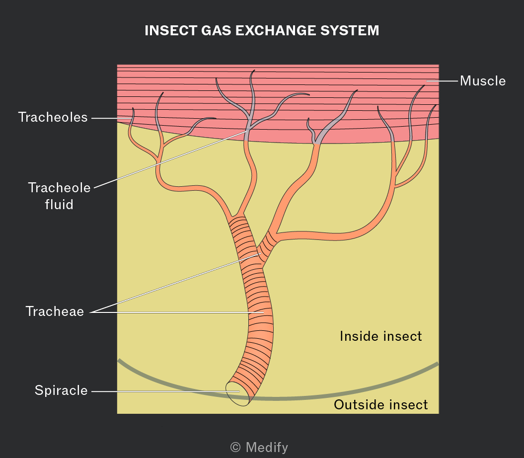
Have exoskeletons made of chitin, which are impermeable so gases are not able to diffuse through.
Do not have a large enough SA:V ratio to rely on diffusion alone.
what is chitin, where is it located in an insect
Chitin is a tough, impermeable polysaccharide that makes up insect exoskeletons.
The tracheae are lined with rings of chitin to keep them open and flexible. The tracheoles do not have rings of chitin.
Explain how insects control gas exchange and increase the efficiency of ventilation.
Insects have sphincters around the openings of their tracheae, which open and close.
This prevents water loss through evaporation from the tracheal fluid.
The sphincters respond to changes in oxygen and carbon dioxide levels.
Some insects use mechanical ventilation by moving their thorax or abdomen to move air through the tracheal system.
Air sacs and enlarged tracheae increase ventilation efficiency.
why do fish need specialised exchange surfaces
Fish have low SA:V ratios, so require specialised exchange surfaces to absorb oxygen and excrete carbon dioxide.
Being aquatic, fish do not need to prevent water loss from their exchange surfaces.
why do fish not require water to move mechanically in and out
Water is more viscous than air and has a lower oxygen content.
Due to its viscosity, moving water mechanically in and out of the body would take too much energy . Fish have developed gills as their specialised exchange surfaces.
what’s the structure of the gills
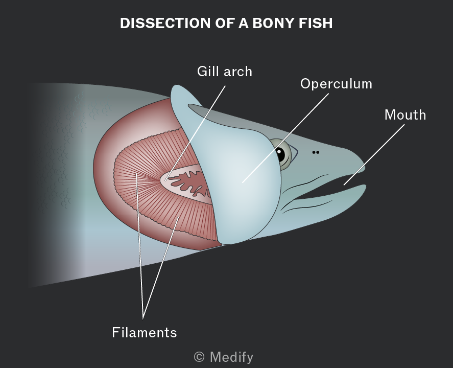
Gills have the following structures:
Operculum: flap covering and protecting the gills.
Opercular valve: controls the opening and closing of the operculum.
Gill arch: support the gill filaments.
Gill lamellae: thin structures where exchange of oxygen and carbon dioxide occurs.
Afferent blood vessel: supplies blood to the gills.
Efferent blood vessel: carries the blood from the gills to the body. (a before e)

how are the gills adapted to diffusion
The gills are adapted for diffusion:
Large surface area.
Good blood flow to maintain the diffusion gradient.
The lamellae have thin walls, only one cell thick, which creates a short diffusion distance for oxygen to diffuse into the blood.
Water flows in one direction over the gills. This leads to a countercurrent system between the water and blood flow in the gills, which increases the rate of diffusion.

Many fish have to keep moving for water to flow over their gills.
Some fish species are adapted to use their buccal cavity and operculum to mechanically move water over their gills when not swimming:
goal; low pressure for water to move in
The mouth opens with the opercular valve closed. This increases the volume of the buccal cavity.
This lowers the pressure, and water moves in and over the gills.
goal ; high pressure so water moves out
The mouth closes and the opercular valve opens (this is to let the water out) This decreases the volume of the buccal cavity.
This increases the pressure and water moves out through the opercular valve.