Osteology and Arthrology of Upper Extermities #7
 Call Kai
 Learn
Learn Practice Test
 Spaced Repetition
 Match
Match Flashcards
 Knowt Play
1/123

There's no tags or description
Looks like no tags are added yet.
Name | Mastery | Learn | Test | Matching | Spaced | Call with Kai | Chat |
|---|
No analytics yet
Send a link to your students to track their progress
124 Terms
1. Shoulder
2. Arm
3. Forearm
4. Hand
What are the Upper Exteremities anatomical divisions? (4)
1. Scapula
2. Clavicle
4. Manubrium
The shoulder overlies half of the pectoral girdle which is formed by what 3 bones?
1. Pectoral (anterior)
2. Scapular (posterior
3. Deltoid (lateral)
What parts of the upper limbs are included in the shoulder
connects the shoulder and the elbow
The Arm is the 1st segment and longest part of the free upper limb. What does it extend between and connect?
Anterior region of arm
posterior region of arm
What are the parts of the Arm (2)
Extends between and connects the elbow and the wrist
The forearm is the 2nd segment and second largest portion of the upper limb. What does it extend between and connect?
Anterior region of forearm
posterior region of forearm
What are the parts of the forearm? (2)
Hand
1. Wrist
2. Palam
3. Dorsum of hand
4. Digits
Wha the most distal upper extremitie? What are the parts?
cervico-axillary canal (passage between neck and arm)
The clavicle forms one of the boundaries of the _____________ together with the scapula and the 1st rib. It protects the neurovasuclar bundle supplying the arm.
Axial skeleton
The clavice transmits shocks from the Upper extermities to the
1. Medial half is convex anteriorly, and lateral half concave anteriorly
2. Sternal end: articulates with manubrium (sternoclavicular joint)
3. Acromial end: articulates with acromion (acromioclavicular joint)
The shaft of the clavicle is double curved and in the horizontal plane, which increases resilience. What are the 3 parts of the shaft of the clavicle?

1. Conoid tubercle: near acromial end of clavicle, support for the medial part of the coracoclavicular ligament
2. Trapezoid line: nearer to the acromial end than the conoid tubercle, support for the lateral part of the coracoclavicular ligament.
The inferior surface of the clavicle is a rough surface with ligaments binding to it. What two processes are found there and what joints attach to them?

Inflection point
medial 2/3 (displaced superiorly by SCM muscle)
lateral 1/3 (displaced inferiorly)
CLINCAL RELIVANCE
Fracture of the clavicle
The __________ is the weakest point of the clavicle and is more prone to fractures. This is a junction between the medial ____ and lateral ______. Which junction is displaces supererioly and which is displaced inferiorly?

Triangular flat bone
What is the shape of the scapula?
posterior part of the thorax, overlying 2nd - 7th ribs
Where is the location of the scapula?
1. humerus (glenohumerical joint) and 2. thoracic wall (physiological scapula-thoracic joint)
What does the scapula articulate with? (2)
1. Posterior
2. Costal (anterior)
3. Lateral
What are the surfaces of the scapula? (3)
1. Spine of scapula: ridge of bone that divides posterior surface in two fossae
2. Acromion: lateral continuation of the spine
3. Supraspinous fossa: above the spine of the scapula
4. Infraspinous fossa: inferior to the spine of the scapula
The posterior surface of the scapula is convex. What (4) features are posterior?

Subscapular fossa: occupies most of the costal surface
The costal (anterior) surface of the scapula is concave and contains what feature (1)?

1. Glenoid cavity: located superolaterally receives and articulates with the head of the humerus (glenohumeral joint)
2. Coracoid process: superior to the glenoid cavity, projects anterolaterally
3. Head of scapula: contains the glenoid cavity
4. Neck of scapula: between the head and body of scapula
5. Suprascapular notch: junction of the superior border with the base of the coracoid process. This notch is converted into a foramen by the superior transverse ligament, and serves for the passage of the suprascapular nerve
The lateral surface of the scapula has what 5 features?
1. Superior: near the suprascapular notch
2. Lateral: near the humerus
3. Medial: runs parallel to spinous processes of the vertebrae
What are the borders of the scapula? (3)

1. Superior: union of superior and medial borders
2. Inferior: union of medial and lateral borders
3. Lateral: contains the head of the scapula
What are the angles of the scapula? (3)

Articulates: scapula (glenohumeral joint) and radius and ulna (elbow joint)
What does the humerus articulate with?
1. Head: articulates with glenoid cavity of scapula
2. Anatomical neck: groove separating the head from the greater and lesser tubercles. Indicates the place of attachment of the glenohumeral joint capsule
3. Surgical neck: narrow part distal to head and tubercles. Common site of fracture
4. Greater tubercle: lateral position from head
5. Lesser tubercle: anterior position from the head
6. Intertubercular groove: located between tubercles and provides passage for the tendon of the long head of the biceps muscle
What are the features of the humerus at the superior part? (6)

Axillary Nerve may become damaged
CLINCIAL RELIVANCE
The fracture of the surgical neck of the humerus may damage what because it wraps around it? The Integrity of this nerve is tested by touching the deltoid, since the nerve provides sensory innervation to this area of the shoulder region.

1. Shaft: long portion of the humerus with a tuberosity and a groove, engrossing distally
2. Deltoid tuberosity: attachment for deltoid muscle
3. Radial groove: oblique demarcation on posterior side. Contains the radial nerve and deep artery of arm
4. Medial and lateral supra-epicondylar ridges: end distally as the medial and lateral epicondyles, involved in muscle attachment
5. Condyle: distal end of humerus that includes the trochlea, capitulum, olecranon, coronoid and radial fossa
6. Capitulum: lateral articular surface. Articulate with head of radius
7. Trochlea: medial articular surface. Articulate with the ulna
8. Coronoid fossa: anteriorly located, receives coronoid process of ulna during flexion of elbow
9. Olecranon fossa: posteriorly located, receives the olecranon of ulna during extension
What are the features of the humerus at the inferior part? (10)

Medial epicondyle
Lateral eipcondyle
Flexor muscles attach to the ______ while extensor muscles attach to the _____ of the humerus.
Radial nerve and deep artery of arm
The radial groove is a oblique demarcation on the posterior side that contains the _______
medial and lateral epicondyles, involved in muscle attachment
The Medial and lateral supra-epicondylar ridges of the humerus end distally as what?
the trochlea, capitulum, olecranon, coronoid and radial fossa
The condyle of the humerus is the distal end that includes what fossa?
Lateral
Head of radius
The capitulum is the ______ articular surface of the humerus and articulates with what?

Medial
Ulna
The trochlea is the ______ articular surface that articulates with the ______

coronoid process of ulna
The coronoid fossa is located anteriorly and receives the ________during flexion of the elbow

Olecranon of ulna
The olecranon fossa is posteriorly located and receives the ______ during extension

Radius (distal)
Ulna (medial)
What are the two bones of the forearm?
Radius pivots on the ulna
What bone pivots on the other due to their parallel position? (Supination and pronation)
proximal end (more bulky) with humerus and head of radius (Wide to narrow). Does NOT form wrist distally
What does the Ulna articulate with?

Articulates with olecranon fossa of humerus and serves as a short lever for extension of the elbow
The olecranon of the ulna is the superior most proximal portion. what does it articulate with?

coronoid fossa of humerus on its anterior side
The coronoid process is in inferior to the olecranon process. what does it articulate with

brachialis muscle
The ulnar tuberosity is attachment for what muscle?

Head of the radius
The radial notch is infero-lateral to the coronoid process and receives what of the radius?

thinner
The shaft of the ulna gets _____ distally
enlargement on distal part of the shaft
The head of the ulna is a
head of the ulna
The ulnar styloid process is the distal ending of the
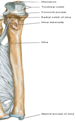
1 Olecranon: superior, most proximal portion. Articulates with olecranon fossa of humerus and serves as a short lever for extension of the elbow
2 Coronoid process: inferior to olecranon process. Articulates with coronoid fossa of humerus on its anterior side
3 Ulnar tuberosity: attachment for the brachialis muscle
4 Radial notch: infero-lateral to the coronoid process, receives the head of the radius
5 Shaft: gets thinner distally
6 Head of ulna: enlargement on distal part of the shaft
7 Ulnar styloid process: distal ending of the head of the ulna
What are the parts of the ulna (7)

ulna (proximally and distally). Carpal bones: on the distal side of radius. Radius forms the wrist joint and articulates with the carpal bones
What does the radius articulate with
1 Head: articulates with the capitulum of humerus and with radial notch of ulna
2 Neck: inferior to head
3 Shaft: gets thicker distally, opposite to ulna
4 Ulnar notch: articulates with the head of the ulna
5 Radial styloid process: distal ending on lateral side of the shaft of radius
What are the parts of the Radius (5)

articulates with the capitulum of humerus and with radial notch of ulna
The head of the radius articulates with what (2)

inferior to the head
The neck of the radius is where?

gets thicker distally, opposite to ulna
the shaft of the radius
the head of the ulna
The ulnar notch articulates with what
distal ending on lateral side of the shaft of radius
The radial styloid process is where?

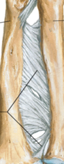
thin fibrous membrane, with oblique fibers running inferiorly and medially from the radius to the ulna
What kind of membrane is the Interosseous membrane How does it run?
Scaphoid: largest bone in the proximal row, articulates with radius
Lunate: moon-shaped, articulates with radius
Triquetrum: pyramidal bone, articulates with the articular disk of the distal radio-ulnar joint
Pisiform: small bone located on the palmar surface of the triquetrum
What are the proximal row of carpal bones? (lateral to medial)
"So Long To Pinky, Here Comes The Thumb"

Scaphoid: largest bone in the proximal row, articulates with radius
Lunate: moon-shaped, articulates with radius
What two proximal carpal bones articulate with the radius?
Trapezium: on lateral side, articulates with 1st and 2nd metacarpals
Trapezoid: articulates with 2nd metacarpal
Capitate: largest bone of carpus (wrist), articulates with 3rd metacarpal
Hamate: articulates with 4th and 5th metacarpal. Hook of the hamate is a remarkable feature of this bone extending anteriorly
What are the distal row carpal bones? (lateral to medial)

Carpal bones phalanges
The metacarpal bones are between what?

Thumb its the thickest and shortest
What is the first metacarpal bone?
Base: proximal part, articulates with carpal bones
Shaft: middle part
Head: Distal part articulates with proximal phalanges
What are the parts of the metacarpal bones?

proximal
middle
distal
***All have proximal, middle, and distal phalanges, except the thumb (1st digit)
What are the three parts of the phalanges?

Sternoclavicular joints
What joint is the only articulation between the axial skeleton and the upper extermities?
act as a strut for keeping the shoulder away from the thorax to give the UL the maximum freedom of motion
What is the function of the sternoclavicular joint?
saddle (type of synovial joint)
What type of joint are Sternoclavicular joints?
Clavicle with manubrium of sternum and medial part of the 1st costal cartilage
What does the Sternoclavicular joints articulate with?
1. Anterior and posterior sternoclavicular ligaments
2. Interclavicular ligament
3. Costoclavicular ligament
What are the ligaments of the Sternoclavicular joints
reinforce the joint capsule anteriorly and posteriorly.
Intrinsic ligaments
What do Anterior and posterior sternoclavicular ligaments reinforce? What kind of ligament are they?

strengthen joint capsule superiorly, runs between the sternal ends of clavicles. Extrinsic ligament
What do Interclavicular ligaments strengthen the joint capsule and runs where? what type of ligament are they?

between inferior surface of sternal end of clavicle to 1st ribs and costal cartilage.
Extrinsic ligament that limits elevation of the pectoral girdle
Where is the Costoclavicular ligament between? what type of ligament is it

60 deg
the Sternoclavicular joints allows movements of elevation and drepession. In full Elevation of the upper limb the clavicle is raised at what angle?
25-30 deg
the Sternoclavicular joints allows movements of Protraction and retraction which moves anterioly and posteroily at what angle?
Plane type of synovial joint, with articular disc dividing the capsule
The Acromioclavicular joint is what kind of synovial joint?
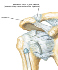
acromial end of clavicle and acromion of scapula
What does the Acromioclavicular joint articulate with?
1. Acromioclavicular ligament
2. Coracoclaviclar ligament
- conoid ligament
- Trapezoid ligament
What are the ligaments of the Acromioclavicular joint?

strengthen capsule superiorly
(intrinsic ligament)
What does the Acromioclavicular ligament strengthen? what type of ligament is it?

strong pair of bands joining the coracoid process to the clavicle. This is a major reinforcement to this joint (extrinsic ligament)
- Conoid ligament: medially
- Trapezoid ligament: laterally
What is the Coracoclavicular ligament? what does it join on the clavicle? what type of ligament is it?

the acromion process, rather than the greater tubercle of the humerus, becomes the most lateral bony structure
CLICNAL RELIVANCE
Dislocation of acromioclavicular joint
A dislocated shoulder loses its normal contour what becomes the most lateral bony structure?

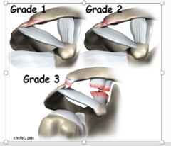
Grade 1: sprain of the intrinsic ligament (acromioclavicular ligament)
Grade 2: tear of the A-C ligament and sprain of the coracoclavicular ligament (tear in medial ligament)
Grade 3 - 4 complete tear of the acromioclavicular ligament and the coracoclavicular ligament
What are the different grades of dislocation?
ball and socket (type of synovial joint)
What type of joint is the Glenohumeral joint?
Head of humerus and glenoid cavity of scapula
(deepened by the glenoid labrum; the cavity only receives less than 1/3rd of the head of humerus)
What does the Glenohumeral joint articulate with

Capsule reinforced by the rotator cuff muscles
(except in the inferior portion, which is the weakest part of capsule)
What is the joint capsule of the Glenohumeral joint reinforced by?

1. Glenohumeral ligament
2. Coracohumeral ligament
3. Transverse humeral ligament
4. Coracoacromial ligament
5. Rotator cuff muscles
What are the ligaments of the glenohumeral joint? (5)

reinforces anterior part of capsule (intrinsic ligament)
What does the Glenohumeral ligament reinforce? What type of ligament is it?

from coracoid process to anterior aspect of humerus
(intrinsic ligament)
The Coracohumeral ligament attaches where? what type of ligament is it?

between greater and lesser tubercle of humerus and holds the tendon of the long head of the biceps brachii muscle in the intertubercular groove
Where is the Transverse humeral ligament?

from coracoid process to acromion. In a superior thrust of the arm, this ligament prevents major damage (extrinsic ligament)
Where is the Coracoacromial ligament? What does this ligament prevent and what type is it?

Glenohumeral joint
aids in the stability of the joint. Maintain head of humerus in glenoid cavity
What joint are rotator cuff muscles in? What do they aid in?
Superiorly: Supraspinatus
Posteriorly : Teres minor and Infraspinatus
Anteriorly: Subscapularis
What are the rotator cuff muscles?

extension, abduction, adduction, rotation (medial and lateral), and circumduction (multi-axial)
What are the movements associated with the Glenohumeral joint?
Subscapular bursa
Subacromial bursa
What 2 bursas are in the Glenohumeral joint?
located between the tendon of subscapularis muscle and neck of scapula.
Protects the tendon
Where is the Subscapular bursa of the Glenohumeral joint located? What does it protect?

located between the acromion and the tendon of the supraspinatus muscle. Protects the tendon
Where is the Subacromial bursa of the Glenohumeral joint located? What does it protect?

Hinge type (flexion and extension) of synovial joint
What type of joint is the Elbow joint?
trochlea and trochlear notch (ulnar-humeral joint) & capitulum and radius (radio-humeral joint)
What does the elbow joint articulate with?
1. Ulnar collateral ligament
2. Radial collateral ligament
3. Annular ligament
What are the ligaments associated with the elbow joint?

located medially and extends from the medial epicondyle of humerus to coronoid process and olecranon of ulna.
Flexor muscles are going to attach to the medial epicondyle
extensor muscles attach to the lateral epicondyle
Where is the ulnar collateral ligament located? What types of muscles attach here?

located laterally and extends from lateral epicondyle to the annular ligament
Where is the radial collateral ligament located

encircles the radial head holding it against the radial notch of the ulna, allowing pronation and supination of the arm. Lifting a kid by the forearm can dislocate this ligament
what does the annular ligament encircle? What movement does it allow?

distal attachment of the annular ligament (around neck of radius)
CLINCAL RELIVANCE
Dislocation of radial head (annular ligament)
Sudden pulling of the upper limb tears what?

Pivot type of synovial joint
pronation and supination. It is the radius who rotates on the ulna
What type of joint is proximal radio-ulnar joint? What movements does it allow?
head of radius and radial notch of ulna
What does the Proximal radio-ulnar joint articulate with?