622 11/2 L pt 3 Immune System and Innate Immunity
 Call Kai
 Learn
 Practice Test
 Spaced Repetition
 Match
 Flashcards
 Knowt Play
1/57

There's no tags or description
Looks like no tags are added yet.
58 Terms
Cells of Innate Immunity
neutrophils (PMNs)
monocytes
macrophages
dendritic cells
mast cells
basophils
eosinophils
natural killer cells
Monocytes
immature circulating macrophages, phagocytosis, leave blood by extravasation when inflammation occurs, differentiate into macrophages or dendritic cells
Monocytes on Wright Stain
large indented or curved purple nucleus, pale blue cytoplasm

Macrophages
tissue resident, longer-lived than monocytes, patrol by crawling, phagocytosis
express PRRs, Fc receptors, and complement receptors
secrete cytokines that regulate inflammation and wound healing, antigen presentation to T cells
Classic Activation of Macrophages
M1 macrophages, induced by PRRs or the cytokine IFN-y, trigger inflammation
fight infections, destroy microbes by phagocytosis
Alternative Activation of Macrophages
M2 macrophages, induced by cytokines IL-4 and IL-13, suppress inflammation
tissue repair, eliminate debris by phagocytosis
Dendritic Cells (DCs)
have numerous cytoplasmic tendrils (dendrites), antigen presentation to lymphocytes, 3 types
3 Types of Dendritic Cells
convetional
phagocytoid
follicular
Conventional Dendritic Cells
most abundant, phagocytosis, antigen presentation to T cells, secrete cytokines that regulate differentiation of T cells
may migrate through lymphatic vessels to secondary lymphoid tissues
in blood, spleen, liver, thymus, MALT gernimal centers, skin (langerhans cells)
Plasmacytoid Dendritic Cells
rarest, secrete antiviral cytokines (type I interferons: IFN-a and IFN-b), poor phagocytosis, antigen presentation to T cells
in blood and secondary lymphoid tissues
Follicular Dendritic Cells
use Fc receptors and complement receptors to trap and display native antigens to B cells without internal processing, no phagocytosis
non-hemtopoietic origin
from B-cell zones of secondary lymphoid tissues (follicles)
Mast Cells
tissue resident (skin, MALT, CT), mediate early effector phase of allergic reactions for type i hypersensitivity, degranulation
immune sentinels, first-line defense, response to helminths
produce chemicals that mediate inflammation
express PRRs, Crs, and FcRs
Chemicals that Mediate Inflammation Released by Mast Cells
degranulation releases stored histamine, heparin, proteases
secrete lipid-derived mediators (platelet-activating factor, prostaglandins, leukotrines)
secrete cytokies and chemokines
Basophils
functionally and phenotypically similar to mast cells but reside in blood, leave blood by extravasion when inflammation occurs, <1% of circulating wbcs
involved in early effector phase of type i hypersensitivity
Basophils Wright Stain
dark, irregular nucleus with numerous dark blue granules

Eosinophils Wright Stain
dark irregular nucleus, numerous red granules (eosin dye)
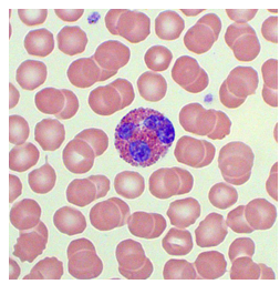
Eosinophils
degranulation and phagocytosis, mediate late effector phase of allergic reactions (type i hypersensitivity), mediate inflammation
present in helminth infections
circulate briefly in blood, mainly found in tissues (skin, MALT)
Eosinophils and Helminths
response to helminths (→ degranulation of cytotoxic proteins that are different from other degranulators)
the problem is that these cytotoxic proteins contribute to tissue damage in chronic inflammation
Natural Killer Cells
kill virus infected cells, some tumor cells, and pathogens
lymphocytes of innate immunity, activation depends on balance between activating signals and inhibitory signals that allows them to determine what is and isn’t a threat
Lymphocytes (B, T, and NK) Cells Wright Stain
smallish cells with large, round, dark purple or blue nucleus and thin rim of pale blue or lavender cytoplasm
cannot differentiate between them on stain

Histology of Innate Immunity Cells

Natural Killer Cells Activating Signals
provided by infected cells, cancer cells, stressed/damaged cells, opsonins
engage activating receptors on NK cells, and trigger activating pathways via ITAMs (immunoreceptor tyrosine-based activation motifs)
Natural Killer Cells Inhibitory Signals
provided by healthy cells (MCH I on all healthy nucleated cells)
engage inhibitory receptors on NK cells, block activating pathways via ITIMs, and prevent NK cells from attacking normal host cell
4 Ways Activated NK Cells Kill Target Cells
secrete IFN-y which activates macrophage (M1) killing
degranulate, releasing perforin (creates holes in target cell’s membrane) and granzymes (enter target through holes and trigger caspase pathway → apoptosis of target)
engage Fas receptor on target cell via Fas ligand on Nk cell → trigger apoptosis of target via caspase pathway
engage antibody opsonin via FcRs → degranulation of NK cell → release of perforin and granzymes → apoptosis of target (antibody-mediated cell-mediated cytocoxiticity ADCC)
Capsase Pathway
culminates in apoptosis, present in nearly all normal nucleated cells
can be triggered by NK activating cells when they release perforin and granzymes or when they engage a Fas receptor
Complement
about 50 proteins produced mainly by hepatocytes but also monocytes, macrophages, and epithelial cells, and fibroblasts complement (or assist) the antimicrobial activity of antibodies
activation involves proteolytic cleavage
Complement Circulation and Activation
proteins circulate in blood and lymphatic fluid in inactive form
acitvation involved proteolytic cleavage, activated complement proteins trigger a chain reaction (complement cascade) amplifying the response
3 Pathways of Complement Activation
classical
alternative
lectin
Classical Pathway of Complement Activation
activated by antigen/antibody complexes
Alternative Pathway of Complement Activation
activated by molecules on microbial surfaces that lack the complement regulatory proteins normally present on host cells
Lectin Pathway of Complement Activation
activated by binding of lectins to carbs on microbial surfaces
4 Basic Functions of Activated Complement
opsonization (enhance phagocytosis)
promote inflammation and chemotaxis of leukocytes
destroy microbes by osmotic lysis (MAC/membrane attack complex)
immune clearance
Immune Clearance
function of activated complement, removal of antigen/antibody complexes and microbes from blood by macrophages in liver and spleen that express matching complement receptors
What do all 3 pathways of complement activation generate and culminate in the formation of?
generate C3 convertase and C5 convertase
culminate in formation of MAC
C3 Convertase (C4b2a or C3bBb)
generated by all 3 complement activation pathways, cleaves C3 into fragments of C3a and C3b
C3a
product of fragmentation of C3 by C3 convertase in complement activation, chemoattractant for neutrophils and monocytes and triggers mast cell degranulation leading to inflammation
C5 Convertase (C4b2a3b or C3bBb3b)
generated by all 3 complement activation pathways, cleaves C5 into C5a and C5b
C3b
product of fragmentation of C3 by C3 convertase in complement activation, an opsonin which enhances phagocytosis (binds to CR1 receptors on macrophages and neutrophils)
involved in immune clearance
C5a
product of fragmentation of C5 by C5 convertase in complement activation, chemoattractant for neutrophils and monocytes and triggers mast cell degranulation leading to inflammation
C5b
product of fragmentation of C5 by C5 convertase in complement activation, initiates formation of membrane attack complex (MAC)
Parts of Membrane Attack Complex
the culmination of all three pathways of complement activation
C5b + C6 + C7 + C8 + C912-18 = C5b678912-18
Membrane Attack Complex Function
inserts onto target’s membrane, creating holes
target dies by osmotic lysis (water in cell makes it explode)
most effective against gram - bacteria (esp neisseria), enveloped viruses, and some parasites
Complement Regulatory Proteins
protect our own cells from complement-mediated damage
most activated complement proteins have a short half-life, preventing activity beyond initial site of activation
3 Complement Regulatory Proteins
c1 inhibitor
decay accelerating factor
cd59
C1 Inhibitor (C1 INH)
complement regulatory protein, soluble protein that inhibits activation of C1 during classical pathway
also separately inhibits production of bradykinin
Bradykinin
powerful trigger of inflammation, production is inhibited by action of C1 inhibitor
Decay Accelerating Factor (DAF)
complement regulatory protein, surface protein that inhibits formation of C3 convertase in all three pathways of complement activation
CD59 (Protectin)
complement regulatory protein, surface protein that inhibits formation of MAC by blocking C9 polymerization
3 Complement Deficiencies
C3 deficiency
hereditary angioedema
paroxysmal nocturnal hemoglobinuria
C3 Deficeincy
complement deficiency, rare autosomal recessive, very low or absent C3
severe recurrent infections, especially encapsulated gram - bacteria (Neisseria and Streptococcus) because MAC struggles to form
C3 Deficeincy Clinical Presentation
causes symptoms resembling systemic lupus erythematosus (SLE) due to failure of immune clearance and deposition of antigen/antibody complexes in blood vessels and kidneys
C3 Deficeincy Prevention
vaccinations and antibiotics reduce risk
Hereditary Angioedema
complement deficiency, rare usually autosomal dominant, C1 INH deficiency leads to elevated bradykinin levels (bc C1 INH usually inhibits bradykinin)
Hereditary Angioedema Clinical Presentation
severe recurrent inflammation of face, limbs, intestines, and airways

Hereditary Angioedema Treatment
bradykinin receptor antagonists, infusions of recombinant C1-INH
Paroxysmal Nocturnal Hemoglobinuria (PNH)
complement deficiency, rare, usually due to spontaneous mutation, defective DAF and CD59
complement-mediated destruction of rbcs by overactive MAC
Paroxysmal Nocturnal Hemoglobinuria Clinical Presentation
dark urine (especially in morning) due to presence of hemoglobin, anemia

Paroxysmal Nocturnal Hemoglobinuria Treatment
infusions of monoclonal antibodies against C5