Musculoskeltal system (health assesment)
 Call Kai
 Learn
 Practice Test
 Spaced Repetition
 Match
 Flashcards
 Knowt Play
1/36

There's no tags or description
Looks like no tags are added yet.
37 Terms
Anatomy
Bones
Joints
Material between joints
Cartilage
Bursa
Supportive structures of the joint
Ligaments
Tendons
Axial Skeleton
Skull
Spine
Ribs
Sternum
Appendicular Skeleton
Upper Extremities
Scapula
Clavicle
Shoulder
Elbow
Forearm
Wrist
Hand
Fingers
Appendicular Skeleton
Lower Extremities
Pelvis
Hip
Knee
Ankle
Foot
Toes
HISTORY GATHERING: MUSCULOSKELETAL
Family history – osteoporosis, OA/RA, or gout
Surgical history – orthopedic surgery or hysterectomy/oopherectomy
Medical history – personal history of musculoskeletal diseases
Medications – glucocorticoids or anticonvulsants
Injuries/ trauma – history of falls, previous injuries/fractures
Personal and psychosocial history
Exercise patterns
Smoking/alcohol us
work and recreational activities
HISTORY GATHERING CONT: MUSCULOSKELETAL
(OLD CARTS)
pain, swelling, decreased ROM, instability trouble w daily activities, muscle weakness/pain
START AT THE TOP AND WORK YOUR WAY DOWN AND REMEMBER TO ASSESS THE FOLLOWING BILTERALLY...
FORM – Inspect And Palpate
FUNCTION – ROM (Range of Motion)
STRENGTH – Provide Opposition To Two Opposing Movements
Physical Exam: TIP
Inspection & Palpate
Symmetry
Alignment
Swelling
Deformities
Redness
Bony crepitusPain or tenderness
Nodules or masses
Bogginess
Warmth
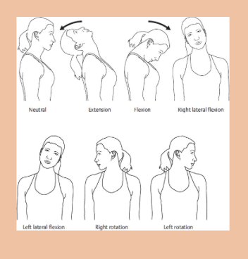
PHYSICAL EXAM:NECK (7 Movements)
Inspect
Palpate
Assess ROM (5 motions)
Flexion
Extension
Hyperextension
Lateral Flexion x 2
Rotation – left and right
Assess Strength (See Strength Scale)
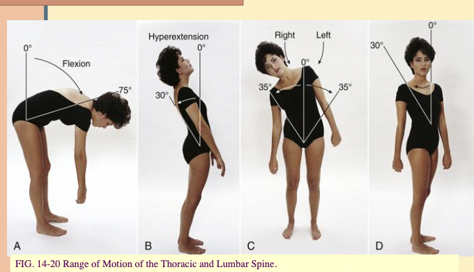
PHYSICAL EXAM: SPINE (7 Movements)
Inspect
Palpate
Assess ROM (5 motions)
Flexion
Extension
Hyperextension
Lateral Flexion x 2
Rotation – left and right
Assess strength
(See Strength Scale)

PHYSICAL EXAM:SHOULDER (8 Movements)
Inspect
Palpate
Assess ROM (5 motions)
Flexion
Extension
Hyperextension
Abduction
Adduction
Circumduction
Internal rotation
External rotation
Assess strength
(See Strength Scale)

PHYSICAL EXAM: Elbow (3 Movements)
Inspect
Palpate
Assess ROM (3 motions)
Flexion
Extension
Hyperextension
Assess Strength (See Strength Scale)
PHYSICAL EXAM: Forearm (2 Movements)
Inspect
Palpate
Assess ROM (2 motions)
Supination
Pronation
Assess Strength
(See Strength Scale)

PHYSICAL EXAM: Wrist (6 Movements)
Inspect
Palpate
Assess ROM (6 motions)
Flexion
Extension
Hyperextension
Radial flexion
Ulnar flexion
Rotation
Assess strength
(See Strength Scale)

PHYSICAL EXAM: Fingers (7 Movements)
Inspect
Palpate
Assess ROM (6 motions)
Flexion
Extension
Hyperextension
Radial flexion
Ulnar flexion
Rotation
Finger opposition
Assess strength (See Strength Scale)

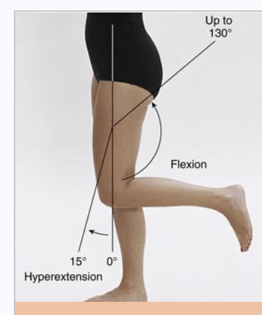
PHYSICAL EXAM: Hip (8 Movements)
Inspection
Palpation
Flexion
Extension
Hyperextension
Abduction
Adduction
Circumduction
External rotation
Internal rotation
Asses Strength (See Strength Scale)

PHYSICAL EXAM: Knee (3 Movements)
Inspection
Palpation
Flexion
Extension
Hyperextension
Assess Strength (See Strength Scale)
PHYSICAL EXAM: Ankle and Foot (5 Movements)
Inspection
Palpation
Dorsiflexion
Plantar flexion
Eversion
Inversion
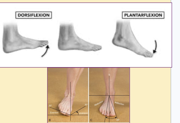
Rotation
Assess Strength (See Strength Scale)
PHYSICAL EXAM: Toes (5 Movements)
inspection
Palpation
Flexion
Extension
Hyperextension
Abduction
Adduction
Assess Strength (See Strength Scale)

MUSCULOSKELTETAL STRENGTH SCALE
Test opposite movements (ex:flexion/extension)
0- no evidence of contractility
1-evidence of slight contractility
2- complete range of motion with gravity eliminated
3- complete range of motion with gravity
4- complete range of motion against gravity with some resistance
5- complete range of motion against gravity with full resistance
Bone Abnormalities
Fractures
Open vs Closed
Trauma
Insufficient integrity
Osteoporosis
loss of bone density increases risk of fracture
Bone Abnormalities risk factors
Age
Gender
Race
Thin/small boned
Family history
Lifestyle issues
Medications
Hormonal deficiency
Eating disorders
sedertary life style decreased calcium intake exessive alc
Joint Abnormalities
Rheumatoid Arthritis
Chronic, systemic autoimmune disorder causing inflammation and erosion of the joints
Fibrosis and shortening of the ligaments and tendons results in contractures and deformities
Pain and stiffness in the AM lasting more than 30 minutes
Systemic symptoms of fever, anemia, and fatigue
Joint Abnormalities
Osteoarthritis
Degenerative joint disease- breakdown of cartilage
Pain in stiffness in the AM less than 30 minutes
Pain decreases with movement
Increased risk with age, females, obesity, joint injury or overuse, family history of OA
Spine Abnormalities
Herniated Nucleus Pulposus
Herniated Disk/ slipped disk
Fibrocartilage ruptures compressing spinal nerves
Pain is dependent on where the disk is affected
Scoliosis
S-shaped deformity - Could be genetic
Kyphosis
Posterior curvature
Lordosis
Anterior curvature
Bursitis
Inflammation of the bursa
Pain, erythema, & tenderness at the joints
Common in shoulder, elbow, and hip but also occurs in knee, heel, and MTP joint
Gout
Increase in uric acid related to increased production or decreased excretion
Typically occurs in MTP joint (base of the big toe) but can occur in any articular joint
Tophi = uric acid crystal deposits in joints
Sprain
Injury to a ligament (connects bone to bone) - common in ankle and knee
Strain
Injury to a tendon (connects muscle to bone) or a muscle – common in Achilles tendon, low back, or quadriceps muscle
Tears
Partial or complete disruption of a structure - meniscus, ligament, tendon, or muscle
Documentation
Gait steady with bilateral contralateral arm swing. No pain, tenderness, swelling, crepitus, bogginess, or inflammation noted in any joints. No deformities. No scoliosis, kyphosis, or lordosis. Full ROM with 5/5 strength testing in all extremities bilaterally.
Health Promotion
Vitamin D/ Calcium intake
Weight bearing exercises
Smoking/ alcohol use
Screenings
Protection
Age Related Changes
Decrease in bone mass
Intervertebral disc space gets reduced
Flexed posture
Loss of muscle mass and strength due to decrease in tone of tendons and muscles
Slower reflexes