The Dynamic Cell
 Call Kai
 Learn
 Practice Test
 Spaced Repetition
 Match
 Flashcards
 Knowt Play
1/78

There's no tags or description
Looks like no tags are added yet.
79 Terms
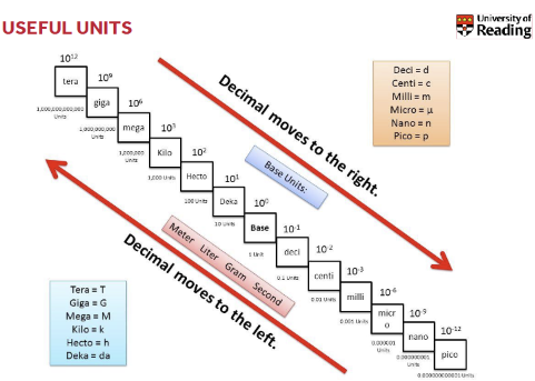
Units
Characteristics of all cells
A surrounding membrane
cytoplasm - cell contents in thick fluid
Organelles
2 basic types of cells
prokaryotic cells:
All bacteria are prokaryotes
No membrane bound nucleus
Nucleoid = region of DNA concentration
organelles not bound by membranes
eg: Spherical (cocci), Rod - shaped (bacilli), spiral
Eukaryotic cells:
Nucleus bound by membrane
possess many organelles
include fungi, protists, plant, and animal cells
eg: blood cells, neurons, skin cells
why do cells have compartments
Eukaryotic cells are larger than prokaryotic cells
larger cells means greater diffusion distance which can make chemical reaction slower.
what is cell compartmentalisation
All cells have a plasma membrane.
Eukaryotic cells have membrane-bound organelles that create separate compartments.
Each organelle has a specific function.
Prokaryotes lack membrane-bound organelles and have one main compartment containing the enzymes needed for basic functions.
Compartmentalisation allows different processes to happen separately and efficiently.
what are the 2 membranes
monolayer and bilayer

Plasma membrane features
composed of phospholipids, cholesterol, proteins and other components
Phospholipids drift within the bilayer
function: regulates what enters and leaves the cell
Cytoplasm components
cytoskeleton (filaments and fibres)
cytosol fluid
organelles (not nucleus)
storage of substances
The nucleus structure and function
Structure: enclosed by a double membrane called nuclear envelope which has pores. Contains nucleolus and chromatin
function: Site of cellular DNA storage, replication and transcription
Ribosomes structure and function
Structure: Composed of RNA and protein
Ribosomes carry out protein synthesis at two locations (in cytosol, free ribosomes or on the outside of endoplasmic reticulum which are bound ribosomes)
Function : protein biosynthesis
Endoplasmic Reticulum
Smooth ER
no ribosomes involved
function = Carbohydrate metabolism, drug metabolism, calcium ion movement
Rough ER
ribosomes involved
Function: involved in the synthesis folding and modification and transport of proteins Site of translation of proteins
Golgi apparatus structure+ function
structure: a stack of flattened membrane bound sacs called cisternae
function - Acts as the packaging and shipping station, packages them into vesicles for transport
what happens in protein trafficking
molecules come in vesicles
vesicles fuse with golgi membrane
molecules may be modified by golgi
molecules go into separate vesicle
vesicle leaves golgi
vesicles combine with plasma membrane to secrete contents
Mitochondria structure and function
Structure - Have their own DNA, Bound by double membrane
Function = Break down organic compounds by aerobic respiration + produces ATP
Lysosomes structure and function
structure: membrane bound vesicles that contain hydrolytic enzymes
Function: Aid in cell renewal, break down old cell parts, involved in intracellular digestion
peroxisomes structure and function
Structure: Membrane-bound organelles containing enzymes, including catalase.
Function: Involved in various metabolic reactions, including the synthesis and degradation of hydrogen peroxide (H2O2H2O2). They also play roles in lipid synthesis, fatty acid oxidation, and detoxification.
structure and function of cytoskeleton
structure = contains microfilaments (actin) , microtubules( tubulin) and intermediate fibres (neurofilament in neurons)
function: Provide the cell with a certain shape and mechanical stability
Cell movement
Distribution of chromosomes in cell division
acts as an anchor to cells
Microtubules have motor proteins which pull them


cells require communication for:
survival, adaptation and changing environmental conditions
How do Multicellular organisms communicate
they respond to their physical environment and other cells. Cells need to know their correct location within the organism . Stimuli include:
growth factors
Hormones
Cell - matrix
Cell to cell contact
light Photons
Touch
Temperature
what is Signal Transduction
signal transduction is the mechanism by which = a cell detects external stimuli and converts this detection into an internal cellular response usually involving a change in cellular activity.
3 phases of signal Transduction
Receptor - the target cell detects a signal from outside the cell. This occurs when a biochemical signal called a ligand, binds to a specific receptor protein, typically located on the cell surface.
Transduction - binding of ligand to receptor, converts initial signal into a different signal (second messenger) Small non - protein water - soluble molecules that are produced or released in response to ligand binding
Response - The transduced signal triggers a specific cellular activity
key concepts of cell signalling
Specific Molecular Recognition- crucial for interactions between ligands and receptors or between signalling molecules
Molecular switches - proteins can be activated or inactivated through phosphorylation/ dephosphorylation
Signal Amplification - The process by which ligand - receptor interaction can lead to larger cellular response
Types of Cell Communication
Cell Junctions:
Gap Junctions: Channels connecting the cytoplasm of adjacent cells, allowing direct passage of small water soluble ions and molecules. This enables rapid communication between neighboring cells.
Cell - Cell recognition - cells communicate through direct contact between molecules on their surfaces.
Chemically Signalling:
Paracrine signalling - A secreting cell releases signalling molecules that act on nearby target cells
Synaptic signalling - A specialised form of paracrine signalling occurring at synapses between nerve cells. An electrical signal triggers the release of neurotransmitters which diffuse across synaptic cleft to act on the target cell
Autocrine signalling - A cell releases signalling molecules that bind to receptors on its own surface, essentially signalling to itself
Endocrine - Signalling cells release hormones into the bloodstream which act on target cells
Ligands and Types of receptors
ligands - external signal molecules that initiate cell signalling, and bind to receptors. They can be proteins, peptides, lipids, small molecules, or environmental stimuli.
Receptors - cellular proteins, They are typical located on cell surface but can be intracellular
Types of receptors;
Transmembrane - proteins that span the plasma membrane having an extracellular domain
Ion channel linked receptors - ligand binding causes a change that open or closes an ion channel alternating the ion concentrating inside the cell
G protein linked receptors - These receptors are coupled to G proteins. Ligand binding activates the G protein, Which then interacts with other enzymes to relay the signal
Enzymes linked receptors - Ligand binding activates the enzyme activity.
Intracellular receptors - receptors located within the cytoplasm or nucleus so ligands are small
What is Phosphorylation
Phosphorylation is the addition of phosphate group to a protein
This process is catalysed by enzymes called kinases
Dephosphorylation the removal of a phosphate group is catalysed by phosphatases
Phosphorylation often acts a molecular switch altering protein activity
examples of signal amplification
ion channel opening - A ligand binding to an ion channel receptor can cause channel to open allowing large flux of ions across membrane changing ion concentration
Enzymatic Generation of Second Messengers - An activated receptor can activate an enzyme that produces many molecules of a second messenger.
Phosphorylation cascades - A series of kinase activating other kinases in sequence. this can activate multiple downstream kinases leading to signal amplification
in phase 3 of signal transduction what are the proteins called
target (response proteins)
proteins at end of signalling pathway
how is gene expression regulated
Transcription factors can bind to DNA and regulate the expression of specific genes
some transcription factors can activates genes that produce proteins that further amplify signalling or inhibit it
regulation of gene expression can lead to change in proteins
chromosome features
consist of double helix DNA
23 pairs of chromosomes in total 22 pairs of autosomes and 1 pair of sex chromosomes
made of base pairs
sister chromatids
each chromosome after DNA replication contains two identical sister chromatids
sister chromatids are joined together at centromere
centromere is a region of DNA hidden beneath proteins

kinetochores
The kinetochore is a protein complex assembled at centromere
it acts as an attachment site for microtubules in cell division

microtubules and centrioles
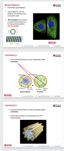
microtubules are part of the cytoskeleton and are responsible for various movements within eukaryotic cells
microtubules form mitotic spindle
animal cells have two small organelles called centrioles
centrioles involved in formation of spindle fibres and consist of cylindrical array of 9 microtubules
mitosis vs meiosis
Mitosis
Division of somatic (body) cells.
Occurs wherever more cells are needed for growth and repair.
Produces two new cells that are genetically identical to each other and to the parent cell.
The process of growth and division is called the cell cycle.
Results in two diploid (2n2n) daughter cells.
Chromosome number remains the same in daughter cells.
Genetic variation does not change.
Consists of 4 main stages (plus interphase
Meiosis
Division of germ cells (gametes: sperm and egg cells).
Occurs for sexual reproduction.
A human body cell has 46 chromosomes (2n2n), while gametes have 23 chromosomes (1n1n).
This reduction in chromosome number is why meiosis is sometimes called "reduction division."
Results in four haploid (1n1n) daughter cells.
Chromosome number is halved in each daughter cell.
Genetic variation is increased due to recombination and independent assortment.
Consists of two successive cell divisions (Meiosis I and Meiosis II), with 8 stages in total (plus interphase).
mitotic cells in the adult human body
eg:
hair follicle cells
fibroblasts
stem cells
skin cells
heart endothelial cells
intestinal epithelial cells
what is hayflick limit
normal somatic cells have limited number of times they can divide known as the hayflick limit. This is around 50 population doublings
mitosis and cancer cells
normal cells have checkpoints that regulate cell division and stop if DNA is damaged
Cancer cells bypass these checkpoints leading to uncontrolled cell division
mitosis and stem cells
2 types of division ]
Symmetric division - produces two identical daughter cells
Asymmetric division - Produces one daughter cell that remains a stem cell and another daughter cell that differentiates into a cell type
cell cycle and mitosis
Interphase: The longest phase, where the cell grows and replicates its DNA.
G1 Phase: Cell growth and preparation for DNA synthesis.
S Phase: DNA replication occurs. The cell goes from a diploid (2n2n) state to a tetraploid (4n4n) state in terms of DNA content.
G2 Phase: Further growth and preparation for mitosis.
M Phase (Mitosis): The period of nuclear division.
Prophase: Chromosomes condense and become visible, each consisting of two sister chromatids. The nuclear envelope disintegrates, and the mitotic spindle begins to form.
Metaphase: Chromosomes align at the metaphase plate (equator of the cell), attached to spindle fibers. Homologous chromosomes do not associate.
Anaphase: Sister chromatids separate and are pulled to opposite poles of the cell.
Telophase: Chromosomes decondense, nuclear envelopes reform around the two sets of chromosomes, and the spindle fibers disappear.
Cytokinesis: The division of the cytoplasm, which usually overlaps with telophase, resulting in two distinct daughter cells.
cell cycle checkpointsG₁ Checkpoint (Restriction Point):
G₁ Checkpoint (Restriction Point):
Question: Is the environment favorable for cell division (e.g., sufficient nutrients, growth factors)?
Outcome: If favorable, the cell enters the cell cycle and proceeds to S phase.
G₂/M Checkpoint:
Question: Is all DNA replicated, and is it free of damage?
Outcome: If conditions are met, the cell enters mitosis.
Metaphase-to-Anaphase Transition (Spindle Checkpoint):
meiosis 1
Prophase 1: Homologous chromosomes pair up (synapsis) to form tetrads. Crossing over occurs, leading to genetic recombination and increased genetic diversity.
Metaphase 1: Homologous pairs of chromosomes line up at the metaphase plate. Spindle fibers attach to the centromeres.
Anaphase 1: Homologous chromosomes separate and move to opposite poles. Sister chromatids remain attached.
Telophase 1: The cytoplasm divides, forming two haploid (1n1n) daughter cells. Each chromosome still consists of two sister chromatids. genetically different
meiosis 2
Prophase 2: Spindle fibers form in each of the two daughter cells.
Metaphase 2: Chromosomes align at the metaphase plate in each cell.
Anaphase 2: Centromeres separate, and sister chromatids (now individual chromosomes) move to opposite poles.
Telophase 2 and Cytokinesis: Nuclei reform at opposite poles, and the cytoplasm divides. This results in four genetically unique haploid (1n1n) daughter cells.
why we need meiosis
For sexual reproduction
Fertilisation
Genetic diversity through crossing over and independent assortment
consequences of Non Disjunction in meiosis
Non disjunction - failure of homologous chromosomes or sister chromatids to separate properly during meiosis
Trisomy - if non disjunction occurs gametes may have extra chromosome eg: down syndrome
monosomy - other gametes may be missing a chromosome
errors in meiosis
chromosome abnormalities, deletions, translocation
cell fates can be broadly categorised into
division - increasing in number
survival - continuing to exist
cell death - undergoing a programmed cell death
differentiation - becoming a specialised cell type
key cell survival pathway IGF1
insulin like growth factor pathway is crucial cell survival pathway
IGF1 binds to IGF - 1 receptor which activates phosphoinositide 3 kinase. that then activates Akt (protein kinase b)
Akt inactivates proteins that promote cell death and thus promotes cell survival
what is Apoptosis
programmed cell death
Characteristics:
cell shrinkage
chromatin condensation
disintegration of organelles
what is cell growth and proliferation
cell growth:
involves an increase in cell size and increase in cell number
synthesis of new proteins, membrane liquids
increase number of organelles
may involve replication of DNA
Proliferation:
Involves replication of DNA and division to produce two cells
Usually follows from a cell growth phase
Positive and negative Growth/Proliferation signals
Growth Stimulating Pathway - Growth factors bind to receptors, activate transcription factors. These transcription factors stimulate the cell cycle
Growth Inhibiting Pathway - Growth inhibiting factors can also signal through receptors
what is differentiation
the process by which cells become specialised cell types
usually but not always involves them ceasing to proliferate
deregulation of cell fate mechanisms and cancer
self suffciency growth signals
insensitivity to anti - growth signals
Evading Apoptosis
Limited Replicative Potential
cell death important roles:
embryogenesis and fetal development
damage control
homeostasis
malignant cancer cell control
immune system regulation
2 types of primary cell death
Necrosis - uncontrolled cell death process often associated with disease or injury
Apoptosis - programmed cell death essential for normal health and development
Importance of Apoptosis
embryonic development - essential for tissue sculpting
immune system - eliminates virus infected cells
homeostasis - balances cell proliferation and removes aged or damaged cells
Cancer treatment - radiotherapy and chemotherapy drugs induce apoptosis in cancer cells
2 main pathways that trigger apoptosis
Extrinsic pathway (Receptor mediated) - Triggered by external signals binding to cell surface death receptors
Intrinsic Pathway (Mitochondria Mediated) Triggered by internal cellular stress or damage
stages of apoptosis
Death signal (intrinsic or extrinsic)
Commitment to die (reversible)
Execution - irreversible stage
Dying cell - cell shrinkage and condensed
Phagocytosis -Apoptotic bodies are engulfed by macrophages and neighbouring cells
what are caspases ( The Chief Executioners)
central to apoptosis
They exist as inactive pro enzymes
Activation occurs through cleavage
Activated caspases can trigger a cascade
Extrinsic pathway explained
Begins with ligands binding to death receptors on cell surface
Ligand binding leads to the recruitment of adaptor proteins eg: procaspase-8 to the receptor.
allows activation of caspase 8
this activates effector caspases
Intrinsic Pathway can be activated by:
DNA damage
UV radiation
Viral infection
Free radical damage
Absence of survival signals
The role of mitochondria intrinsic pathway
Cytochrome C release - under stress, pores form in outer mitochondrial membrane releasing cytochrome c from intermembrane space into the cytosol
Apoptosome formation - in cytosol, cytochrome c binds to other proteins to form multi protein complex called apoptosome
caspase 9 activation - apoptosome recruits pro caspase 9 this activates effector caspases
Hallmarks (characteristics) of Apoptosis
Cell shrinkage
Maintenance of organellar integrity (initially)
Maintenance of ATP levels
Maintenance of ion homeostasis
Membrane surface blebbing
Nuclear chromatin condensation and fragmentation
Requires the synthesis of death effector proteins (active process)
Can be prevented by blocking steps in the death cascade
Does not adversely affect neighboring cells
No inflammation
causes of necrosis
infection (pathogens)
Direct toxic threats (chemicals, toxins)
Extreme temperatures (freezing, burns)
Excitotoxicity (excessive neurotransmitter stimulation)
Trauma (mechanical injury)
Hypoxia (lack of oxygen)
Ischemia (lack of blood flow)
Necrosis process
Insult - trauma, toxic, chemicals, heat
loss of homeostasis - loses ability to regulate internal environment
ion influx and swelling
Enzyme activation and damage
production of toxic compounds
cell death
Inflammation - leakage of cellular contents trigger inflammatory response
Hallmarks (characteristics) of Necr
Cell swelling
Organellar swelling and damage
Depletion of ATP
Loss of ion homeostasis
Membrane rupture
Nuclear lysis
Cessation of protein synthesis
Irreversible process
Promotes the death of neighboring cells
Causes inflammation
apoptosis vs necrosis

what is necroptosis
Necroptosis is a more recently identified form of programmed cell death that shares features of both apoptosis and necrosis.
It is a regulated, controlled form of necrosis.
It involves molecular "switches" that allow cells to decide between apoptosis and necroptosis.
in tissues cell can be connected together via:
cell to cell
cell to extracellularmatrix
microfilaments (actin filaments)
functions
Determine cell shape
essential for cell movement
involved in muscle contraction
role in cytokinesis (cell division)
intermediate filaments
composition
structure
function
composition: diverse group of proteins
structure: appear as rope like fibres
functions: provide mechanical strength and resilience to cells and tissues. Helps connect cells together, maintain cell shape
Microtubules
structure
functions
structure - hollow cylinders
function: position organelles within cells, contribute to cell shape and movement, crucial for forming mitotic spindle during cell division, internal transport system for vesicles and organelles
what is cell migration process
Protrusion - actin filaments polymerise, pushing cell membrane forward
adhesion - adhesion sites form between cell and extracellular matrix
retraction - myosin motors pull cell body forward and old adhesion sites disassemble, actin enables rapid shape changes and directional movement in response to chemical changes
Extracellularmatrix components
Long protein fibres:
Collagen - most abundant protein in human body, high temsile strength and crucial for bones
elastin - provides elasticity to tissues to stretch and recoil
proteoglycans - long chain sugar molecules, lubricate ecm
specialised molecules:
fibronectin - a large glycoprotein receptor that binds to cell surface receptors
Extracellularmatrix functions
provides structural and mechanical support to tissue
acts as a scaffold influencing cell behaviour and differentiation and survival
forms basement membranes
describe all the cell - cell connections
tight junctions - seal adjacent cells together forming a barrier. They provide mechanical strength
adherens junctions - provide mechanical strength and involved in cell - cell adhesion
desmosomes - strong adhesion points that resist mechanical stress, anchor cells together
gap junctions - channels that directly connect the cytoplasm of adjacent cells allowing passage of small molecules and ions
describe all cell - ECM connections
focal adhesions - sites where cells attach to the ECM
Hemidesmosomes - anchor cells to the basement membrane
Role of cytoskeleton
Mechanical Strength
coordinated action
cellular behaviour - differentiation, proliferation, migration
cell movement
what diseases can occur in:
cytoskeleton?
cell - cell interactions?
ECM?
Neurodegenerative diseases
autoimmune diseases
chronic inflammation
what other movement is there outside ECM
motor proteins attached to microtubules propel cell forward -eg: sperm
substances move along cell surface eg: cilia