RBC Morphology: Grading, Poikilocytes, and Clinical Associations
 Call Kai
 Learn
 Practice Test
 Spaced Repetition
 Match
 Flashcards
 Knowt Play
1/64

There's no tags or description
Looks like no tags are added yet.
65 Terms
What CBC parameter is primarily evaluated to detect anocytosis?
Red Cell Distribution Width (RDW)
What is the normal reference range for Red Cell Distribution Width (RDW)?
11.5% to 14.5%
What hematologic term describes a variation in red blood cell size?
Anocytosis
What hematologic term describes a variation in red blood cell shape?
Poikilocytosis
In red blood cell morphology grading, what percentage range of abnormal cells corresponds to a 1+ grade?
10% to 25%
In red blood cell morphology grading, what percentage range of abnormal cells corresponds to a 2+ grade?
25% to 50%
In red blood cell morphology grading, what percentage range of abnormal cells corresponds to a 3+ grade?
50% to 75%
Why should a 4+ grade for red blood cell morphology be avoided unless virtually all cells (>75% or 100%) exhibit the abnormality?
Extreme 4+ morphology is rare and typically restricted to specific severe hereditary conditions.
When deciding between two adjacent morphology grades (such as 1+ vs. 2+ or 2+ vs. 3+), what general rule of thumb should be followed?
Choose the more conservative (lower) grade.
What percentage threshold of abnormal cell morphology is generally reported as 'few' or 'occasional' rather than assigned a numerical grade?
Below 10% (0% to 10%)
What term describes a peripheral blood smear population containing a mixture of both microcytic and macrocytic red blood cells?
Dimorphic population (or dual population / bimodal population)

Which clinical scenario frequently causes a dimorphic red blood cell population?
Dual anemias, such as concurrent iron deficiency (microcytic) and folate or vitamin B12 deficiency (macrocytic).
How does a dimorphic red blood cell population affect the RDW index on an automated analyzer?
It significantly increases (skews) the RDW to the right.
What defines a normocytic, normochromic red blood cell on microscopic examination?
Normal red blood cell size with a central area of pallor occupying approximately one-third of the cell diameter.
What red blood cell classification applies when central pallor occupies more than one-third of the cell's diameter?
Hypochromic
In true red blood cell morphology, which specific cell type can be classified as hyperchromic?
Spherocytes (which are microcytic cells lacking central pallor)

What are pseudo-macrocytes, and what causes their appearance on a blood smear?
Artifactually enlarged, dark red blood cells lacking central pallor caused by smudging or poor smear preparation technique.
What do polychromatophilic red blood cells on a Wright-stained peripheral smear represent?
Immature red blood cells (reticulocytes)
What is the normal reference range for circulating reticulocytes (polychromatophilic cells) in human blood?
0.5% to 1.5%
At what minimum numerical grade does polychromasia become clinically significant enough to report on a peripheral smear?
2+ grade or above

What is the scientific name for target cells?
Codocytes
Which organ disorder is most characteristically associated with the presence of codocytes (target cells)?
Liver disease (obstructive liver disease)
Besides liver disease, what major genetic disorder of hemoglobin synthesis commonly presents with abundant codocytes?
Thalassemia (or hemoglobinopathies such as HbS and HbC)

What structural feature defines a spherocyte on a peripheral blood smear?
A small, dark, round red blood cell completely lacking central pallor.
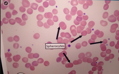
A high count (>3+) of spherocytes on a peripheral smear strongly suggests which hereditary disorder?
Hereditary spherocytosis
What acquired pathological mechanism commonly causes spherocyte formation?
Immune-mediated hemolytic anemia (or microangiopathic hemolytic anemia)
Which follow-up laboratory test is performed to evaluate red cell membrane fragility when spherocytes are noted?
Osmotic fragility test (osmolality test)

What visual feature characterizes a stomatocyte?
A red blood cell with a slit-like, mouth-shaped, or lip-shaped central pallor.
Non-hereditary stomatocytosis on a peripheral smear is most frequently associated with which liver-related condition?
Chronic alcohol abuse (excessive alcohol consumption / cirrhosis)
What distinguishes an ovalocyte from an elliptocyte in terms of morphology?
Ovalocytes are wider, fat-oval macrocytic cells, whereas elliptocytes are narrow, elongated, slender cells with smooth ends.
Marked presence of ovalocytes (>3+) is diagnostic for which inherited membrane condition?
Hereditary ovalocytosis
In addition to hereditary elliptocytosis, what nutritional deficiency anemia can present with prominent elliptocytes?
Megaloplastic anemia (vitamin B12 or folate deficiency)
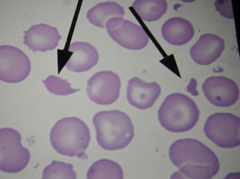
What general term refers to fragmented red blood cells resulting from mechanical disruption?
Schistocytes
Which severe intravascular coagulation disorder characteristically features schistocytes formed by fibrin strands breaking passing RBCs?
Disseminated Intravascular Coagulation (DIC)
What cardiac medical device can cause mechanical damage resulting in schistocyte formation in circulation?
Prosthetic heart valves

What is the scientific name for horn cells or helmet cells?
Keratocytes
What systemic metabolic condition is associated with the presence of keratocytes (helmet cells)?
Uremia (elevated blood urea nitrogen / renal failure)
What physical trauma condition frequently leads to keratocyte (horn cell) formation?
Severe thermal burns
What is the scientific name for bite cells?
Degmacytes
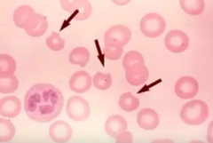
Bite cells (degmacytes) are the classic poikilocyte associated with which enzyme deficiency?
Glucose-6-Phosphate Dehydrogenase (G6PD) deficiency
What red blood cell inclusion is generated alongside bite cells in G6PD deficiency?
Heinz bodies
What is the scientific name for sickle cells?
Drepanocytes

Unlike most poikilocytes which are assigned numerical grades, how must sickle cells (drepanocytes) be reported on a peripheral smear?
By exact numerical count per field or smear, even if only a single sickle cell is identified.
Sickle cell formation (drepanocytosis) results from the polymerization of which abnormal hemoglobin variant?
Hemoglobin S (HbS)
What is the scientific name for burr cells?
Echinocytes

How do echinocytes (burr cells) differ morphologically from acanthocytes (spur cells)?
Echinocytes have uniform, evenly spaced, small blunt projections, whereas acanthocytes have irregular, unevenly spaced, pointed projections.
What common pre-analytical artifact causes echinocyte (burr cell) formation on a blood smear?
Specimen aging or prolonged blood exposure to EDTA anticoagulant.
What severe eating disorder is clinically associated with the presence of acanthocytes (spur cells)?
Anorexia nervosa (due to severe starvation)

What is the scientific name for teardrop-shaped red blood cells?
Dacrocytes
What memory cue links the scientific name dacryocyte to its morphological appearance?
The root word dacry- (or cry-) refers to crying or tears (teardrop cell).
Dacrocytes (teardrop cells) are prominently seen in severe iron deficiency, severe megaloplastic anemia, and which primary bone marrow disorder?
Myelofibrosis

What is the biochemical composition of Howell-Jolly bodies?
DNA (nuclear remnants)
How do Howell-Jolly bodies typically appear inside a red blood cell on a Wright-stained smear?
As a single (or occasionally double), small, round, dark purple dot located near the cell periphery.
Under what clinical circumstance are Howell-Jolly bodies most commonly observed in peripheral blood?
Post-splenectomy (or hyposplenism / asplenia)
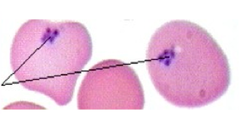
What is the biochemical composition of Pappenheimer bodies?
Iron granules (ferric iron / siderotic granules)
How do Pappenheimer bodies differ in appearance from Howell-Jolly bodies on a Wright-stained smear?
Pappenheimer bodies appear as clusters of small purple granules near the edge of the cell, rather than a single isolated dot.
Which specific anemia classification is characteristically associated with Pappenheimer bodies?
Sideroblastic anemia (or conditions causing iron overload)
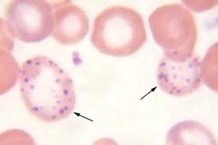
What cellular organelle composition makes up basophilic stippling in red blood cells?
Aggregated ribosomes (RNA)
How does basophilic stippling present morphologically across a red blood cell cytoplasm?
Fine or coarse dark blue/purple granules distributed throughout the entire cell.
Basophilic stippling is a classic diagnostic marker for exposure to which environmental toxin?
Lead (lead poisoning / heavy metal poisoning)

What are Cabot rings composed of inside a mature red blood cell?
Remnants of the nuclear membrane or mitotic spindle
What morphological shapes can Cabot rings form inside an RBC?
Figure-eight loops or circular ring structures

What are Heinz bodies composed of?
Denatured, precipitated hemoglobin (methemoglobin)
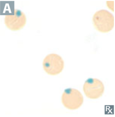
Why can Heinz bodies NOT be seen on a standard Wright-stained peripheral blood smear?
Wright stain does not stain denatured hemoglobin; a supravital stain (such as Crystal Violet or New Methylene Blue) is required.
What underlying enzyme deficiency triggers Heinz body precipitation upon oxidative stress?
Glucose-6-Phosphate Dehydrogenase (G6PD) deficiency