Tibia and Fibula & Knee
 Call Kai
 Learn
 Practice Test
 Spaced Repetition
 Match
 Flashcards
 Knowt Play
1/136

There's no tags or description
Looks like no tags are added yet.
137 Terms

Identify this image
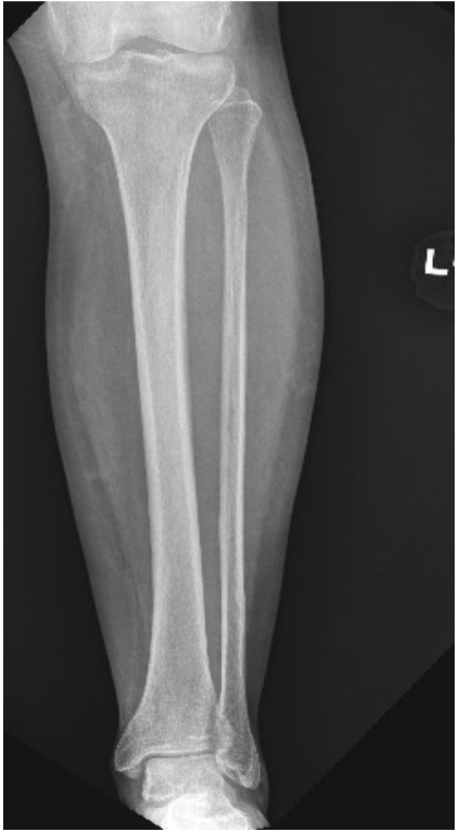
AP Tibia and Fibula (Lower Leg)

What is the positioning for this image
Patient supine on the table with the knee under the cathode end of the tube
Ensure the patient is supine with no rotation of the pelvis
Extend the affected leg
IR to go to the long axis of the lower leg if possible
If the leg is too long the IR may need to be turned diagonally to go with the leg so that both the knee and ankle joint can be included – Merrill’s also suggests that a technologist could use a 48” SID to reduce magnification and more of the leg will fit on the IR
If the leg is still too long to fit both joints on even in a diagonal position you may need to take two images – one to include the knee joint and one to include the ankle joint
Dorsiflex the foot to place the plantar surface of the foot perpendicular to the IR – a sandbag can be placed against the plantar surface of the foot to help the patient hold the position
The femoral condyles need to be parallel to the IR
What is the proper CR location for this image
CR is perpendicular to enter at the center of the lower leg

Referring to this image, where is CR going
Side to side: Going too far to the lateral aspect of the lower leg- move the CR medially about ¼”
Top to bottom: Going to about the center of the lower leg, maybe 1/8” below the actual center- move CR up towards the knee
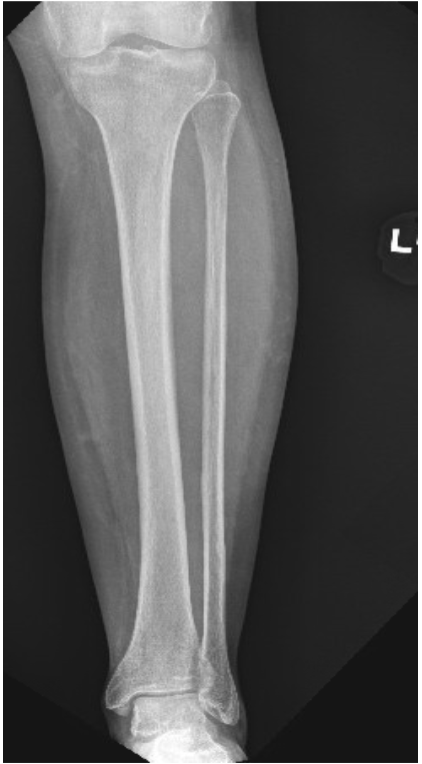
What is the rotation criteria for this image
Proximal and distal articulations of the tibia and fibula will be slightly overlapping
Fibular midshaft free of tibial superimposition

Referring to this image, is this image positioned correctly with no rotation
Looks to be in a good position without any rotation

What technical factors are used for this image
70 kVp @ 2

What FSS is used for this image
SFS
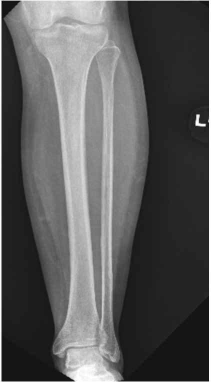
What FOV is used for this image
14 × 17 LW
Collimated to 1” on the sides and 1 ½” beyond the ankle and the knee joints
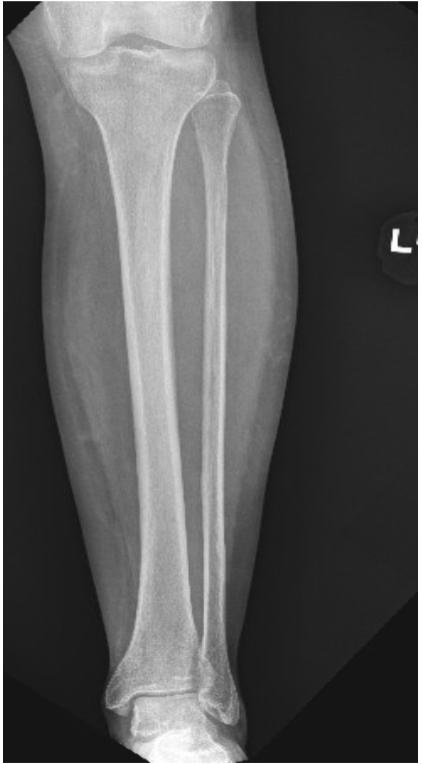
What SID is this image taken on
40” SID

What photocells are used for this image
N/A done tabletop

What phase of respiration should this image be taken on
N/A just ask the patient to hold still

List all structures demonstrated on this image
Entire tibia
Entire fibula
Knee joint
Ankle joint

What are the parameters for good visibility of detail
Bony trabecular detail
Surrounding soft tissue

Are all markers that should have been utilized seen on this image
A personal tech marker is needed
There is a personal tech marker but the intials are cut off

Does this image need to be repeated`
No, it has the knee joint, ankle joint, entire tibia, entire fibula demonstrated and it has decent visibility of detail

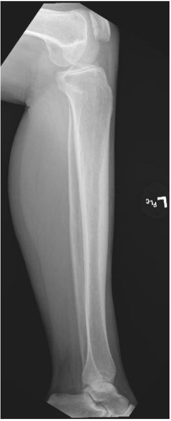
Identify this image
Lateral Tibia and Fibula (Lower Leg) (Mediolateral Projection)
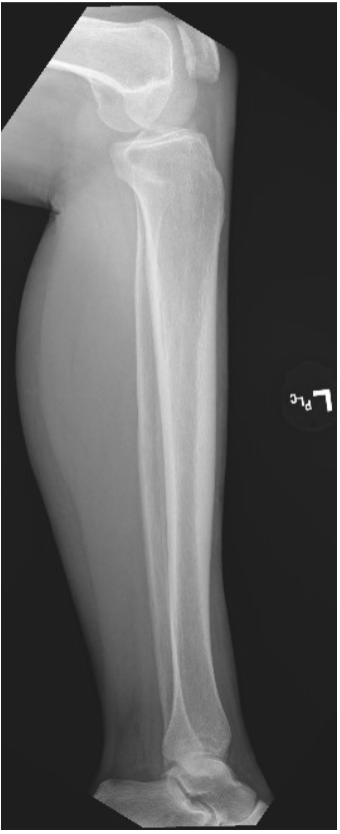
What is the positioning for this image
From the supine position have the patient roll onto their affected side
Adjust the patella so that it is perpendicular to the IR (may need to support the foot with a sponge or sandbag to help the patient hold this position).
Femoral condyles need to be perpendicular to the IR
Flex the knee slightly to help get a true lateral position
Place the IR to go with the long axis of the lower leg
If the leg is too long the IR may need to be turned diagonally to go with the leg so that both the knee and ankle joint can be included – Merrill’s also suggests that a technologist could use a 48” SID to reduce magnification and more of the leg will fit on the IR
If the leg is still too long to fit both joints on even in a diagonal position you may need to take two images – one to include the knee joint and one to include the ankle joint
Dorsiflex the foot to place the plantar surface of the foot perpendicular to the IR – a sandbag can be placed against the plantar surface of the foot to help the patient hold the position

What is the CR for this image
CR to enter perpendicular to the midpoint of the leg
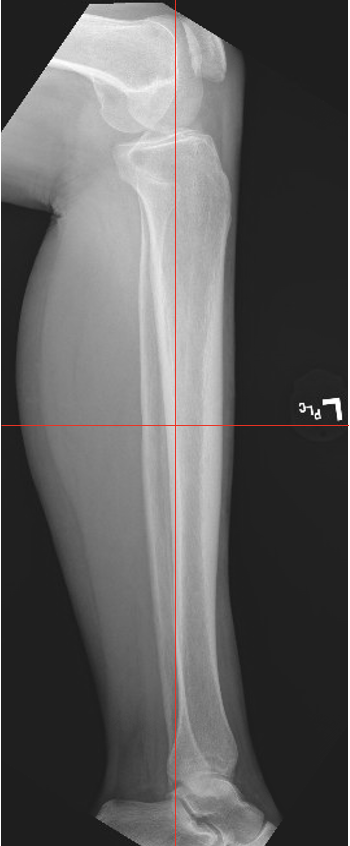
Referring to this image where is the CR going
Side to side: Looks nice and centered to the lower leg bones
Top to bottom: Slightly superior (closely towards the knee) than it needs to be about ¼” or so - come down towards the ankle

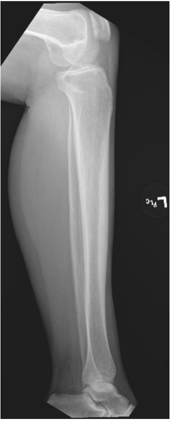
What is the rotation criteria for this image
Distal fibula will be superimposed over the posterior half of the tibia
Slight overlap of the tibia on the proximal fibular head
Moderate seperation of the tibial and fibular bodies or shafts (except at the articular ends)

Referring to this image, is there rotation
The patient may be a bit over rotated when looking at the knee, we see a lateral condyle more posterior and the medial condyles more anterior
The posterior fibula is superimposing the posterior half of the tibia, we dont see separation of the tibial and fibular bodies
What technical factors are used on this image
70 kVp @ 2 mAs

What FSS is used for this image
SFS

What FOV is used for this image
14 × 17 LW
Collimated to 1” on the sides and 1 ½” beyond the ankle and the knee joints

What SID is this image taken on
40” SID
What photocells are used for this image
N/A it is done tabletop

What phase of respiration is used for this image
N/A just ask the patient to hold still

List all structures that are demonstrated on this image
Lateral view of the:
Entire tibia
Entire fibula
Knee joint
Ankle joint

What are the parameters for good visibility of detail
Bony trabecular detail
Surrounding soft tissues

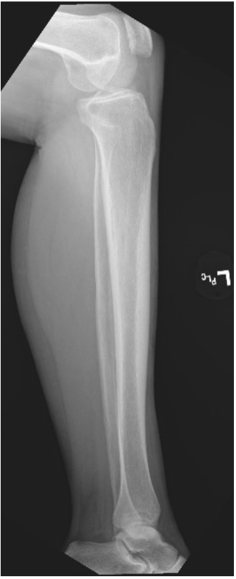
Are all markers that should have been utilized seen on this image
A personal tech marker is seen on this image
Does this image need to be repeated
No, it is not a perfect lateral but we do see what we need to see well enough without needing to repeat it
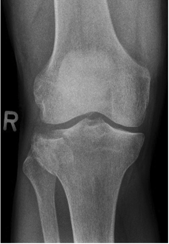
Identify this image
AP Knee (Right)

What is the positioning for this image
Pt supine on the radiographic table – ensure that the patients pelvis is not rotated
Fully extend the affected leg
Femoral epicondyles need to be parallel to the IR
Shield your patients gonads

What is the proper CR location for this image
CR to enter perpendicular to a point that is ½” below the apex of the patella
Is there a required angulation on the tube for this image
According to Merrills, this view may require an angle depending on the measurement between the ASIS and the table top

Referring to this image, where is the CR going
Side to side: ¼” too lateral on the knee, move CR more to the medial aspect
Top to bottom: ¼” above where it should be- bring CR down towards the lower leg

What is the rotation critria for this image
Femoral condyles symmetric
Intercondylar eminence of the tibia centered
Slight superimposition of the fibular head on the tibia is normal
Patella completely superimposed on the femur
Open femorotibial joint space with equal width on both sides of the knee
Referring to this image, is there evidence of proper positioning and no rotation
The image does not appear to have any rotation of the knee
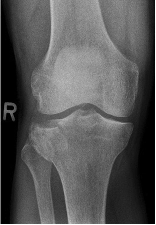
What are the technical factors for this image
80 kVp @ 4 mAs

What FSS is used for this image
SFS

What FOV is used for this image
10 × 12 LW
Then collimate to an 8 × 10 and adjusts the collimation to 1” beyond the sides of the knee

What phase of respiration should this image be taken on
N/A just ask the patient to hold still

What SID is this image taken on
40” SID
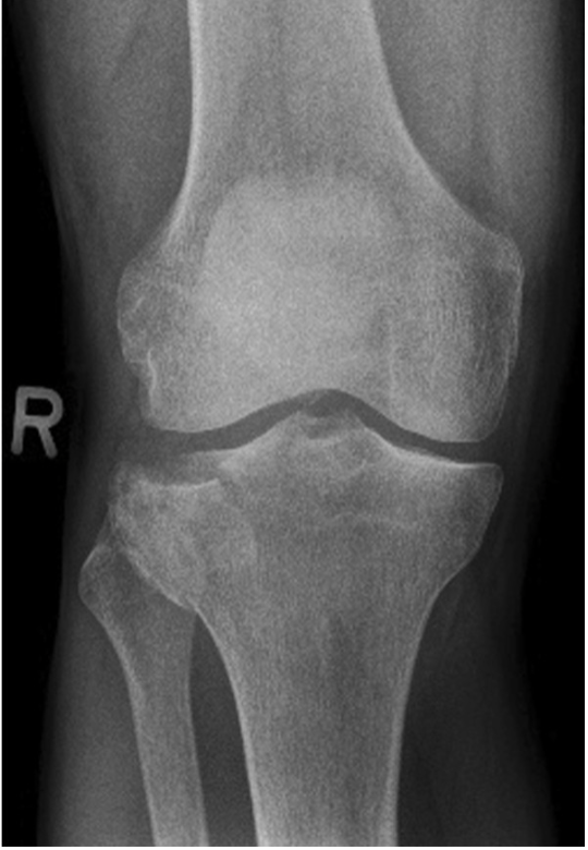
What photocells are used for this image
Center Cell

What phase of respiration should this image be taken on
N/A just ask the patient to hold still

List all structures demonstrated on this image
AP projection of the knee structures
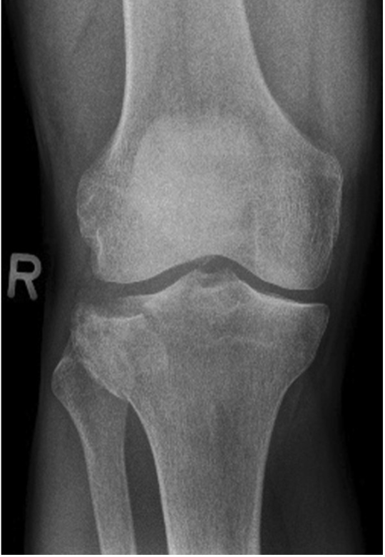
List the parameters for good visibility of detail
Bony trabecular detail
Surrounding soft tissue

Are all markers that should have been utilized seen on this image
No a personal tech marker is missing

Does this image need to be repeated?
No, it is an acceptable image even if it may have slight underexposure and a little longer scale of contrast

Identify this image
Intercondylar fossa (Beclere Method)

What is the positioning for this image
Patient supine on the radiographic table
Flex the knee so the long axis of the femur and the long axis of the tibia and fibula form a 60 degree angle
Adjust the leg so the femoral condyles are equidistant from the IR
Shield your patients gonads

What is the proper CR for this image
CR angled to enter perpendicular to the long axis of the lower leg to enter at the knee joints which is ½” distal to the apex of the patella

Is there an angulation required on the tube for this image
Yes, CR angles until it is perpendicular to the long axis of the lower leg

Referring to this image, where is the CR going
Side to side: ¼” to the medial aspect on the leg- move CR over towards the lateral aspect
Top to bottom: 1/8” too superior - move CR down
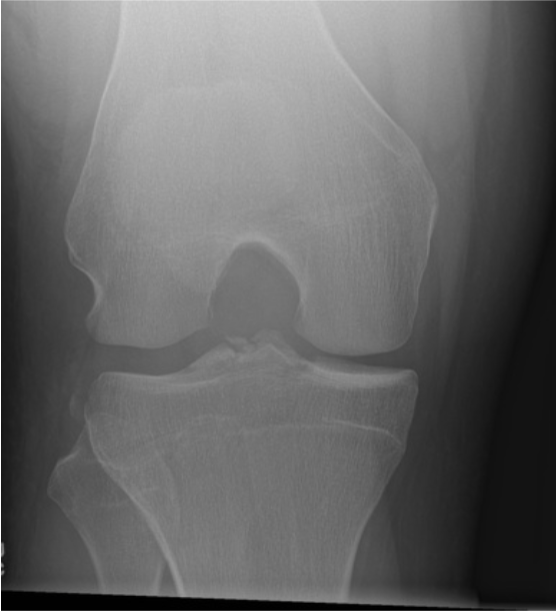
What is the rotation criteria for this image
Symmetrical appearance of the femoral condyles and tibial plateaus
Slight tibiofibular overlap

Referring to this image, is there evidence of proper positioning and no rotation
The patient looks to be positioned good, the femoral condyles appear symmetrical and so do the tibial plateaus
Doesnt look to be any rotation
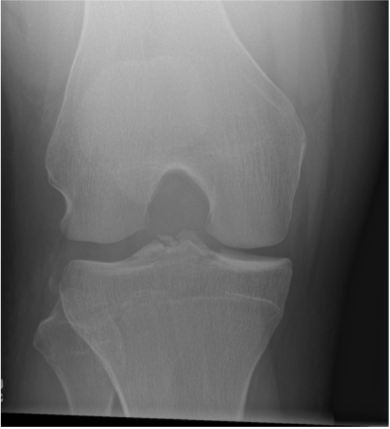
What are the technical factors for this image
80 kVp @ 2 mAs
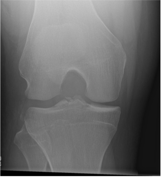
What FSS is used for this image
SFS

What FOV is used for this image
10 × 12 CW
Then collimate to 8 × 10 CW and adjust the FOV to 1” beyond the sides

What SID is this image taken on
40” SID
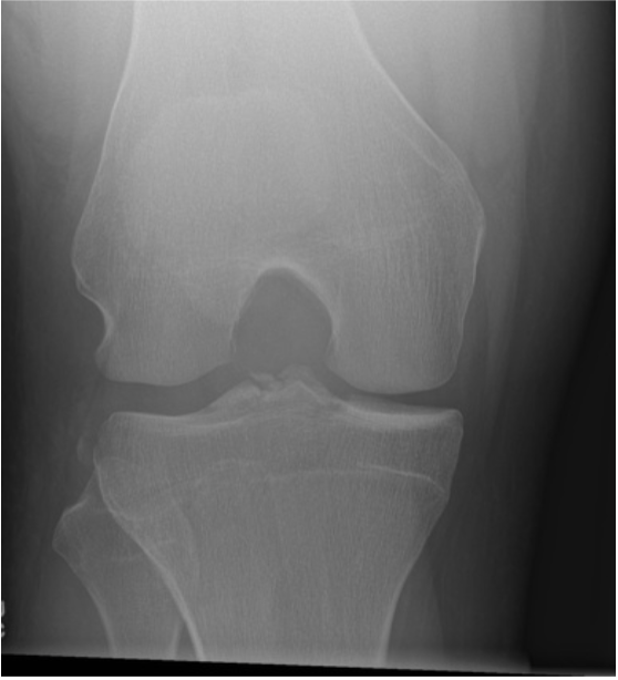
What photocells are used for this image
N/A it is done tabletop

What phase of respiration should this image be taken on
N/A just ask the patient to hold still

List all structures demonstrated on this image
Intercondylar fossa (open)
Intercondylar eminence (tibial spine)
Femerotibial joint space
Posteroinferior surfaces of the femoral condyles

List the parameters for good visbility of detail
Bony trabecular detail
Surrounding soft tissue

Are all markers that should have been utilized seen on this image
No there is no personal tech marker

Does this image need to be repeated
No it is an acceptable image

Identify this image
Lateral Knee

What is the body position for this image
Patient supine on the radiographic table
Roll the patient to the affected side
Flex the knee 20 – 30 degrees (relaxes the muscles and shows the maximum volume of the joint cavity)
If the patient has a new or unhealed patellar fracture, do not flex the knee more than 10 degrees to prevent fragment separation
You may bring the opposite leg forward over the affected leg or extend the unaffected leg behind the patient
Femoral epicondyles perpendicular to the IR
Place a small sponge for support under the ankle
Shield your patients gonads
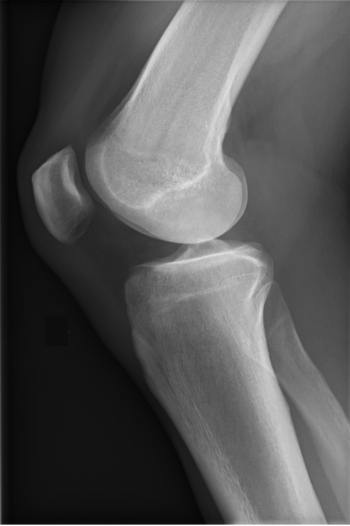
What is the proper CR location for this image
CR angled 5-7 degrees cephalic to enter 1” distal to the medial femoral condyle

Is there a required angulation on the tube for this image
Yes a 5-7 degree cephalic angle
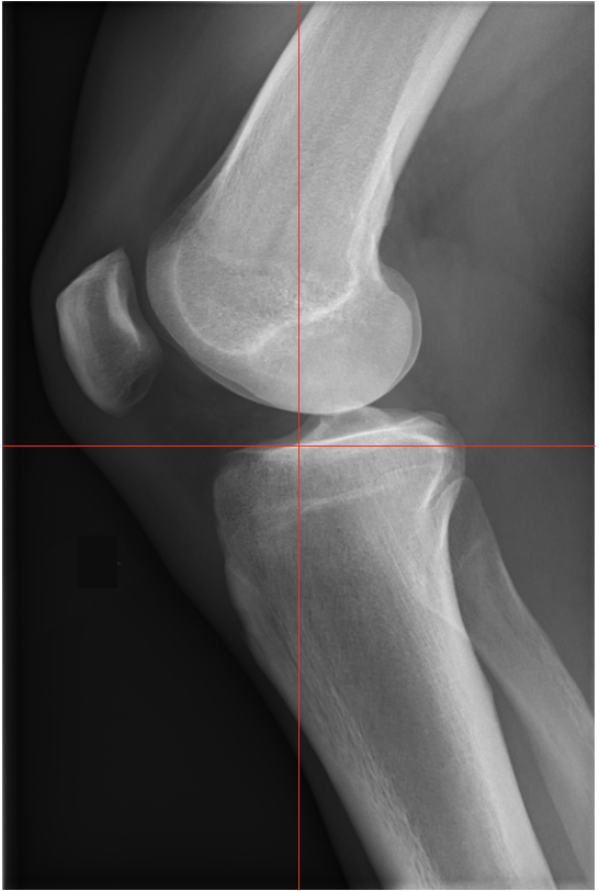
Referring to this image where is the CR going
Side to side: Pretty close to the middle of the knee - could go posterior a tiny bit
Top to bottom: looks to be 1” distal to the medial femoral epicondyle

What is the rotation criteria for this image
Femoral condyles are superimposed
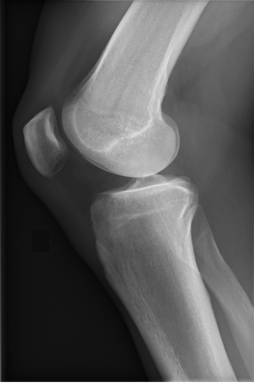
Referring to this image, is there evidence of proper positioning and no rotation
There is no rotation and there is adequate angle on the tube

What are the technical factors for this image
80 kVp @ 4 mAs

What FSS is used for this image
SFS

What FOV is used for this image
10 × 12 LW
Collimate to an 8 × 10 LW and adjust the FOV to 1” anterior to the patella and 1” beyond the posterior shadow

What SID is used for this image
40” SID

What photocells are used for this image
Center cell

What phase of respiration should this image be taken on
N/A just ask the patient to hold still
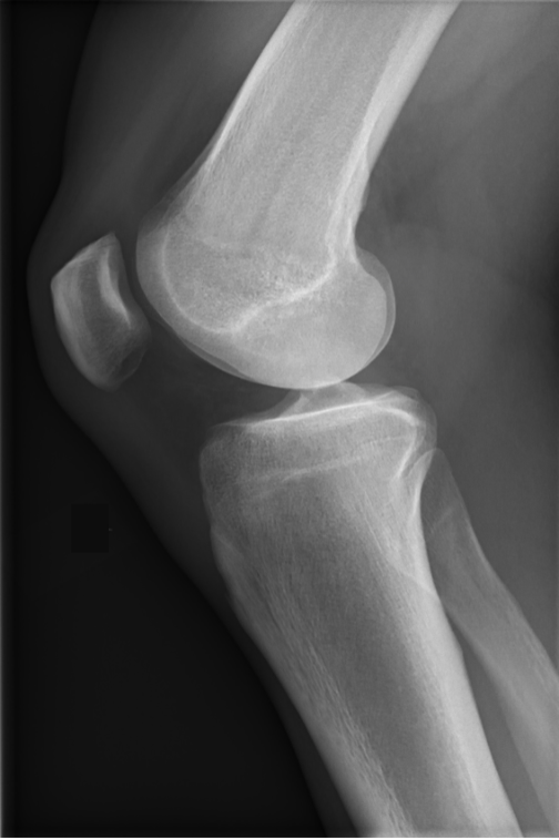
List all structures that should be demonstrated on this image
Lateral projection of:
Distal femur
Patella in profile (transverse fx of the patella if present)
Open femorotibial joint (knee joint)
Open patellofemoral joint space
Proximal ends of the tibia/fibula
Fibular head slightly superimposed by the tibia
Adjacent Soft tissue

What are the parameters for good visibility of detail
Bony trabecular detail
Surrounding soft tissue

Are all markers that should have been utilized seen on this image
No a personal technologist marker is needed

Does this image need to be repeated
No this is a very good image

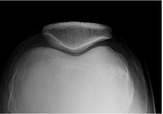
Identify this image
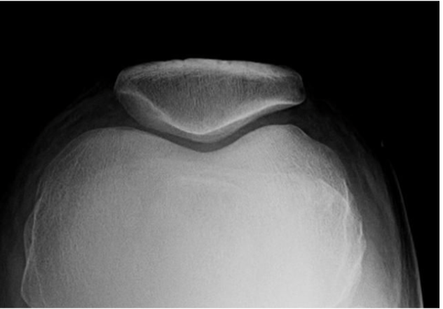
Tangential Patella (LT)
Sunrise View
Settegast Method

What is the positioning for this image
This can be done with the patient seated or prone (refer to Merrills page 366 to see the prone positioning)
Patient seated on the radiographic table
Flex the knee as much as the patient can tolerate until the patella is perpendicular to the IR (about 90 degrees if possible)
If they are experiencing a lot of pain have them flex slowly so that the patient can tolerate the flexion
Center the IR against the anterior distal thigh and have the patient hold the IR
Shield your patients gonads

What is the proper CR for this image
CR angles to be perpendicular to the IR and the patellofemoral joint space entering the middle of the patellofemoral joint

Is there a required tube angulation for this image
Yes, CR angled cepahlic until it is perpendicular to the middle of the patellofemoral joint space (about 15-20 degrees cephalic)

Referring to this image, where is the CR going
Side to side: ¼” too lateral of the midpoint of the patellofemoral joint space- move the CR medially
Top to bottom: Just a little below the patellofemoral joint space by 1/8”- move CR up towards the patella

Referring to this image, is there evidence of proper positioning and no rotation
Patient is positioned well and we can see an open patellofemoral joint space

What technical factors are utilized for this image
80 kVp @ 2 mAs

What FSS is used for this image
SFS
What FOV is used for this image
10 × 12 CW then collimate the FOV to a 4 × 4
What SID is used for this image
40” SID

What photocells are used for this image
N/A done tabletop

What phase of respiration is used for this image
N/A just ask the patient to hold still

List all structures demonstrated on this image
Articulating surfaces of patellofemoral joint space
Open patellofemoral joint space
Vertical fracture of patella if present
Patella in profile
Femoral condyles
Intercondylar sulcus (Trochlear groove)

List the parameters for good visibility of detail
Bony trabecular detail
Surrounding soft tissues
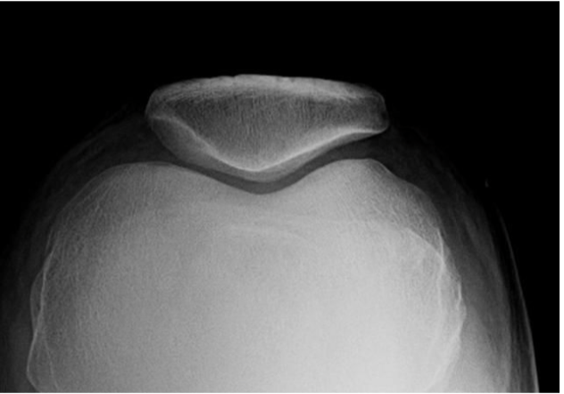
Are all markers that shoud have been utilized seen on this image
No there is no personal technologist marker
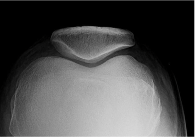
Does this image need to be repeated?
No all structures that need to be seen can be seen and there is good visibility of detail