angina and acute coronary syndrome (ACS)
 Call Kai
 Learn
 Practice Test
 Spaced Repetition
 Match
 Flashcards
 Knowt Play
1/32

There's no tags or description
Looks like no tags are added yet.
33 Terms
angina overview
•Chest pain resulting from an imbalance between oxygen supply and demand
•May be due to:
•Obstruction of coronary blood flow (atherosclerosis)
•Coronary artery spasm
•Conditions increasing myocardial oxygen consumption

PQRST of angina
•Precipitating Events
•What events or activities precipitated the pain/discomfort
•Quality of pain
•What does the pain/discomfort feel like? (crush, ache, pressure, heaviness)
•Region/radiation of pain
•Where is the discomfort?
•Does the pain radiate?
•Severity
•Rate pain on scale of 0-10
•Timing
•When did the pain/discomfort start?
•Does the pain come and go versus constant?
•How long has it lasted?
•Has this type of pain happened before?
atypical symptoms of chest pain
•More common in women and older adults
•Dyspnea
•Nausea
•Abdominal/epigastric discomfort
•fatigue
Precipitating Factors of Angina
•Circadian Rhythm Changes
•Symptoms of chest pain occur in the early morning and within the first few hours of awakening
•Consuming a Heavy Meal
•Can increase workload of the heart
•During digestion, blood diverted to GI system decreasing blood flow in coronary arteries
•Physical Exertion
•Increases heart rate
•Arm exercises (raking, shoveling snow) can cause exertional angina
•Sexual Activity
•Increases cardiac workload and SNS stimulation
•Stimulants
•Increase HR and BP leading to increased myocardial O2 demand
•Vasoconstriction causes decreased myocardial O2 demand
•Strong Emotions
•Stimulate SNS, activating stress response
•Increase workload of heart
•Temperature Extremes
•Increase workload of the heart
•Blood vessels constrict in response to extreme cold
•Blood vessels dilate and blood pools in the skin in response to heat
•Tobacco Use
•Causes vasoconstriction and an increased HR
•Decrease available O2 by increasing level of carbon monoxide
coronary artery disease

Microvascular Angina
•Ischemia from microvascular disease affects the small, distal branches of coronary arteries
•Not associated with severe CAD or spasm of a major coronary artery
•More common in women
•Chest pain is brought on by physical exertion
•Usually have a positive stress test and a negative cardiac catheterization
•Manage is the same as CAD
Prinzmetal Angina
•Results from coronary artery spasms
•Spasms can cause ischemia and lead to MI
•May be associated with ST-segment elevation on the ECG
•Occurs most often at rest
•May or may not be associated with CAD
•Risk factors:
•Smoking, Raynaud phenomenon, migraine
•Treatment:
•Calcium channel blockers and/or nitrates
•PRN SL nitro
•May disappear on own
Chronic Stable Angina
•Also called exertional angina
•Often related to atherosclerosis; leads to ischemia
•Occurs with exertion, stress or emotional upset
•Chest pain has a stable pattern of onset, duration, severity, and relieving factors
•Chest pain lasts for a few minutes
•Characteristics of chest discomfort varies among individuals
•Chest pain is relieved with nitro
chronic stable angina diagnostic
•12-lead EKG
•Observe for ST elevation/depression and/or T wave inversion
•Stress test
•Evaluate for ischemia
•Cardiac catheterization
•Definitive diagnosis of coronary artery patency
•Echocardiogram
•Cardiac enzymes
•Troponins
•Other labs
•Lipid panel, CBC, BMP, homocysteine
management chronic stable angina
acute: Place patient upright (unless contraindicated), Obtain a 12-lead EKG, Apply supplemental oxygen, Assess vital signs, Complete respiratory & cardiac assessments, Place on cardiac monitor, Troponin series, Start an IV, Administer Nitro, Administer IV pain medication, Obtain chest x-ray
long term: Patient teaching, Modify CAD risk factors,Educate patients about CAD & angina, Promote healthy diet choices, Maintain a healthy weight, Encourage increased activity level
•Surgical intervention→ Coronary Artery Bypass Graft (CABG)
Drug therapy: **Nitrates, ACE Inhibitors, Beta- blockers, Calcium channel blockers, Lipid-lowering agents, Low-dose aspirin
blood supply to myocardium
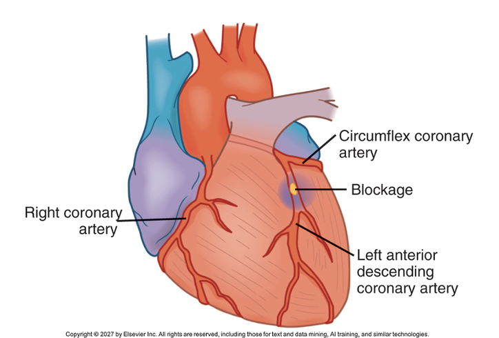
•Left Coronary Artery branches:
•Supply blood to the left atrium, left ventricle, interventricular septum, part of right ventricle
•Left anterior descending artery (LAD)
•Left circumflex artery (LCA)
•“Widowmaker”
•High rate of mortality!
•Right Coronary Artery (RCA)
•Supplies blood the right atrium, right ventricle, part of posterior wall of the left ventricle
•Severe cardiac conduction effects if obstruction occurs here
•Nitroglycerin contraindicated
acute coronary syndrome overview
•Occurs when there is a rupture or erosion of an atherosclerotic plaque with partial or complete coronary artery thrombosis
•Decreased blood flow to the myocardium leads to a myocardial infarction
•Non-ST Segment elevation myocardial infarction (NSTEMI)
•ST Segment elevation myocardial infarction (STEMI)
•Damage occurs over several hours

Unstable Angina
•Associated with worsening ischemia
•Can occur with exertion and/or at rest
•Increases in occurrence, duration, and severity
•Chest pain lasts longer than 15 minutes
•Characteristics of chest discomfort varies among individuals
•Pain may not be relieved with nitroglycerin OR rest
Non-ST Elevation Myocardial Infarction (NSTEMI)
•Caused by a non-occlusive thrombus
•Leads to prolonged myocardial ischemia
•12- Lead EKG changes:
•ST depression
•T wave inversion
•More common in women
•Less severe
•Elevated cardiac enzymes
•Cardiac catheterization within 24 hours
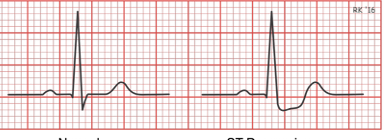
ST Segment Elevation Myocardial Infarction (STEMI)
•Caused by a complete occlusive thrombus
•Confirmed when ST segment elevation seen on 2 different leads of a 12-lead EKG
•Medical Emergency

manifestations of ACS
•Chest pain
•Persistent heavy pressure/tightness, crushing, squeezing, burning sensation
•Substernal and/or epigastric pain
•Pain may radiate
•SNS Stimulation
•Diaphoresis
•Ashen, cool, clammy skin
•Changes in BP/HR
•Cardiovascular Symptoms
•Abnormal heart sounds
•Symptoms of heart failure
•Gastrointestinal Symptoms
• Nausea/vomiting
ACS diagnostics
•Initial 12-lead EKG
•Repeat EKGs as ordered
•STEMI = ST elevation
•NSTEMI = ST depression and/or T wave inversion
•UA = ST depression and/or T wave inversion
•Cardiac enzymes (troponins)
•Proteins released into blood from necrotic heart tissue
•Obtained initially then at 3 and 6 hours
•STEMI = significant elevated troponins
•NSTEMI = elevated troponins
•UA = negative troponins
management of ACS
•MONA
•Morphine
•Oxygen
•Nitroglycerine
•Aspirin-anti-platelet
•Emergent Percutaneous Coronary Intervention (PCI)
•Immediately opens blocked artery
•STEMI patients
•Premedicate with ASA and Brillinta
•May also receive SQ heparin
•Thrombolytic Therapy
•STEMI- within 30 minutes of arrival if PCI not available
•NSTEMI- Not an appropriate treatment option
•Coronary Artery Bypass Graft (CABG)
•May be necessary if area of blockage is high risk for PCI
•May be necessary if several areas of occlusion are involved
percutaneous coronary intervention
•Can be done through the femoral artery or radial artery
•Prep client for cardiac cath with possible angioplasty with stent.
•Dye is used to visualize the coronary arteries
•Not available in all hospitals
stent care
•Low dose aspirin + P2Y12 inhibitor (Plavix or Brilinta) for a full year
•After a year, may only need one or the other
•ASA therapy

Percutaneous Coronary Intervention Post-Procedure
•Monitor ECG for ST & T wave changes
•Assess for angina
•Monitor VS
•Administration of IV fluids (excretion of dye)
•Instruct on restrictions in activity & extremity movement
•Assess catheter insertion site
•Monitor for bleeding & hematoma
•Assess peripheral circulation distal to catheter insertion site
•Monitor retroperitoneal hematoma
•Bedrest protocol (femoral site)
•Care of TR band (radial site)
PCI bed rest protocol
•Determine hemostasis
•Sandbag remains in place
•Lay flat for one hour
•Elevate HOB no more than 30 degrees and maintain bedrest per physician order
•Assess site per protocol
•If bleeding noted, hold firm pressure and call a Rapid Response
PCI TR band
•Determine hemostasis
•Determine total amount of air (cc)
•Begin removing 2-3cc of air every 30 minutes until all air removed
•Assess site for oozing
•Palpate site and feel for ulnar pulse

thrombolytic therapy
•Dissolves clot to restore blood flow through coronary artery
•Reserved for STEMI patients when PCI is not available
•Goal is to administer within 30 minutes of arriving to ED
•Monitor closely for bleeding complications
•Inclusion criteria:
•Chest pain <12 hours with STEMI confirmed with EKG
•No absolute complications
post MI care
•Ace Inhibitors
•Drug of choice
•Help prevent cardiac remodeling
•Antiplatelet therapy
•Nitroglycerin
•Beta blockers
•Lipid-lowering agents
•Aldosterone antagonist
•Spironolactone
•Stool softeners
complications of MI
•Dysrhythmias
•Frequent PVCs warning sign of potential impending VT and VF
•Bradycardias
•Heart failure
•Left-side symptoms first seen
•Cardiogenic shock
•Severe perfusion
•High mortality rate
•Pericarditis
•Occurs 2-3 days after MI
•Dressler Syndrome
•Rare form of pericarditis
•Possibly autoimmune origin
•Occurs 1-8 weeks after MI
•Aspirin is drug of choice
cardiac remodeling
•The heart changes its size, shape, or structure after an MI
•Occurs over several months
•The heart muscle may thicken, or the chambers may stretch and enlarge.
•These changes may help at first, but over time the heart can become stiff or weak.
•The heart pumps less effectively, which can lead to or worsen heart failure.

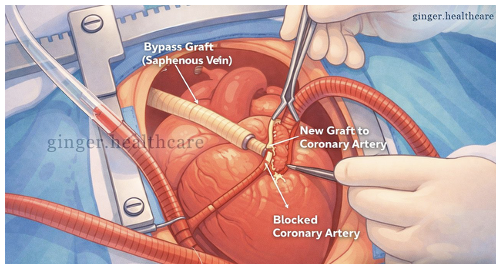
Coronary Artery Bypass Graft Surgery
•Bypass blocked artery to allow for perfusion to cardiac muscle below blockage
•Graft may be harvested from:
•Saphenous vein (lower leg)
•Radial artery (forearm)
•Internal mammary artery (chest)

CABG cont.
•Sternotomy is performed
•Blood is diverted from the heart to a machine, oxygenated and returned to the patient
CABG post op
•Monitor for bleeding
•Chest tube drainage
•Incision sites
•Monitor for fluid and electrolyte imbalance
•Maintain close hemodynamic monitoring
•Replace fluids/blood/electrolytes
•Monitor for infection
•Assess incision sites
•Evaluate fever
•Manage pain
•Medications
•Splinting techniques
•Prevent DVT
•Early ambulation
•SCDs
•Respiratory Complications
•Incentive spirometer
•Dysrhythmias
•Atrial fibrillation is common
cardiac rehab
•Phase I
•Occurs while patient is in hospital
•Activity level varies among individuals
•May sit up in bed or chair, perform ROM and self care
•Progresses to ambulating in halls and using stairs
•Focus on management of pain, anxiety dysrhythmias, and complications
•Phase II
•Begins after discharge
•Usually lasts 2-12 weeks
•Increase activity level determined by HCP
•Risk factor reduction teaching
Phase III
•Long-term maintenance program
•Transition to independent exercise and exercise maintenance
•Lifestyle changes should become lifelong habits
sudden cardiac death (SCD)
•Abrupt, unexpected cardiac death
•Usually occurs within an hour of symptom onset
•Life threatening dysrhythmia causes abrupt loss of cardiac output and cerebral blood flow
•VF or VT
•SCD may be first sign of CAD in up to 50% of patients
•Manifestations- chest pain, palpitations, dizziness, or lightheadedness
•Treatment- Cardiac enzymes to rule out MI, EKG, cardiac catheterization, PCI, CABG
•Electrophysiology study
•Holter monitor (48 hours/ 30-day event monitor
•Implantable loop recorder (3 years)
•Internal cardiac defibrillator (ICD)
•Gold standard