619 9/30 GLA pt 2 Functional UL
 Call Kai
 Learn
 Practice Test
 Spaced Repetition
 Match
 Flashcards
 Knowt Play
1/46

There's no tags or description
Looks like no tags are added yet.
47 Terms
Shoulder Imaging
typically done secondarily for pain and trauma
obtained AP or PA with external and internal rotation
occasionally axillary and axillary y views are done, especially for dislocation/subluxation/scapular fracture
clavicle is a separate radiographic study
When you perform an axillary or axillary y imaging?
shoulder dislocation, shoulder subluxation, scapular fracture
Imaging Modality Strengths

Normal Shoulder AP Film
look at long component if they have chest pain, as that could be pneumothorax
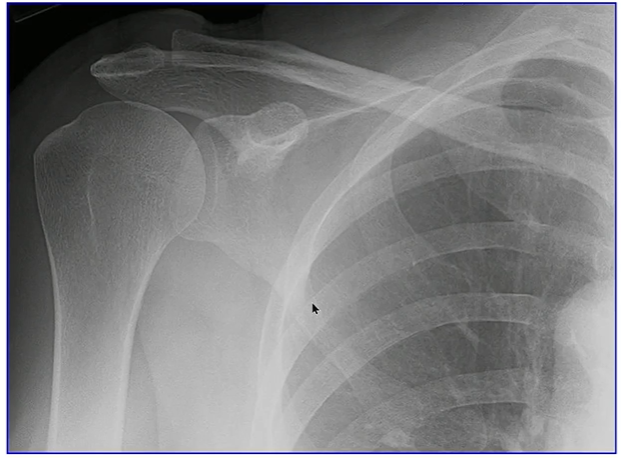
Shoulder AP X-Ray Labeled

Shoulder Y-View
lots of mach overlap, sometimes difficult interpretation but can be useful for dislocation/sublocation

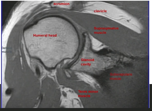
Coronal T1 Shoulder MRI
Calcific Tendonitis
chronic pain from calcification

Osteoarthritis
especially common on the shoulder joint of the side of the dominant hand
can have bony spurs

PA and Lateral Humerus Radiograph

Axial T1 MRI Through Arm

Elbow AP Labeled

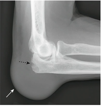
Lateral Elbow X-Ray

Olecranon Bursitis
often due to gout
Wrist AP X-Ray

Hand PA X-Ray

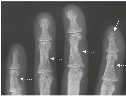
Subperiosteal Resorption in Phalanges
commonly due to hyperparathyroidism
loss of bones in phalanges
Distal Forearm/Wrist T1 MRI Axial
median nerve is especially important

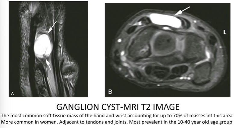
Ganglion Cyst
most common soft tissue mass of the hand and wrist, more common in women adjacent to tendons and joints
most prevalent from 10-40 yo
Carpal Tunnel Borders
transverse carpal ligament spanning from the pisiform medially to scaphoid bone laterally
base is formed by remaining carpal bones of hand
Contents of Carpal Tunnel
median nerve
flexor tendons (flexor pollicis longus, flexor digitorum profundus tendons, flexor digitorum superficialis tendons)
Carpal Tunnel Syndrome
compression of median nerve as it travels though carpal tunnel
pain or paresthesia in area innervated by median nerve, weakness of hand or clumsiness, worse at night or when wrist is flexed/extended
Sensory Innervation of Median Nerve

Carpal Tunnel Syndrome Diagnosis
initial is based on clinical presentation, electrodiagnostic studies can be used to comfirm
Ultrasound for Carpal Tunnel
it is now being used a little, and could detect abnormalities even in the presence of normal nerve conduction studies
Carpal Tunnel Ultrasound Procedure
supinate wrist on comfortable and stable surface, place linear probe at crease of wrist
Wrist Ultrasound Labeled

Short Axis Carpal Tunnel Medial

Short Axis Carpal Tunnel Lateral

Short Axis Carpal Tunnel Distal

Structures to Identify on the Carpal Tunnel Ultrasound
pisiform and scaphoid bones
transverse carpal ligament
flexor tendons
median nerve
radial artery
ulnar artery
Erb’s Palsy Damage
C5-6, over-separation of head and shoulder
Motor Effects of Erb’s Palsy
localized to superior part of upper limb
weakened shoulder abduction (deltoid m) → adducted at rest
weakened shoulder flexion (anterior arm)
loss of shoulder lateral rotators → medially rotated at rest
Sensory Effects of Erb’s Palsy
C5 & 6 dermatomes
lateral shoulder loss (axillary n)
lateral arm loss (from branch of radial n)
lateral forearm loss (from lateral cutaneous n of forearm)
Wrist Drop
when additional injury caused by erb’s palsy impacts C7, resulting in loss of forearm supinators and weakened extension by radial n, also loss of sensation on C7 dermatome
Wrist Drop Test
test radial n, hold wrist normal, wrist and digits will flex
Klumpke’s Palsy Damage
loss of C8-T1 becasue of over-seperation fo shoulder and trunk
Klumpke’s Palsy Motor Effects
total claw hand
forearm digit flexors unaffected
Klumpke’s Palsy Sensory Effects
loss of sensation over majority of hand (medial, ulnar, radial n)
Total Claw Hand
damage to the inferior trunk (with klumpke’s palsy) causes the dip and pip joints to flex, while the mcp joints extend
Additional Issues that May Occur with Claw Hand
sensory loss to C8-T1 dermatomes
damage associated with proximal branches that arise from the inferior trunk
Ulnar Claw/Partial Claw
injury to ulnar nerve, patient is asked to extend fingers
mcps will hyperextend, pip and dip will flex (often more prominently in digits 4 and 5)
Additional Issues That Might Occur with Ulnar Claw
wasting (atrophy) of hypothenar eminence
loss of sensation to medial hand
“Ape Hand” Deformity
damage to recurrent branch of median n, patient is asked to touch fingertips
they will be unable to oppose and have an adducted thumb
Additional Issues That Might Occur with “Ape Hand” Deformity
wasting (atrophy) of thenar eminence, if the injury is proximal then there will also be loss of sensation on some of the lateral hand
Benediction Sign (Hand of Benediction)
damage to proximal median n, patient is asked to make a fist
digits 4&5 will flex, digits 1-3 will extend
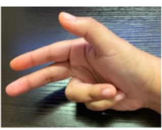
Additional Issues That Might Occur with Benediction Sign (Hand of Benediction)
wasting of thenar eminence, loss of sensation to lateral palmar hand