Ankle & Tib/Fib
 Call Kai
 Learn
 Practice Test
 Spaced Repetition
 Match
 Flashcards
 Knowt Play
1/34

There's no tags or description
Looks like no tags are added yet.
35 Terms
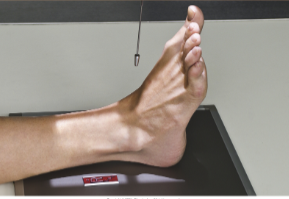
AP ankle
10x12 lw
Center ankle joint to IR
CR perpendicular to IR directed midway between malleoli
Dorsiflex foot
AP Ankle Image Analysis Criteria
Structures Shown:
Distal 1/3 of the tibia-fibula, lateral and medial
malleoli, talus and proximal half of the metatarsals.
Position:
The long axis of the leg should be aligned to collimation
field and to IR.
No rotation
medial mortise joint is open
lateral mortise closed
Some superimposition of the distal fibula by the distal tibia
and talus
AP Ankle Image Analysis Criteria Collimation and CR:
CR to the tibiotalar joint midway between the medial and
lateral malleolus.
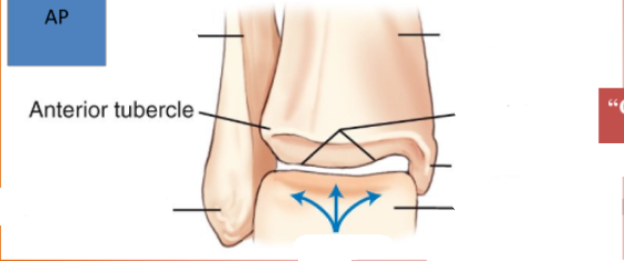
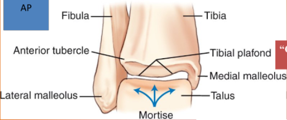
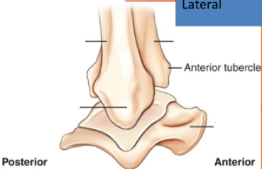
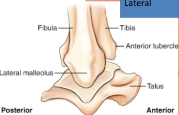
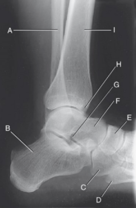
Label
A: Body/shaft of Fibula
B. Calcaneus/Os Calcis
C. Cuboid
D. Base of tuberosity of the 5th metatarsal
E. Navicular/scaphoid
F. Talus/Astragalus
G. Sinus Tarsi/ Tarsal sinus
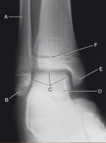
Label
A. Distal Fibula
b. Lateral Malleolus
c. Lateral/superior/Medial Mortise
d. Talus/Astragalus
e. Medial Malleolus
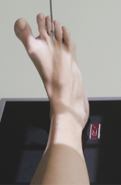
AP Medial Oblique “Mortise”
10x12 lw
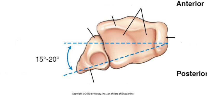
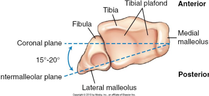
Intermalleolar line parallel to IR (15-20 degrees)
CR midway between malleoli
AP Medial Oblique (Mortise) Ankle Image Analysis Criteria
Structures Shown:
Distal 1/3 of tibia and fibula, tibial plafond, lateral and
medial malleoli, talus, and proximal half of the metatarsals.
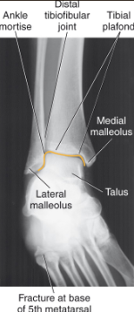
Entire ankle mortise open
3 to 4 mm space over entire talar surface =normal
extra 2 mm widening = abnormal
Position:
Proper obliquity for the mortise joint:
Open lateral and medial mortise joints
Malleoli in profile
Minimal superimposition of the distal tibiofibular joint.
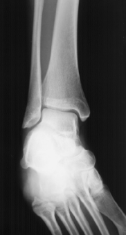
Label medial oblique Mortise
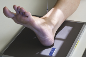
AP 45 degree Medial Oblique
Medial rotation 45 degrees.
Need to rotate entire leg
Dorsiflex foot so planter surface is perpendicular
CR midway between malleoli
Some facilities require the base of 5th metatarsal included.
Why? Might be a fracture there
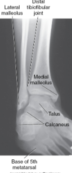
AP Oblique (45 degree) Ankle Image Analysis Criteria
Position:
A 45° medial oblique:
distal tibiofibular joint open, with no or minimal
overlap.
The lateral malleolus and talus joint:
no or slight superimposition
medial malleolus and talus partially superimposed.
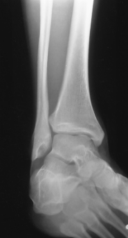
Label AP 45 degree medial oblique
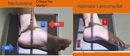
Mediolateral Lateral Ankle
Center ankle joint to IR.
CR perpendicular to medial malleolus.
Dorsiflex foot so plantar surface is at a right angle to leg, do not force
Lateral Ankle Image Analysis Criteria
Structures Shown:
Distal 1/3 of tibia and fibula with the distal fibula superimposed by the distal tibia
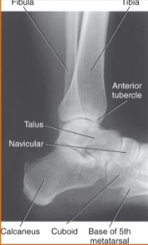
The talus, and calcaneus will appear in lateral profile.
The tuberosity of the 5th metatarsal, the navicular, and cuboid also visualized.
Position:
No rotation:
Distal fibula superimposed over the posterior half of the tibia.
The tibiotalar joint open.
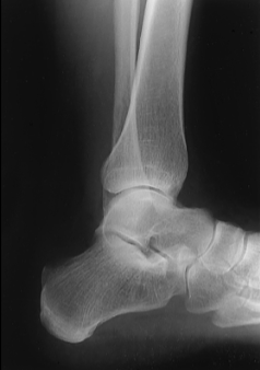
Label Mediolateral lateral ankle
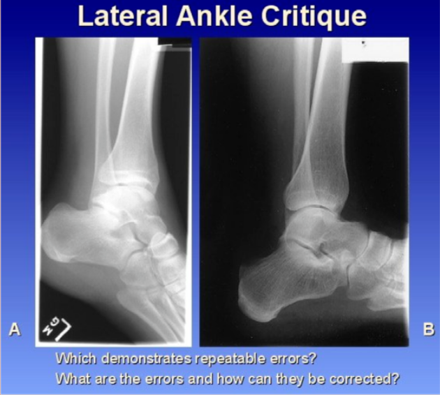
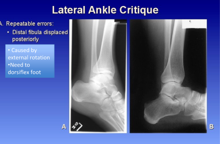
Positioning error for lateral position
Positioning error:
– Talar domes should be superimposed.
• Separation of domes anterior/posterior = rotation
– Fibula should be on posterior half of tibia. If not, indicates rotation. (lateral ankle is not parallel to the IR = rotation
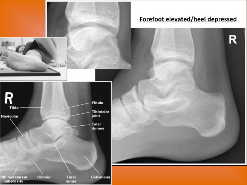
Forefoot (toes) away from IR = leg internally rotated ( Lateral Ankle)
Distal fibula on the mid/anterior portion of the tibia
Lateral talar dome visible anteriorly
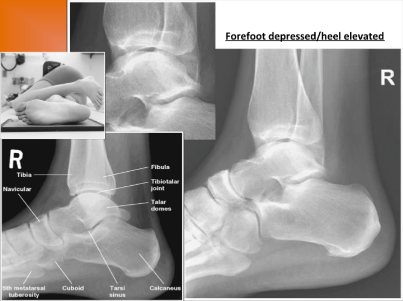
Heel away from IR = leg externally rotated ( Lateral Ankle)
Distal fibula posterior to the tibia
Lateral talar dome visible posteriorly
Lower Leg Tilt Error ( Lateral ankle)
Positioning error: Tibiotalar joint closed
– Talar domes should be superimposed.
• Separation of domes superior/inferior = tilt
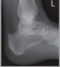
Knee elevated (proximal lower leg) above ankle (distal
lower leg)
Lateral dome superior
– Cuboid superimposing with navicular
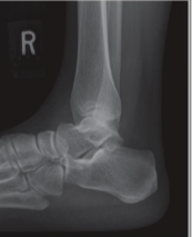
Knee depressed (proximal lower leg) below ankle (distal
lower leg)
Medial dome superior
– Lots of cuboid separating from navicular
Axial view Label
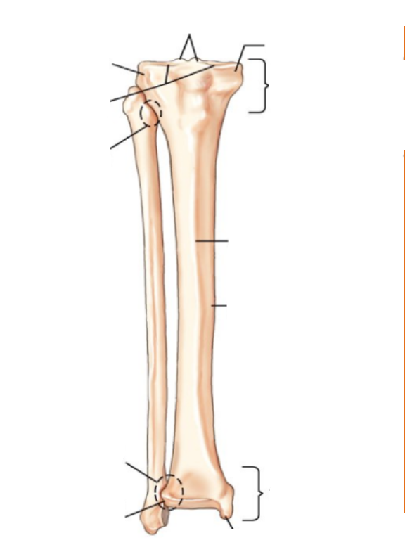
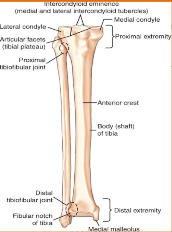
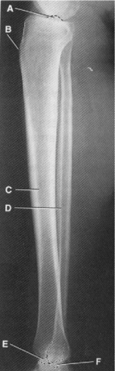
Label AP Tib/fib
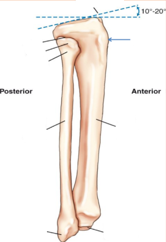
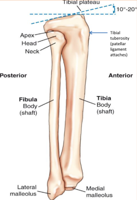
Label Mediolateral Lateral Tib/fib
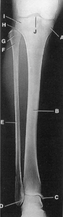
Label AP Tib/fib
A. Medial condyle of the tibia
B. Body or shaft of tibia
C. Medial malleolus
D. Lateral malleolus
E. Body or shaft of fibula
F. Neck of fibula
G. Head of fibula
H. Apex (styloid process of
head) of fibula
I. Lateral condyle of tibia
J. Intercondylar eminence
(tibial spine
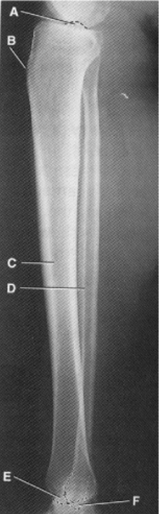
Label Mediolateral Lateral Tib/fib
A. Intercondylar
eminence (tibial spine)
B. Tibial tuberosity
C. Body or shaft of tibia
D. Body or shaft of fibula
E. Medial malleolus
F. Lateral malleolus
Positioning the Tib/Fib
Protocol
– AP
– Mediolateral Lateral
No Grid
Small focal spot
AP Tib/Fib
14x17 diagonal (requires 44 in.
SID minimum. WHY? Divergence of beam
Include both ankle and knee joints on IR (this means entire
tibia and fibula).
CR perpendicular directed to midpoint of leg.
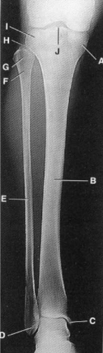
AP Lower Leg Image Analysis Criteria
Entire tibia and fibula included with both the ankle and knee joints demonstrated.
Position:
No rotation:
Femoral and tibial condyles in profile
Intercondylar eminence centered within the intercondylar fossa.
Overlap of fibula and tibia visible at both ends.
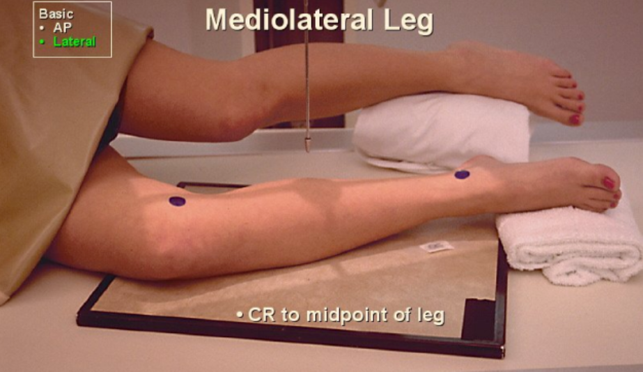
Mediolateral Lateral
Flex knee 45 degrees
Plane of patella perpendicular with IR
Same SID as AP
Both knee and ankle joint included on IR – this means
to include entire tibia and fibula)
Lateral Lower Leg Image Analysis Criteria
Structures Shown:
Entire tibia and fibula with both ankle and knee joints on one (or two if needed) IR(s). (Exception is alternate routine on follow-up exams.)
Position:
True lateral without rotation:
– Tibial tuberosity in profile
– Portion of the proximal head of the fibula superimposed by the tibia
– Outlines of the distal fibula seen through the posterior half of the tibia.
Posterior borders of femoral condyles appear superimposed
AP Tibia and Fibula Rotation
Internal rotation: Tib/fib separating
External Rotation: Tib/fib superimposing
Lateral Tibia and Fibula Rotation
Internal rotation: Tib/Fib superimposing
External rotation: Tib/fib separating