Medsurg Exam III
 Call Kai
 Learn
 Practice Test
 Spaced Repetition
 Match
 Flashcards
 Knowt Play
1/204

There's no tags or description
Looks like no tags are added yet.
205 Terms
Neurological Assessment
Cerebral Lobes
Frontal
Parietal
Temporal
Occipital

Front Lobe Function
Located in the front area of the brain and is largely responsible for motor movement and personality.
Motor cortex. voluntary movement
Broca’s expressive speech center - dominant hemisphere
Personality
Behaviors: social, sexual, judgement, and problem solving
Parietal Lobe
Found behind the frontal lobes
Controls sensation and perception - sight, taste, hearing, smell, touch
Plays a role in social relationship such as body position
Integration of sensory input, such as visual input
Being able to assess size, shae, and orientation in space of both visible stimuli and objects you see.
Temporal Lobe
Located on the sides of the brain
Auditory sensation and perception
Being able to hear and understand speech, it gives the ability to understand and give meaning to language
Being able to control unconscious and apparently automatic reactions such as appetite, thirst, and hunger.
Long term memory
Wernicke’s receptive speech center
Allows for the production of speech
Occipital Lobe
Located at the back of the brain, and is responsible for vision
Perception of color and shapes
Glascow Coma Scale
Used to assess level of consciousness, identifying patients responsiveness and orientation to person, place, or time.
the GCS assigns a score for eye-opening, best motor response, and verbal response.
Highest possible score: 15
With a score of 8 or less indicating severe neurological issues.
Components of the Glasgow Coma Scale
Best Eye Opening
Best Motor Response
Best Verbal Response
Best Eye Opening
Spontaneous - 4, responds no neccessary to command
To speech - 3, Opens eye only to verbal stimuli
To pain - 2, opens eyes only to painful stimuli
None - 1, doesn’t open eyes
Best Motor Response
Obeys commands - 6, obeys commands
localized - 5, purposeful movement to pain
withrdraws (flexion) - 4, withdraws from painful stimuli
abnormal flexion - 3, flexion in response to pain
abnormal extension - 2, extension in response to pain
flaccid - 1, no response to pain
Best Verbal Response
Oriented - 5, Orientated to person, place, and time
Diagnostic Tests
CT (computed tomography) or CTA (CT angiography)
Magnetic Resonance Imaging (MRI) or MR angiography (MRA)
Lumbar Puncture (LP)
Electroencephalography (EEG) & Biopsy
CT or CTA
Uses cross-sectional images that are viewed as cuts or slices that allow the provider to view the brain, spine, and orbits in a quick, NONINVASIVE manner.
Used to assess for bleeding, edema or masses
It is a required screening test for someone with suspected ischemic stroke - CT is used to rule out hemorrhagic stroke which is a contraindication to thrombolytc therapy.
Nursing Implication - CT or CTA
Provide patient education for the procedure
Mention that it is noninvasive and painless
Ensuring a patent IV line is in place
Checking for allergies such as shellfish or dyes, because contrast ontaines iodine
Monitoring BUN and creatinine to assess kidney function before contrast because it is toxic to the kidney and can impair renal function
Assess for diabetes DM and use of metformin in the management of type 2 diabetes, if yes - hold metformin 48 hours before to avoid renal impairment
Providing post procedure care
Administering IV fluids or increasing intake of oral fluids to clear contrast mediun from the system
Monitoring if allergic reactions occur due to the contrast.
Magnetic Resonance Imaging (MRI) or MRA
Type of MRI that uses radio-wave signal characteristics of flowing blood to get images of the body’s blood vessels.
MRA - Used to determine the presence of aneurysms, clots, dissections, or vessel stenosis.
Diagnosis of tumors, infections, and bleedings
Diagnosis of cerebrovascular accident (CVA)
Nursing Implications of MRI and MRA
Patient education prior about the procedure
It is noninvasive and painless
Non Iodine contrast - there is no need to worry about kidney damage
Ensure that the patient doesn’t have any metal objects
Check patient for any cardiac implantable electronic device, mechanical metallic heart valve(s), metallic foreign bodies, implantable neurostimulation systems, etc.
Assess the patient for claustrophobia and anxiety (long-test and causes lots of noises) - patient may require sedation.
Ensure the patient receives adequate hydration post procedure
Lumbar Puncture (LP)
Common procedure done to obtain a sample of cerebrospinal fluid (CSF) for the analysis and to measure intracranial pressure.
Increased pressure can indicate CSF obstruction or overproduction.
Presence of abnormalities in glucose or protein as well as a cloudy color = infection.
Presence of breakdown products of RBCs, known as xanthochromia or frank blood can indicate subarachnoid hemorrhage or a traumatic tap.
Lumbar Puncture can also be used for spinal anesthesia and intrathecal medication or to remove CSF from the spinal cord to reduce pressure.
Pt positioning during Lumbar Puncture
Requires the patient to be positioned laterally (on the side) in bed, drawing the knees up toward the tress. Or the patient can be leaning cross a bedside table with the back arched.
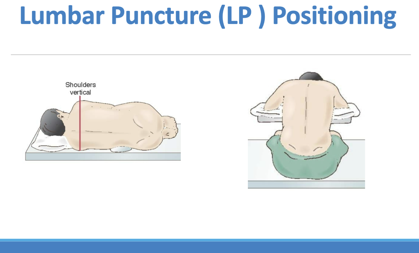
the procedure occurs with a puncture of a spinal need into the L3 to L4 space of the spinal column below the level of the spinal cord.
Nursing Implication of LP
Provide education for the patient about the procedure
Providing preprocedure care:
Ensuring that antiplatelet or anticoagulation medication are held prior to the test to reduce risk of bleeding.
Checking coagulation studies prior to test
Informed consent is obtained
Post LP Care
Ensuring flat bedrest for 4-6 hours after the test to help prevent CSF leakage, which can cause severe headrest
Encouraging fluids post procedure to decrease headache intensity if present.
EEG & Biopsy
EEG is an inexpensive, nonvasive procedure that uses 8 to 24 scalp electrodes to trace the spontaneous electrical activity in the brain.
Diagnostic test for seizure activity
Used during sleep study.
Nursing Implication for EEG
No caffeine 8-12 hours prior
Wash hair, no hair products
Brain Biopsies
Tissue sample to identify tumor type infection, or abscess
Types:
Needle
Stereotactic
Open
Needle
drilling a small hole in the skull and a needle is inserted to obtain tissue
Stereotactic
Uses specialized computer imaging that utilizes a 3D coordinate system to pinpoint area
Open
Performed OR, removing portion of bone from the skull.
Brain Disorders
Headaches
Also referred to as cephalalgia
There are two types of headaches: primary and secondary
Epidemiology of Headaches - clinical manifestation
Common Manifestations of pain, accounting for more missed days of work and school, and more visits to healthcare providers.
20-40% of the US population reports having migraines (women>men, greater than 2:1)
WHO mentiones that it is in the top 10 ranking for disabling conditions, ranking headaches in the top 5 for women
Primary Headache
Not associated with underlying causes
Typically is a migraine, tension, or cluster.
Secondary Headache
Caused by an underlying pathology
Typical presentation: sudden onset, severe pain
Due to infection (meningitis), neoplasm (tumor), vascular (blood vessel aneurysm or rupture)
Secondary Headache Diagnosis
CBC and Blood Culture to detect infection
CRP and ESR (C-reactive protein, erythrocyte sedimentation rate) - checks for inflammation
CT and MRI (check for masses, cysts, vessel, and osseous skull abnormalities)
EEG used to check for seizures, tumors, inflammation, and brain injury.
Sleep studies (check for fatigue & sleep apnea)
Brain Tumors
Tumors that originate in the brain and range from slow-growing, benign tumors to highly malignant, aggressive tumor
Types of Brain tumors:
Primary Brain Tumors
Metastatic Brain Tumors
Primary Brain Tumors
Originiate from brain cells, brain meninges (membranes covering the brain), nerves, and glands.
Male individuals typically having a higher number
Can be both malignant and benign meningioma
Malignant Tumors
Grade I/II - Due to Glioma
Grade III - Anaplastic Glioma
Grade IV - Glioblastoma Multiforme
Oligodendrogliomas
Gliomas
Generally originiate in the cerebrum - glial cells provides the physical structure of the brain and supports endothelial cells of the blood-brain barrier.
Glial cells provide nutrients and ionic balance that are involved with repair and scarring process.
Gliomas are typically developed along the curved areas of the brain, making the frontal lobes more susceptible, typically found in patients between the ages of 20 and 50 years old, they are slow growing tumors.
Anaplastic Glioma
Grade III tumors cells out of control, lacking differentiation and/or orientation to one another and to surrounding vessels.
Glioblastoma Multiforme
Grade IV astrocytoma and is the most aggressive and lethal type of tumor, especially when located in or near the brainstem.
Oligodendrogliomas
Arises from oligodendrocytes, or oligodendroglia and their main functions are to provide support and insulation to axons in the CNS, equivalent to the function performed by Schwann cells in the PNS and they are slow-growing tumors that generally don’t spread to surrounding tissue.
Typically found in middle-aged patients.
Benign Tumor Types
Meningioma
Acoustic Neuromas
Schwannomas
Pituitary Tumors
Meningioma
Most common form of brain cancer - 90% are typically benign but cause significant damage based on its location.
Acoustic Neuromas
CN VIII, which are slow-growing benign tumors that generally don’t invade other tissues, however compression on other cranial nerves and tissue (cerebellum and brainstem) can manifest in severe complication.
They are also known as schwannomas, and they originate from the protective covering around nerve fibers (CN VIII) at the anatomical location of the cerebellopontine angle.
Pituitary Tumors
Generally found in the anterior lobe of the pituitary.
Most common of which is the adenoma and the clinical manifestations are pituitary adenomas related to hypersecretion of hormones by the pituitary gland including acromegaly from hypersecretion of growth hormone, amenorrhea-galactorrhea from prolactin hypersecretion.
Brain tumor Management
Chemotherapy
Gliadel Wafers
Radiation Therapy
Cyberknife
Chemotherapy
Cross the BBB (blood brain barrier):
Temozolomide ( radiosensitizer)
Carmustine, Lomustine, Methotrexate
Temozolomide ( radiosensitizer)
Used to treat brain cancer, it inteferes with the growth of cancer cells, which are then destroyed by the body.
Carmustine
Chemotherapy medication that works by damaging the DNA cancer cells to stop them from growing and dividing.
Giladel Wafers
Impregnated with chemotherapy and put in the surgical bed at the time of surgical resection
Side effects to be aware of:
Neutropenia (susceptibility to infection), nausea, vomiting, fatigue, mucositis, hair loss, sterility.
Radiation Therapy
Concentrated on the affected area and it destroys both tumor and normal cells by damaging cellular DNA.
Side effects includes:
fatigue, redenning of the skin (sunburn appearance), headache, swelling, visual changes, nuerological disturbances, hearing loss, and facial numbness.
CyberKnife
A technology that is used to direction radiation to the tumor, avoiding healthy brain tissue
Seizure
An uncontrolled, sudden, excessive discharge of electrical activity which results in a range of manifestations from changes in behaviors to loss of consciousness.
Types of Seizures:
Epilepsy
Cryptogenic Seizure
Nursing Assessment for Seizures
Check for Airway and breathing
Maintaining airway patency during seizure and postictal state
Decreased level of consciousness
Vital Signs
Oxygen Sat and RR during a seizure.
HR and BP may increase during a seizure
Tonic movements (muscle activity characterized by a sudden, sustained increase in muscle tone and stiffness)
Clonic Movement (rhythmic, rapid, and repetitive contraction and relaxation of a muscle, typically resulting in unstoppable shaking or jerking)
Loss of Consciousness
Aura (being fully conscious and alert - warning sign before a larger seizure)
Automatisms (involuntary, stereotyped, and repetitive behavior performed without conscious self-control, intention, or awareness)
Loss of Motor Tone
Nursing Interventions during Seizure
Setting up suction equipment at the patient’s bedside - important to provide assistance with airway.
Have oxygen available at the patient’s bedside
During the postictal phase, the pt may require supplemental oxygen to maintain the oxygenation saturation.
Safety Measures
ensuring bed is at lowest position, suction at bedside, and oral airway is at the bedside.
For someone who just experienced a seizure, protection includes not forcing any object into the mouth (for fear of biting tongue, breaking teeth, causing injurt, etc), loosening of restrictive clothing, and turning the patient to the side (preferably the left to reduce the risk of aspiration)
Place an IV catheter per orders
IV access may be needed to stop the seizure activity.
Document specific of seizure activity
date, time, and duration of the seizure; clear description of the seizure, sequence of the seizure progression, and precital and postictal observation.
Pt education (seizure)
Medication regimen
Control of seizure activity is determined by therapeutic levels of prescribed antiseizure medications.
Medic Alert Bracelet
Important for pts with seizures to wear a Medic Alert braclet in event of seizure activity and it facilitates prompt interventions in the event of a seizure.
Driving Restrictions
Important for pt to research driving restrictions.
Parkinson’s Disease
Progressive, neurodegenerative disease of the CNS manifesting primarily in motor dysfunction
Caused due to idiopathic origin (unknown), environmental toxins - pesticides and herbicides, brain injury, brain tumors, and use of antipsychotic medication.
PD is typically found in men more and generally begins the ages of 40 and 70 years.
in US it is typically prevalent within Amish communities.
Pathophysiology of Parkinson’s Disease
It is a motor system disorder, which involves the substantia nigra. this is responsible for transmitting signals to allow for dopamine and GABA release, and it allows for motor functions to occur, along with regulating muscle tone and posture.
Motor activity is the result of the release of dopamine and Ach
Due to the a mutation defect in the gene, there is a deterotiation of the substantia nigra and it decreases the amount of dopamine in the brain.
Clinical Manifestations of Parkinsons
Resting tremors
Muscle Rigidity
Bradykinesia
Postural instability
Bradykinesia
Slowness of voluntary movement
“limbs feel heavy”
everything becomes a lot of work
causes a masked face - slow blinking
Resting Tremor
Usually shows in hand as steady, rhythmic shaking
Disappears with movement
tremors can also affect legs, jaws, or lips it is worse when stressed or anxious
Rigidity
Muscle stiffness and resistance to passive movement
Postural Instability
Due to weakness of the body, which can also lead to falls
It is a loss of balance
Progressive Stages of Motor Dysfunction
Initial Phase:
Presents with unexplained unilateral weakness and upper extremity tremors.
Later Phase:
Slow shuffling gate
Postural Instability
Stooped Posture
Rigidity
Generalized tremor
Maslike face
Parkinson’s Diagnosis Factors
When there are two or more observable symptoms with asymmetry on presentation:
Bradykinesia
Resting Tremor
Rigidity
Postural Instability
No defintive test to confirm PD
ch 37: Spinal Cord Disorders
Spinal Column
Responsible for the spinal cord and the nerve pathways that carry info between the brain and rest of the body.
parts of the spine

Butterfly of the Spinal Cord
Gray Matter of the Spinal Cord and is surrounded by white matter
This is the processing center bu recieving and integrating information.
Gray Matter
contains motor neurons that control movements
White matter
contains the conduction tracts for nerve impulses and the tracts of the nerve fibers consists of axons, which are going to carry impulses away from the cell body.

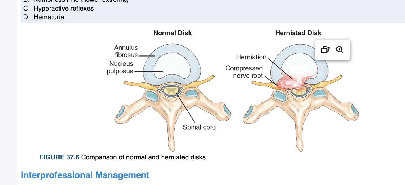
Herniated Disc
Vertebral disk rupture or tearing that causes leaking into the vertebral area resulting in back pain

Epidemiology of Herniated Disc
As people age, the disk between the vertebrae degenerates and weakens, so the disk becomes more inflexible and more prone to tearing or rupturing with movement.
a herniation occurs when the leaking out of the interior nucleus disk contents into the vertebral areas
Tends to occur more in men.
Risk Factors of Herniated Disc
Age + gender
Smoking, obesity, and occupations.
Clinical Manifestation
Presents with pain caused by the inflammatory process and compression to nerve roots.
Radiculopathy (a nerve root compression) which can result in numbness in the affected extremity as well as pain, weakness, and inability to control motor m
ovements in the affected area.
Diagnosis of Herniated Disc
History and Physical, with focused neurological examination to test:
reflex
Muscle strength
patients ability to feel touch and vibration to localize the affected lvel
Tests used:
Imaging studies such as - x-ray, CT scans, MRIs, and myelograms.
Additional neurological tests used:
EMG and nerve conduction studies
These are used to pinpoint the location of nerve damage.
Spinal Cord Injuries
Unexpected catastrophic event that results in the loss of function such as mobility or sensation.
Spinal cord trauma can be due to direct injury to the cord or indirect injury to surrounding bones, tissues, and blood vessels,
Risk Factors of Spinal Cord Injuries
Typically occurs in males between the ages of 29 and 43.
Activities such as participating in high-risk physical activities such as speeding and drinking while under the influence of alcohol, substance use, and not using protective gear in sports or recreational activities.
Most common places for spinal cord injuries
C4-C6
T12
Clinical Manifestation of Spinal Cord Injury
The level of injury predicts what part of the body is affected:
Cervical injuries
Quadriplegia
C4 and above → inability to breathe, phrenic nerve innervates the diaphragm at this level
Thoracic Injuries
Paraplegia with trunk and leg involvement, normal arm and and movement
T6-T12: paraplegia with fair ability to control balance and trunk, little or no voluntary bowel or bladder control.
Lumbar and Sacral Injuries:
Leg control, bowel and bladder function, sexual function (functional losses)
Other Effects
Hypotension
Chronic Pain
Decreased temperature control
Conservative Treatment for Spinal Cord Injuries
Once spinal cord damage occurs, it is not reverisble
Acute Stage
Treatment is focused on maintaining airway patency, adequate breathing and oxygenation, preventing spinal shock, restoring and maintaining blood pressure, preventing further cord damage, spinal immobilization, and avoiding possible complications.
Rapid vital signs!!
Nursing Assessment of Spinal Cord Injuries
Respiratory Function
Check for hypoventilation (intercostal muscle paralysis)
C4 and higher (no diaphragmatic innervation)
Vital Signs
Loss of sympathetic input
Spinal shock
Neurogenic Shock
Respiratory or cardiac arrest
Autonomic dysrexia
Patients are unable to regulate their temperature , and hypothermia can occur due to the loss of control of blood vessels.
Nursing Intervention for Spinal Cord Injuries
Maintain suction equipment at patient’s bedside (with decreased cough effectiveness - the patient may require suctioning to clear the airway)
Facilitate cough effectiveness
Maintain spinal immobilization and stabilization
Passive ROM
Reposition and maintain good alignment
prevent pressure injuries and decreasing risk of DVT due to immobility
Perform routine pin site care
Patient Education for Spinal Cord Injury
Ensure information about respiratory distress
Clinical manifestations of autonomic dysreflexia
Skin care/management
prevent skin breakdown
Amyotrophic Lateral Sclerosis
Also known as Lou Gehrig’s disease where there is rapid degradation of the motor nerves.
It is a fatal CNS (brain and spinal cord) disease that affects voluntary muscle control and it leads to muscle atrophy.
It is more prevelant in males.
Clinical Manifestation of ALS
Cause is unknown.
Clinical manifestation typically appear in males between ages of 55 and 75.
Early Symptoms:
Muscle cramps or stiffness
Muscle weakness in one part of the body
Difficulty swallowing (dysphagia)
Slurred speech
As the disease progresses it can affect the diaphragm, and an artificial airway is needed
Complications of ALS
Aspiration of fluid or food due to dysphagia
Respiratory failure
Pneumonia
Pressure injuries
DVT
PE
Constipation
Contractures
Depression
Weight loss
Loss of the ability to care for self.
Nursing Assessment
Check the airway
Motor weakness involves muscle of the face, mouth, and neck, maintaining an intact airway is compromised.
Airway comprise is greater as the patient demonstrates weakened cough and impaired swallowing
Check O2 saturation
Motor Strength
Muscle weakness increases as the disease progressing due to neuronal degeneration and cell death
Ability to swallow
increased risk of aspiration
Skin
immobility promotes skin breakdown due to the compresison of soft tissue between a bony prominence and an external surface.
Coping skills
assess effectiveness of current skills and identify support syste,
Nursing Intervention ALS
Check for ROM
Administer Medications to treat clinincal manifestations
alleviate muscle cramps, pain, constipation, fatigue, depression, etc.
Elevate the head of the patient’s bed when eating, drinking, or brushing teeth
Difficulty with swallowing
Turn, cough, deep breathing
Turn scheduled every 2 hours
Emotional Support
Renal Failure
Partial or complete inability of the kidney to filter waste products and water from the blood through glomerular and tubular filtration.
Types of Renal Failure
Acute Kidney Injury
Chronic Kidney Disease
Renal Replacement Therapies
Acute Kidney Injury
Sudden, rapid decrease in renal function, which leads to the build up of waste, fluid and electrolyte imbalance.
It can be reversible if it is managed in a timely manner.
The injury can be in different places of the kidney
Types of AKI
Pre-renal
Infra renal
Post renal
Prerenal Injury
Most common cause of AKI
It is caused due to an external factor, where there is an issue with perfusion to the kidney, which decreases the function of the kidney
There is decreased blood flow to the kidney, which prevents it from filtering and it deprives kidney nutrients.
Prerenal Causes
Typically a cardiac issue, external bleeding, dehydration, burns. This all leads to causes of
Hypovolemia
Decreased Cardiac Output
Decreased Peripheral Vascular Resistance
Vascular Obstruction
While this occurs, the renal system senses poor perfusion and activates the RAAS to maintain BP. If perfusion remains low, there is damage to the renal issue.
Infrarenal AKI
Impaired nephron functioning and causes decreased ability to filter blood, remove waste and this leads to excessive water and lack of maintain electrolyte levels.
Infrarenal Causes
Prolonged ischemia
Nephrotoxic drugs - NSAIDS, aminoglycosideantibiotics (tobramycin, gentamicin, neomycin)
Contrast dye using imaging studies
Chemo drugs
Infection - glomerulonephritis
Hemoglobin released from RBC hemolysis
Myoglobin released from necrotic muscle cells.
Post Renal AKI
Blockage in the urinary tract after the kidney to the urethra, which prevents urine from draining out of the system.
It will lead to build up of pressure in the kidneys and waste.
Postrenal Causes
Obstruction of the lower urinary tract:
Ureters
Bladder
Urethra
BPH - men having issues with bladder
Prostate CA
Calculi
Trauma
Tumors
AKI Management
The goal is to ensure circulating volume, correcting fluid overload, and correcting biochemical abnormalities.
important to ensure adequate hydration.
Medications/Diuretic therapy
Lasix (loop diueretic)
Bumetanide (loop diuretic)
Mannitol (osmotic diuretic)
Treatment of hyperkalemia - increasing cellular uptake of K+
IV glucose followed by IV regular insulin
Albuterol
Bicarbonate
Hyperkalemia can lead to cardiac arrest
Enhancing K+ elimination
Sodium polystyrene sulfonate (kayelxalate) binds K+ and eliminates K+ in the stool.
lasix
HD
Nursing Assessment AKI
Vital Signs
Initially check for BP and HR
Hypotension and Tachycardia (FVD)
Later: Hypertension (FVO)
Nursing Interventions AKI
Manage fluid balance and daily weights
Measure intake and output
Provide fluids to ensure volume
Restrict fluids in FVO
Administer medications as orders
Diueretics
Potassium lowering therapy
Mobility and skin care
Patient Education - AKI
Knowledge of the cause and treatment of AKI
Helps manage anxiety
Encourages adherence with treatment
Avoiding nephrotoxic substances
Information about finding rationale for fluid and dietary restrictions.
Chronic Kidney Disease
Significant decrease in renal function and it occurs over a long period of time
It is irreversible
It will lead to buildup of proteinuria, nothing is filtering and it will cause to fluid overload.
The whole kidney structure will be affected
Causes of CKD
Diabetes Mellitus and Hypertension
Diabetes
Uncontrolled hyperglycemia causes glucose to stick to artery walls (sugar is sticky), so it affects the artery that is supplying.
HTN
Asymptomatic
Constant high pressure hitting the arterial wall, so the kidney becomes damaged and there is less blood to nephrons