metab ii: exam 3
 Call Kai
 Learn
 Practice Test
 Spaced Repetition
 Match
 Flashcards
 Knowt Play
1/65

There's no tags or description
Looks like no tags are added yet.
66 Terms
what are micelles
lipids arranged in a spherical form in aqueous solution
contains free fatty acids, cholesterol, & fat soluble vitamins
do not contain proteins
requires bile acids for fat emulsification
what are chylomicrons
lipoprotein particles containing triglycerides, phospholipids, cholesterol, & proteins
formed in the enterocyte
contain lipoproteins that mediate transport & uptake in tissues

how are fats metabolized
all fats are absorbed via micelles in small intestine
enterocyte produces chylomicrons containing all dietary fats & fat soluble vitamins
during transport of chylomicrons to the liver: LPL digests TGs releasing FFAs to tissues
this process reduces size of chylomicrons producing chylomicron remnants
(CM-R)CM-R are taken by the liver → liver produces VLDLs → VLDLs are digested by LPL → forms LDL → LDLs are taken up by peripheral tissues
chylomicrons are produced in:
the blood stream
the enterocytes
the liver
in the lumen of the small intestine from dietary fats
the enterocytes

how are fats (dietary fat, TGs, cholesterol, & fat-soluble vitamins) absorbed in small intestine
absorbed in micelles
micelles are formed with bile acids & taken into enterocyte by NPC1, SR-BI, CD36
inside enterocyte: fats are taken into chylomicron → CM are secreted into lymphatic vessels
what are the functions of CM
transport dietary fats & TGs from small intestine to liver
CM are rich in TGs
contains all fat soluble vitamins from diet
LPL produces:
free fatty acids
cholesterol
HDL
chylomicron remnants
VLDL remnants
free fatty acids

how do CM get absorbed in blood circulation & form CM remnants
after lymphatic vessels: CM reach blood circulation
CM travel through blood vessel (which have LPL)
LPL digests TGs and forms free FAs
FFAs can be used by peripheral tissues & can be stored in adipose tissue
after digestion by LPL: CM are reduced in size & become CM remnants
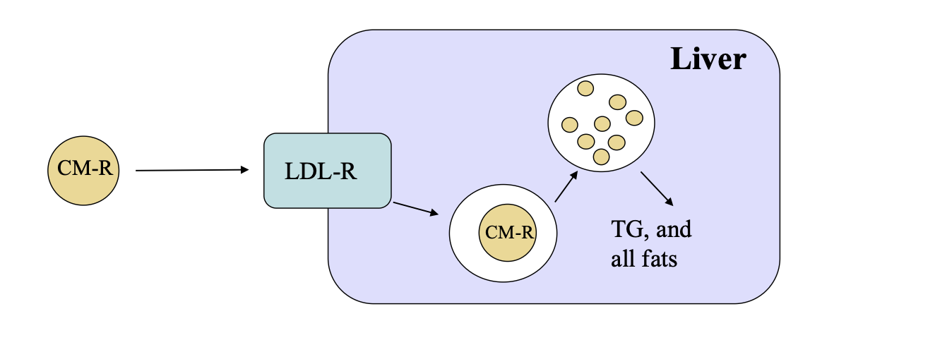
how do CM get absorbed in liver
CM remnants are taken up by the LDL receptor (LDL-R)
the receptor binding to CM-R are internalized in vesicles that fuse w/ lysosomes
lysosomes contain digestive enzymes that degrade all proteins of the particle → release FFAs & fat soluble vitamins into the liver
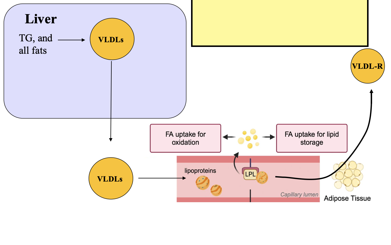
how does the liver produces VLDLs
fats & fat soluble vitamins are assembled into VLDLs
in circulation: VLDLs are digested by LPL becoming VLDL remnants
VLDLs are rich in TGs

how are VLDLs absorbed in peripheral tissues
VLDLs & VLDL remnants are taken up by peripheral tissues using VLDL receptor (VLDL-R)
receptor binding to VLDL are internalized & all content is released into cells
VLDL contains:
triglycerides
all fat soluble vitamins
vitamin E & K
vitamin D & A
carotenoids
triglycerides

how are VLDLs converted in LDLs
VLDL remnants can be taken up by cells or be converted to LDL
LPL takes more TG from VLDL remnants until the particle becomes LDL
LDLs are rich in cholesterol
LDL takes cholesterol to peripheral tissues
describe VLDL → LDL & what happens to LDL
VLDLs converted to VLDL-Rs
VLDL-Rs converted into LDL
VLDL & VLDL-R can be internalized via VLDL receptors
LDL can be internalized using LDL receptors
VLDL, VLDL-R, & LDL taken to peripheral tissues
LDLs are produced by the following mechanism:
synthesized in the liver from endogenous cholesterol
produced from HDL
produced by digestion of VLDLs via LPL
produced in the small intestine from dietary cholesterol
produced by digestion of VLDLs via LPL
what is reverse cholesterol transport
transport excess cholesterol from peripheral tissues to the liver for excretion via HDL

describe reverse cholesterol transport
cholesterol is taken from peripheral tissues by HDLs
HDLs are formed by ApoA1 (secreted by the liver)
HDL precursors goes to peripheral tissues
cholesterol is excreted from tissues via exporter ABCA1
HDL loads the cholesterol becoming mature HDL
mature HDL goes to liver & interacts with SR-BI receptor
the cholesterol is transported to liver leaving HDL empty → becomes immature HDL again
HDL goes back to peripheral tissues to get more cholesterol
HDL is rich in cholesterol
HDL takes cholesterol from peripheral tissues to liver & interacts with SR-BI
HDL releases cholesterol in the liver → used for synthesis of bile acids/excreted in bile
what is the function of chylomicrons
transport dietary fats & cholesterol from intestine to tissues
what is the function of VLDLs
transport of endogenous fats (TGs) & cholesterol from liver to tissues
what is the function of LDL
transport of endogenous cholesterol from liver to tissues
what is the function of HDL
transport of endogenous cholesterol from tissues to liver
HDLs are produced in (choose all that apply):
the liver
the kidneys
peripheral tissues
the small intestine
the liver
peripheral tissues
small intestine
describe vitamin E:
forms of vitamin
best source
RDA
functions
8 different forms (“vitamers)
tocopherols & tocotrienols
only alpha tocopherol has biological activity
in the liver: alpha-TTP takes only alpha tocopherol into VLDLs
best source: wheat germ oil
RDA: 15 mg/day
naturally occurring sources are better than supplements
stored in membranes & acts as an antioxidant for lipids
reduces lipid peroxidation
what diseases are associated with vitamin e
defect in the alpha-TTP gene: causes ataxia with vitamin E deficiency
affects motor control & movement
symptoms: slurred speech (dysarthria), difficulty coordinating movements (ataxia), numbness in the hands & feet (peripheral neuropathy), & vision loss
deficiency causes hemolytic anemia
toxicity causes reduced vitamin K absorption
causes excessive bleeding

how is vitamin e absorbed in small intestine
same pathway as all fats
(form micelles w/ bile acids & transported to small intestine via NPC1, SR-BI, & CD36 transporters)
secreted from enterocytes via chylomicrons
small intestine produces ApoA1 & HDL
HDL gets cholesterol & vitamin e from enterocytes
CM & HDLs are transported to liver
LDL has highest vitamin e content

what happens to vitamin e at apical & basolateral membranes of small intestine
apical:
lipid uptake
receptors: SR-BI, NPC1, CD36
basolateral:
excretion of chylomicrons in secretory vesicles
cholesterol efflux via ABCA1 transporters into HDL particles
only liver & small intestine produce ApoA1 & HDLs

how is vitamin e taken to liver
chylomicron remnants, HDLs, & LDLs deliver vitamin e to liver
uptake of chylomicrons by receptors (like LDL-R)
hepatic alpha-tocopherol transfer protein (alpha-TTP) mediates the loading of RRR-a-tocopherol (active form) into VLDLs
aTTP protects alpha-tocopherol from degradation
all other vitamers are metabolized & excreted in feces (bile) or in urine
liver produces VLDLs & HDL containing vitamin e
the molecule involved in the targeting of vitamin e to VLDL is:
alpha-TTP
LPL
Apo-A1
LDL-R
alpha TTP

what are:
reactive oxygen species (ROS) & superoxide dismutase
& how is H2O2 destroyed
superoxide anion (one extra electron), very reactive
superoxide dismutase (SOD): converts superoxide into H2O2
H2O2 can generate hydroxyl radical
H2O2 can also generate hypochlorous acid (HOCl)
H2O2 is destroyed by peroxidase enzyme
glutathione peroxidase (GPx)
peroxiredoxins (Prx)
catalase
what is lipid peroxidation
polyunsaturated fatty acids (PUFAs) react with O2 or are attached by free radicals → generates lipid radicals & lipid peroxides
these molecules interact with the surrounding PUFAs propagating the oxidation of the cell membrane → membrane ruptures inducing ferroptosis (a regulated form of cell death caused by lipid peroxidation)
how does vitamin e protect from lipid peroxidation
vitamin e eliminates the lipid radical producing a lipid peroxide
lipid peroxide is then eliminated by glutathione peroxidase
vitamin e becomes oxidized & is regenerated by vitamin c
vitamin c becomes oxidized to DHA
DHA is regenerated by DHA reductase using glutathione
glutathione becomes oxidized to GSSG
GSSG is regenerated by glutathione reductase using NADPH
final product = reduced vitamin e, c, glutathione, & NADPH
vitamin e eliminates lipid radicals by:
donating electrons to lipid radical producing a lipid peroxide
converting the lipid radical back to its original state
eliminating the lipid radical from the membrane
inducing the synthesis of new lipids
donating an electron to the lipid radical producing a lipid peroxide
describe vitamin a:
forms
carotenoids
best source
RDA
forms:
retinol
retinal: 11-cis-retinal (night vision)
retinoic acid: gene expression
retinyl esters: storage mainly in liver in stellate cells
carotenoids:
vitamin a precursors (alpha & beta carotene)
all other carotenoids are antioxidants
RDA: 900 microgram/day
best sources:
liver - vitamin a
red/orange fruits & vegetables - carotenoids
what other vitamin deficiencies/toxicities are associated with vitamin a
vitamin e toxicity: inhibits conversion of beta carotene to retinol
vitamin a toxicity: reduces vitamin k absorption causing excessive bleeding
carotenoid toxicity: reduces vitamin e absorption causing hemolytic anemia
zinc deficiency: causes night blindness due to reduced regeneration of rhodopsin
what does vitamin a deficiency cause
reduces iron uptake in RBCs causing iron-deficiency anemia
night blindness
bitot’s spot (eye)
anorexia
what does an increase/deficiency in each vitamin cause:
vitamin e
vitamin a
b-carotene
toxicity of vitamin a or e → decreased vitamin k absorption → decreased blood coagulation → increased bleeding
toxicity of b-carotene → decreased vitamin e → hemolytic anemia
can also cause jaundice
deficiency of vitamin a → decreased iron absorption → iron deficiency anemia
deficiency of zinc → decreased rhodopsin regeneration → night blindness

what happens to retinyl esters inside enterocytes
retinyl esters (storage form) → retinol + carotenoids (by esterase) → form micelles → transported to small intestine
retinol → retinol-CRBP (by CRBPII) → CRBP-retinyl palmitate (by LRAT) → chylomicrons
carotenoids directly converted to chylomicrons in SI
chylomicrons exit SI → CM-R (digested by LPL) → transported to liver
only retinyl esters can go into chylomicrons

what happens to b-carotene inside enterocytes
retinyl esters → retinol + carotenoids (by esterase) → forms micelles → transported to small intestine
b-carotene in SI → 2x retinal → CRBP-retinal (by CRBPII) → CRBP-retinol (by retinal reductase {requires NADH}) → CRBP-retinyl palmitate (by LRAT) → forms chylomicrons
retinal → also converted to retinoic acid → retinoic acid leaves SI + binds to albumin → transported to liver
chylomicrons leave SI → transported to liver
retinol + retinal can bind CRBP
the correct order for the formation of retinoic acid from b-carotene is from b-carotene to:
retinol → retinal → retinoic acid
retinal → retinyl ester → retinoic acid
retinyl ester → retinol → retinoic acid
retinal → retinoic acid
retinal → retinoic acid

describe transport out of SI for vitamin a & carotenoids
CM for retinyl esters & carotenoids → transported out of enterocyte to lymphatic vessels
HDL for retinol → transported out of enterocyte to bloodstream
retinoic acid binding to albumin → transported out of enterocyte to bloodstream
how do carotenoids act as antioxidants
b-carotene → 2x retinal (by BCO1) → 2x retinol
carotenoids → apo-carotenes → Nrf2 → antioxidant enzymes
carotenoid function as antioxidants: eliminate lipid radicals & singlet oxygen
what is Nrf2
a transcription factor
master regulator of antioxidant enzymes (i.e. GPxs, SDOs, etc)
how is vitamin a absorbed in the liver
chylomicron remnants enter liver → retinyl esters → retinol (via CRBPII) → CRBP-retinol → CRBP-retinal (via retinal dehydrogenase) → retinoic acid → retinoic acid-CRABP (via CRABP)
CRBP-retinol → Holo-RBP (via RBP) → Holo-RBP exits liver
the synthesis of holo-rbp from b-carotene is as follows:
chylomicron remnants enter liver → retinyl esters → retinol (via CRBPII) → CRBP-retinol → Holo-RPB (via RPB) → Holo-RPB exits liver
list transporter for each vitamin in small intestine & liver:
retinyl esters
carotenoids
retinol
retinioic acid
carotenoids
small intestine:
chylomicrons: retinyl esters & carotenoids
HDLs: retinol
retinoic acid
liver
VLDL: carotenoids
holo-rbp-ttr-t4 trimolecular complex: retinol
how does retinoic acid play a role in gene expression
retinol → enters trimolecular complex → transported to SI (by STRA6) → retinol CRBP → retinal CRBP → retinoic acid CRABP → RAR-RXR & DNA → gene expression
what is the rhodopsin cycle
rhodopsin → opsin + cis-retinal → opsin+ trans-retinal → opsin + trans-retinal → rhodopsin + cis-retinal (then to rhodopsin) → opsin + cis-retinal
describe vitamin d:
forms
RDA
best source
absorption & transport (brief)
forms
D2 & D3 from the diet and D3 from the skin
synthesized from 7-dehydrocholesterol (cholesterol derivative) in the skin
activation in the liver (25-hydroxylation) & in the kidney (1 hydroxylation)
1,25-OH vitamin D or calcitriol is the active form
low calcium increases serum PTH which increases expression of 1-hydroxylase to increase calcitriol synthesis
calcitriol is a hormone - binds to VDR & regulates gene expression
skin is the major source of vitamin d
RDA
15 microgram/day
best source:
fatty fish & cod liver oil
absorption & transport: follows fat absorption up to the liver
what diseases are associated with vitamin d
vitamin d/calcium deficiency:
rickets (children)
osteomalacia (adults)
vitamin d deficiency:
hypercalcemia
calcification of soft tissues (kidney/liver/blood vessels)
hypertension
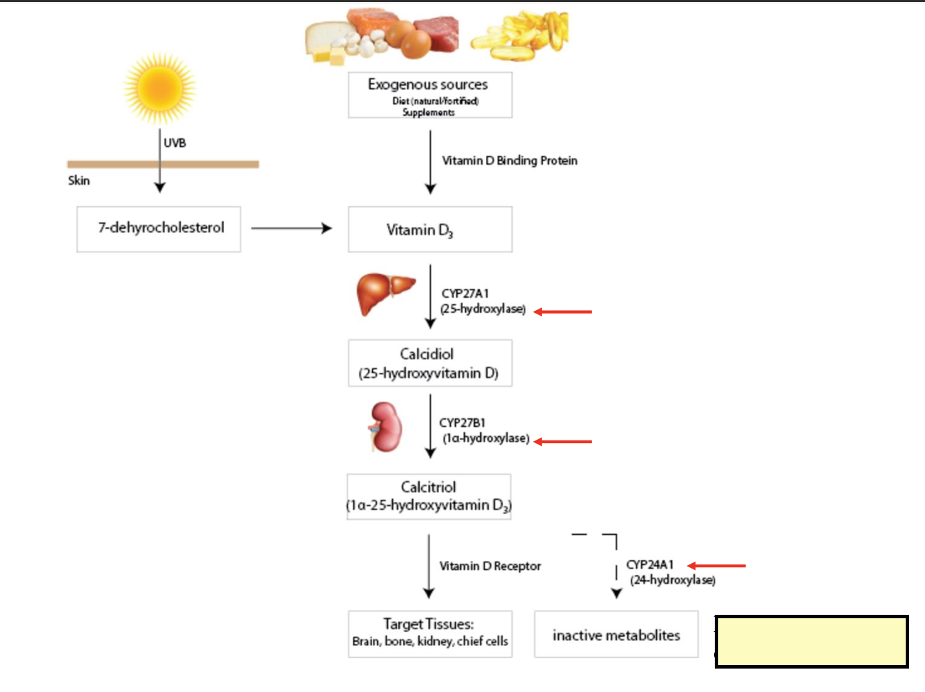
how is vitamin d metabolized
from sun (uvb): 7-dehydrocholesterol → vitamin D3
from diet: foods/supplements → vitamin D3 (by vitamin d3 binding protein)
vitamin D3 → calcidiol (25-hydroxyvitamin D) in liver (by 25-hydroxylase) → calcitriol (1a-25-hydroxyvitamin D3) in kidneys (by 1a-hydroxylase) → tissues (brain, bones, kidneys, chief cells) (by vitamin d receptor)
calitriol → also converted to inactive metabolites (by 24-hydroxylase)
tissues + metabolites induces degradation of vitamin d

how is vitamin d activation regulated
uptake of 25-OH D in the kidney & other tissues is upregulated by an increased concentration of PTH
PTH is secreted when calcium + phosphorous concentrations are low
PTH increases the synthesis of 1 hydroxylase in the kidney which produced 1,25-OH D
hypocalcemia stimultes the production of PTH to increase Ca+ levels by:
increasing Ca+ resorption in bones
increasing Ca+ resorption in kidneys
increasing 1,25-OH D in kidneys which increases Ca+ absorption in SI

how does vitamin d regulate its own synthesis
calcitriol increases FGF23 secretion from bone
FGF23 inhibits 1-hydroxylase enzyme in kidneys reducing calcitriol formation
calcitriol inhibits secertion of PTH
explain 1,25-OH D deficiency vs 1,25-OH D excess
deficiency:
stimulates synthesis of 1 hydroxylase in the kidneys increasing synthesis of the active form
excess:
inhibits synthesis of 1 hydroxylase reducing the synthesis of the active form of vitamin d
stimulates expression of 24 hydroxylase that inactivates vitamin d producing 24,25-OH & 1,24,25-OH vitamin d that are excreted
excessive PTH production can cause osteoporosis because:
PTH reduces the levels of osteoblasts
PTH increases osteoclasts degrading bones
PTH inhibits calcitriol reducing calcium absorption
PTH competes with calcitriol in the bones
PTH increases osteoclasts degrading bones

what is the function of calcitriol in bones
calcitriol increases the transcription of RANKL in osteoblasts (genomic)
RANKL interacts with receptor RANK in the surface of immature osteoclast precursors inducing diferentiation to osteoclast (maturing osteoclasts via signal transduction)
activated osteoclasts secrete HCl, alkaline phosphatase, & collagenases that eat away the bone releasing calcium + phosphorous
summarize vitamin d metabolism

describe vitamin k
forms
RDA
best source
overview of absorption & transport
forms
K1 (phylloquinone) & K2 (menaquinone), K3 (synthetic form)
K1 found in liver
K2 found in peripheral tissues
RDA:
80 micrograms/day
best source:
K1: green veggies
K2: animal sources + gut bacteria
absorption & transport
follows fat absorption up to liver
in liver: vitamin k goes to VLDLs
stored in cell membranes
what are the functions of vitamin k
k1: activates coagulation factors in liver
k2: activates osteocalcin in bones & MGP in blood vessels
vitamin d increases calcium absorption & k2 mediates binding of calcium to bone
k2 inhibits binding of calcium to blood vessels
where does each vitamin start & end up:
vitamin a, d, e, & k
vitamin a: small intestine → chylomicrons → liver → holo-rbp → trimolecular complex
vitamin d: small intestine → chylomicrons → liver (25-OH-DBP) → kidney → 1,25-OH-DBP
skin → D-DBP → liver (25-OH-DBP) → kidneys → 1,25-OH-DBP
vitamin e & k: small intestine → chylomicrons → liver → VLDL

what is the vitamin k cycle
for activation vitamin k is first reduced by vitamin k reductase
reduced vitamin k works on the carboxylation of glutamic acid on target proteins
after carboxylation reaction vitamin k is oxidized forming vitamin k epoxide
enzyme epoxide reductase regenerates vitamin k & new cycle begins
what is vitamin k mediated carboxylation
damaged blood vessel: injury to vessel lining triggers the release of clotting factors
formation of platelet plug: vasoconstriction limits blood flow + platelets form a sticky plug
development of clot: fibrin strands adhere to the plug to form an insoluble clot

explain primary vs secondary hemostasis
coagulation factors are produced in the liver where k1 is stored
primary hemostasis: regulated by activation + aggregation of platelets → creates an unstable plug
secondary hemostasis: coagulation creates a stable fibrin mesh
extrinsic pathway or initiation is mediated by the external damage to the vessel → endothelial cells secrete tissue factor (TF)
intrinsic pathway or amplification is activated by internal damage to the endothelium that amplifies the clotting response
explain fibrinolysis
coagulation cascade from hemolysis requires calcium → calcium binds to carboxylated glutamic acid
fibrinolysis: degradation of fibrin mesh to dissolve the clot (from hemolysis)
endothelial cells release tissue plasminogen activator (t-PA) into clot
t-PA converts plasminogen into plasmin
plasmin degrades fibrin
what genetic diseases are associated with vitamin k
hemophilia (increased bleeding)
a bleeding disorder caused by a mutation in coagulation factors (factor VIII, IX, & XI)
more common in men
hereditary thrombophilia (increased coagulation)
factor V leiden - a mutation that makes factor V resistant to inhibition increasing blood clotting
protein C & protein S deficiency (increased coagulation)
these proteins reduce clotting - its deficiency increases clotting
what disease are associated with vitamin k deficiency
vitamin k deficiency bleeding (VKDB) in infants:
a severe + life threatening bleeding disorder in newborns & young infants due to insufficient vitamin k at birth
preventable with a vitamin k shot given to newborns
in adults:
reduced coagulation & excessive bleeding
long term deficiency
osteoporosis (reduced ostecalcin activity)
cardiovascular disease (reduced MGP activity), increased arterial stiffness + calcification, increased risk of heart disease
what are the causes of each anemia:
hemolytic
pernicious
megaloblastic
microcity hypochromic
iron deficiency
hemolytic
vitamin e deficency
carotenoid toxicity
pernicious
caused by lack of intrinsic factor (B12 injection)
megaloblastic macrocytic
caused by B9 or B12 deficiency
treated with B12 and/or folate supplement
microcity hypochromic
vitamin B6 deficiency reduces heme synthesis
iron deficiency
vitamin a deficiency