Cardiac M for physiology
 Call Kai
 Learn
Learn Practice Test
 Spaced Repetition
 Match
Match Flashcards
 Knowt Play
1/42

There's no tags or description
Looks like no tags are added yet.
Name | Mastery | Learn | Test | Matching | Spaced | Call with Kai | Chat |
|---|
No analytics yet
Send a link to your students to track their progress
43 Terms
functional syncytium
a group of individual, separate cells that are connected to work together as a single, unified unit
connected by intercalated disks
gap junctions, adhering junctions, desmosomes
how are cardiac myocytes arranged
in series and parallel

2 syncytium:
atria
ventricles
action potential
avg. 105 millivolts each beat
0.2 sec depolarization (15x longer than skMSC)
Phase 0 of depolarization
depolarizes to -20 from -80mv
by voltage activated fast sodium channels
open for 1/1000th of a second
same for Sk m.
L Type ca channels (calcium channels)
open for longer (1/10th of a second)
slower
Phase 1: initial repolarization
fast sodium channels close, Ca channels stay open, and K leaves the cell
Phase 2: plateau
Ca remains open
K channels close
reduced permeability of membrane to K
Reduction of efflux of K prevents early repolarization
Phase 3: Repolarization
Calcium channels close
Calcium ceases to enter cell
Membrane permeability to K is restored, channels re-open, and cells rapidly repolarize Membrane permeability
Phase 4:
Resting membrane potential

explain this chart

explain this chart and how it correlates to other charge
Na is phase 0
ca is phase 2
K+ balanced out after decreases is phase 3
Velocity of action potential
0.3-0.5 m/sec
Refractory in nature?
Effective refractory period (ERPA)
from phase 0-2, cell cannot depolarize again until repolarization
0.23-0.30 sec
Relative refractory period
typically occur in phase 2
+0.05 sec
if stimulus is strong enough u can overcome RRP to get an early premature contraction
only during RRP
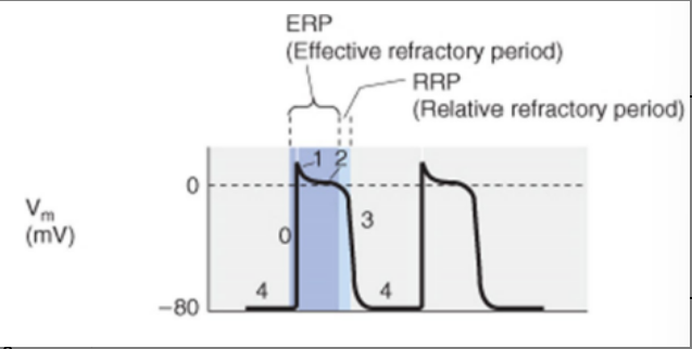
explain

can u get premature contractions to make wave summation?
no
Excitation-contraction coupling and relaxation in cardiac muscle: explain
AP travels along membrane to transverse (T) tubules
Activation of voltage-dependent Ca channels
Travels along longitudinal sarcoplasmic tubules
Release of calcium from SR via ryanodine receptor (RyR) channels
Calcium in sarcoplasm binds troponin
Activation of actin-myosin complex
Relaxation occurs via Ca ATPase (SERCA) and Ca/Na exchanger
T tubules:
Ca required from T tubules for full contraction strength.
T tubules retain calcium through electronegative mucopolysaccharides.
Diameter 5x and volume 25x compared to SkMsc.

No ca what?
NO CA no HEAWRT BEAT
Sarcomere
basic contractile unit of the myocyte

Myosin combines (what is combined myosin called) o form?
combined myosin is called filament
combines to form thousands of cross bridges with actin

myosin is made of?
2 heavy and 4 light chains
head has 2 flexible hinges at either side of the arm
contains ATPase enzyme
anchored by titin
actin contains:
2 F actin and 2-tropomyosin molecules
F actin contains G actin + ADP (active site)
Tropomyosin lies on top of active sites at rest
structure of actin and myosin?
both have twisted helix structure

troponin complex
troponin I has high affinity for actin
troponin T has high affinity for tropomyosin
Troponin C has high affinity for calcium
what happens at rest on troponin-tropomyosin complex
there is an inhibitory effect
4 Ca ions induce a conformational change and the troponin complex pulls tropomyosin away from active sites
Active sites bind to myosin cross-bridges and contraction occurs
Cross-bridges are independent thus number in contact with actin filament correlates to force of contraction

specific sequence of propagation of cardiac action pot.?
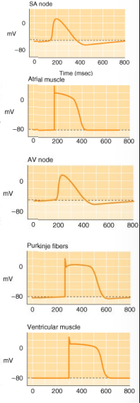
Propagation of cardiac action potential is simultaneous: T/F
False
Influence of voltage-gated Na channels absent in SA and AV node: T/F?
T
which node is the primary pacemaker?
SA Node
connects directly to the atrial muscle fibers
sets normal sinus rhythm

phases in SA node :
Phase 0: Depolarization
L-type Ca channels open
Phase 1-2: Absent in SA
Phase 3: Repolarization
Ca channels close and K channels open
Phase 4: Return to resting membrane
can SA node self excitate?
yes,
Resting potential is less negative
Leaky to sodium (funny currents) and calcium
Slight delay with K channels remaining open (hyperpolarization)
what effect does neural control of SA node have
alters rate of depolarization, HR
sympathetic control
(mostly release NE, activates beta1 adrenergic receptors, increases rate of depolarization (note: circulating Epi also activates beta1-AR)
Parasympathetic control
Right = SA and Left = AV
Release Ach, activates muscarinic receptors and decreases rate of depolarization
Neural control is needed to initiate action potential?
FALSE
AP spreads thru atrial muscle cells and atria contract?
True
what phases do atrial m AP phases exhibit?
typical phases of cardiac muscle
Basal level of neural control
Strong vagal stimulation can decrease the strength of heart muscle contraction by 20% to 30%.
Can stop heart beat for a few seconds
Vagal fibers are distributed mainly to the atria so primary effect is on heart rate

Contraction movement of ventricle
by purkinje system and left ventricle rotation

Purkinje System
Causes synchronous contraction of the ventricular muscle
Rapid conduction results in almost all parts of the ventricles contracting within a narrow time
First to last muscle fiber only 0.03-0.06 sec apart
Functional syncytium
Run ~1/3 of the way into ventricular muscle
Merge with ventricular muscle fibers
Muscle fibers conduct the action potential to the rest of the ventricular muscle mass
Left ventricular rotation
During systole, the left ventricle contracts in a twisting motion
Counterclockwise rotation of the apex of the heart
Clockwise rotation of the base of the left ventricle during the ejection phase
Pulls base downward towards apex during systole (wrings it out)
Like a loaded spring, recoils (untwists) during diastole
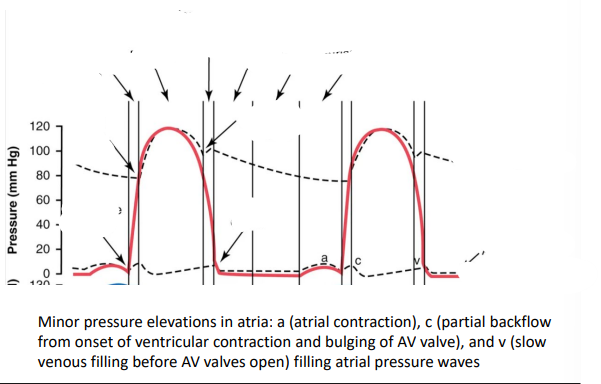
Pressure across the Cardiac Cycle
Pressure changes in the aorta (top dotted), left ventricle (red), and left atrium (bottom dotted)
Iso = equal or same
Atrial contraction usually causes an additional 20% filling of the ventricles
Approximately 60% of the blood in the ventricles at the end of diastole is ejected during systole

systole vs diastole
Systole:
60% volume ejected during period of rapid ejection (first 1/3 systolic time)
30% volume ejected during period of slow ejection (last 2/3 systolic time)
Diastole
Ventricle fillin
