Microbiology Exam 1
 Call Kai
 Learn
Learn Practice Test
 Spaced Repetition
 Match
Match Flashcards
 Knowt Play
1/152

There's no tags or description
Looks like no tags are added yet.
Name | Mastery | Learn | Test | Matching | Spaced | Call with Kai | Chat |
|---|
No analytics yet
Send a link to your students to track their progress
153 Terms
microbiology
study of living things too small to be seen without magnification
microbes/microorganisms
organisms that are too small to be seen without magnification
ex: bacteria, viruses, fungi, helminths, protozoa
pathogen
microbe that causes a disease
infectious disease
disease caused by a pathogen
prokaryotes
“prenucleus”
organisms that don’t have nuclei or other organelles; predominantly unicellular (bacteria)
eukaryotes
“true nucleus”
organisms that have nuclei and other organelles w/ special functions; mostly multicellular (fungi, helminths, protozoa)
bacteriology
study of bacteria
virology
study of viruses
parasitology
study of protozoa + helminths
mycology
study of fungi
binomial nomenclature
standardization for naming microbes that has 2 parts: genus and the species
genus is capitalized; species is lowercase
ex: Staphylococcus aureus
Louis Pasteur (1800s)
developed pasteurization (heat-treatment process that kills harmful microbes + prevents spoilage in liquids w/out ruining the taste)
contributed to germ theory of disease (specific microbes cause fermentation, spoilage, and infectious diseases
Robert Koch (1800s)
established “Koch’s postulates” (series of steps that could establish whether an organism was pathogenic + what disease it caused)
Alexander Fleming (1900s)
discovered the antibiotic activity of penicillin (a stray mold spore contaminated a petri disc of Staphylococcus bacteria and killed the surrounding growth)
culture
introducing microorganisms to a medium that supports their growth + multiplication
classification
process of arranging microbes to groups based on similar characteristics
the five I’s
1) inoculation
2) incubation
3) isolation
4) inspection
5) identification
inoculation
adding a pathogen, antigen, or microbial sample to a medium to stimulate immunity or encourage growth
incubation
maintaining controlled environments (heat, humidity, oxygen) to promote the growth of a culture
isolation
separating a single species/strain of microorganism from a mixed population to create a pure culture
inspection
performing tests and observing
identification
using the information from inspection to determine the exact species or type of microorganism in a sample
medium
provides nutrients to microorganisms, allowing them to grow outside their natural habitat
types of cultures
pure: contains 1 species
mixed: contains 2 or more known species
contaminated: contains contaminants (unwanted/unknown species)
colony
discrete mound of cells
isolated colony
colony that was formed from a single cell; contains only 1 species
bacteria
independent, single-celled, prokaryotic organisms w/out a nucleus
1) nuclear material (DNA) is free inside the cell
2) walls are sturdy and made of peptidoglycan
3) have no complex, membrane-bounded organelles
5 main bacterial shapes
1) coccus - spherical shape
2) bacillus - rod/cylindrical shape
3) curved - shaped/curved rods
4) spirillum - spiral and rigid
5) spirochete - spiral and flexibile
coccobacillus shape
short, oval rod that looks halfway between a coccus (round sphere) and an elongated rod (bacillus)
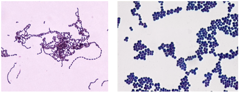
what bacterial shape is this?
coccus (plural cocci)
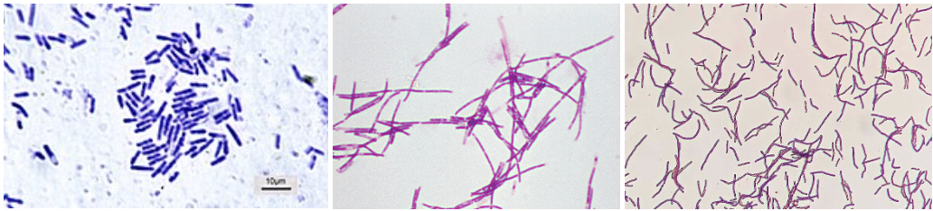
what bacterial shape is this?
bacillus (plural bacilli)
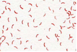
what bacterial shape is this?
curved
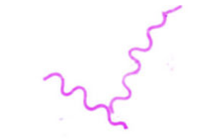
what bacterial shape is this?
spirillum
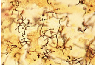
what bacterial shape is this?
spirochete
external structures of bacteria
appendages:
1) flagella: motility
2) fimbria: attachment
3) pilus: connection
surface coatings:
1) capsule
2) slime layer
flagellum
long, whip-like structures used for cell motility via self-propulsion; allows bacteria to swim freely through aqueous habitats

fimbria
small, bristle-like fiber sprouting off the surface of a cell
used for attachment (stick to each other + surfaces) + establishing infection or colonization

pilus
long, rigid tubular structure used for connection + sharing genetic material

conjugation pili
partial transfer of DNA from 1 cell to another
slime layer
loose shield that protects bacteria from loss of water + nutrients
capsule
a layer tightly bound to the cell; denser + thicker than a slime layer
encapsulated bacteria
bacteria that has a capsule; more likely to cause pathogenesis b/c the capsules protect the bacteria against phagocytosis
phagocytosis
when phagocytes (immune cells) engulf bacteria to destroy them
biofilm
feature of the slime layer
mixed community of bacteria + other microbes attached to a surface + each other
can complicate treatments by acting as a shield that blocks medications + hides microorganisms from the immune system
cytoplasmic membrane
innermost layer
lipid bilayer (containing phospholipids) w/ proteins embedded within the membrane
cell wall
strong, structural support that keeps bacteria from collapsing
protects from lysis (disintegration/rupture of a cell)
made of peptidoglycan
gram positive vs. gram negative stain
gram positive - purple
gram negative - pink
differences in staining has to do w/ the peptidoglycan in the cell walls
gram-positive cell envelope
2 layers:
1) cytoplasmic membrane
2) cell wall: thick, homogenous sheath of peptidoglycan + lipoteichoic acid (LTA)
LTA functions as the endotoxin of gram positive bacteria (not technically an endotoxin like LPS but works similarly)
gram-negative cell envelope
3 layers:
1) cytoplasmic membrane
2) cell wall: single, thin sheet of peptidoglycan and NO LTA
3) outer membrane: lipopolysaccharide (LPS)
LPS is an endotoxin; stimulates immune reactions like fever/shock
benefit of an outer membrane for gram-negative bacteria
outer membrane acts as an extra barrier for gram-negative bacteria, making them impermeable to some antibiotics + therefore more difficult to inhibit/kill than gram-positive bacteria
exceptions of cell walls
1) some bacteria aren’t characterized as gram-positive or negative; must use an acid-fast stain (mycobacterium)
2) some bacteria don’t have a cell wall at all (mycoplasma)
3) some bacteria are so small we can’t determine the gram stain (rickettsia + chlamydia)
bacterial chromosome
single circular strand of DNA that contains most of the bacterial DNA
every bacterial cell has a bacterial chromosome
plasmids
non-essential pieces of DNA; double stranded circles
not every bacterial cell has plasmids
prokaryotic vs. eukaryotic ribosomes
eukaryotes have larger ribosomes than prokaryotes
endospores
dormant bodies produced by bacteria
hardiest of all life forms; can withstand extreme heat, drying, freezing, radiation, and chemicals
sporulation
process by which bacteria form endospores to survive harsh environmental conditions
not reproduction; only 1 endospore is made
germination
reverse process of sporulation where the spore wakes up and turns back into an active, growing cell when conditions are favorable
fungi
type of eukaryotic microbe w/ a defined nucleus + specialized organelles
can either be unicellular or multicellular
extracellular matrix of fungi
helps protect, adhere, and receive signals from other cells and the environment
cell wall of fungi
all fungi have cell walls to give them structural support/shape
built mostly from chitin + glycans; doesn’t have peptidoglycans like bacterial cell walls
cell membrane of fungi
phospholipid bilayer intermixed w/ sterols (give the membrane more structure)
internal structures of fungi
1) nucleus - contains DNA
2) lysosome - contains variety of enzymes
3) vacuole - storage
4) mitochondria - generates ATP for the cell
2 forms of fungi
1) yeasts - round/oval shape, asexual production
2) hyphae (molds) - long, threadlike cells
dimorphic fungi
can transition b/w yeast and mold forms depending on the growth conditions
yeasts at body temperature; molds at room temperature
morphology of yeasts vs. molds
yeasts: unicellular w/ smooth, creamy colonies
molds: multicellular w/ soft, hairy, or velvety colonies

mycelium
intertwining mass of hyphae that makes up a colony
different morphology of molds
1) septate hyphae - segmented
2) nonseptate hyphae - continuous cell
3) vegetative hyphae - growth phase, absorb nutrients from substrate
4) reproductive hyphae - branches from vegetative hyphae + forms spores (fungal reproductive bodies)
how do yeasts replicate?
“budding”: reproduce asexually by making identical copies of itself through mitosis
how do molds replicate?
asexual spore formation: result of mitotic division from 1 parent cell
sexual spore formation: spores formed through meiosis; increases genetic variation
fungal spores
microscopic reproductive units that help the fungus disperse throughout the environment
saprobes vs. parasites
saprobe fungi obtain nutrients from dead plants/animals
parasite fungi live in or on the body of a living host to acquire nutrients
parasite
requires a living host to complete its lifecycle
no benefits to the host; detrimental
ex: protozoa + helminths
protozoa
single-celled, microscopic eukaryotic organisms
most diverse class of microbes
all are unicellular
require living hosts to complete their life cycle
external structures of protozoa
1) flagella - used for mobility (overall movement)
2) cilia - used for motility (internal movement
3) pseudopodia - used for motility + phagocytosis
boundary structures of protozoa
1) cytoplasmic membrane
no cell wall; cell shape can be constant or always changing
internal structures of protozoa
1) ectoplasm - clear outer layer of cytoplasm used for pseudopod formation
Are protozoa similar to each other, or different?
protozoa are similar in their basic cellular design as unicellular eukaryotes but they are different in their shapes, movements, and ways of life
how do protozoa move?
by using the flagella (whip-like tails), cilia (hair-like structures), or pseudopodia (temporary extensions of the cell’s cytoplasm)
trophozoites
active, feeding, and multiplying stage of a protozoan parasite that causes disease inside a host
cyst
dormant, thick-walled, and resistant stage of a protozoan parasite that survives outside of the host (harsh environments) and spreads infection
What life stage do most protozoa infect humans at?
protozoa infect humans in the cyst phase (usually through fecal-oral contamination) and cause human disease in the trophozoite stage (once inside the body)
helminth
large, multicellular worms
a type of parasite that can infect humans
eggs and larvae are microscopic but the adult stage can be seen w/out magnification
2 body types: flatworms + roundworms
roundworms
also called nematodes
cylindrical body shape
usually infect the human gastrointestinal tract

2 types of flatworms
1) cestodes (tapeworms): long, ribbonlike, segmented worms; usually infect the gastrointestinal tract
2) trematodes (flukes): flat, ovoid bodies that can infect many human tissues

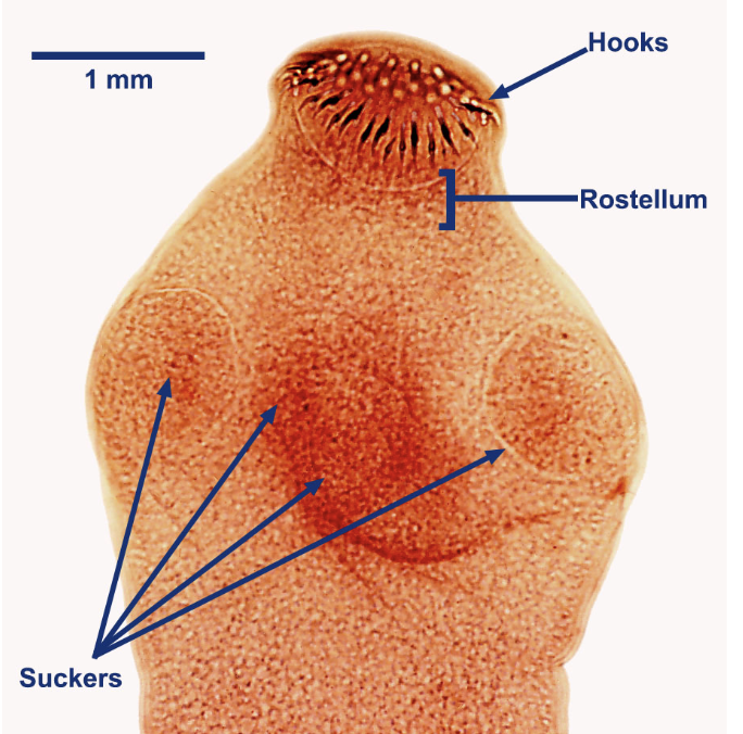
scolex
tapeworm (cestode) mouth parts
hooks are used to attach the tapeworm to the intestinal tract and the suckers are used to feed off the tissue
proglottid
tapeworm segments; can be full of eggs

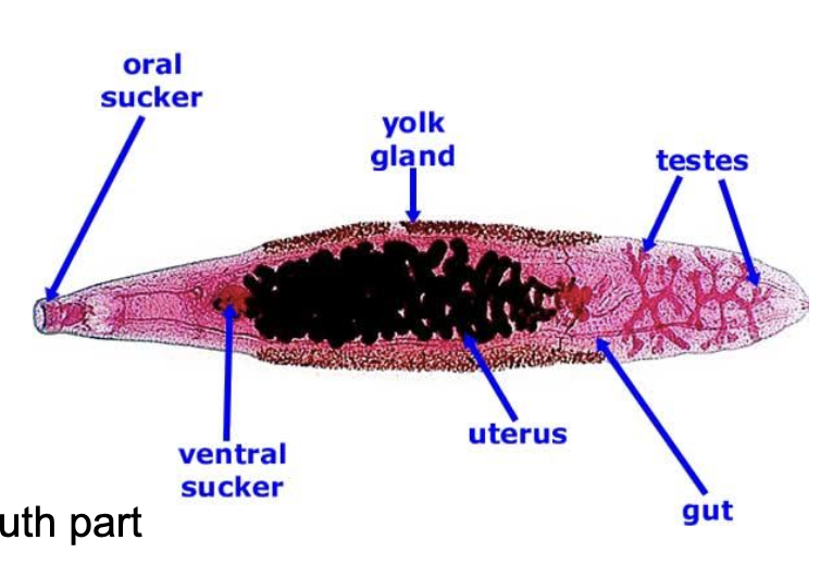
ventral sucker
fluke attachment part that suction cups to the tissue; prevents detachment
cuticle
outer layer of roundworms that functions as protection

What are the life cycle stages for most helminths?
3 stages:
1) fertilized egg
2) larval
3) adult
infection of humans usually occurs in the egg or larval stage
pathogenesis (disease progression in an organism) occurs in the adult stage
What is the difference between an intermediate and definitive
host?
intermediate (secondary) host: host in which the larval development occurs
definitive (final) host: host in which the adulthood and mating occur
Do worms use sexual or asexual reproduction?
reproduce sexually in the host’s body
acellular
refers to something that lacks a cellular structure (lack cell membranes, cytoplasm, ribosomes)
usually made up of proteins, nucleic acids, lipids, carbohydrates, etc.
acellular pathogens
infectious agents that don’t have a cellular structure
ex: viruses and prions
relative scale of microbes
helminths > fungi > protozoa > bacteria > viruses > prions
What are the 3 essential components of a virus?
1) genome
2) capsid
3) viral attachment proteins
viral attachment proteins
bind specifically to host receptor proteins
functions: attachment and entry
each viral species has a unique viral attachment protein
determines tropism: which cells can be infected by a virus
capsid
protects the genome from damage
can be helical (rod-shaped), icosahedral (spherical), or complex (irregular shape)
What is the purpose of an envelope? Do all viruses have
this?
envelope is made up of a host cell membrane functioning to protect and disguise the virus from the host immune system
not all viruses have envelopes (naked)
genome
total genetic material of a virus (DNA or RNA) packed inside the protective capsid
negative sense RNA (complementary to mRNA that must be transcribed before translation) or positive sense RNA (acts directly as mRNA for protein translation)
What else might viruses carry into the cell with them?
enzymes or other proteins
polymerases for replicating nucleic acids
RNA Dependent RNA Polymerase (RDRP) to translate RNA to RNA
reverse transcriptase (RT) to convert RNA to DNA