Anatomy Ch 10 Nervous system
 Call Kai
 Learn
 Practice Test
 Spaced Repetition
 Match
 Flashcards
 Knowt Play
1/69

There's no tags or description
Looks like no tags are added yet.
70 Terms
nervous system
detects changes, makes decisions, stimulates muscles and glands to respond, and maintains homeostasis
2 cell types: neurons (communication), neuroglia (support)
2 divisions: central (brain and spinal chord) and peripheral (connection to other body parts)
neurons
nerve cells
excitable cells that transmit electrical signals, react to changes, and communicate
structural units of the nervous system
large, highly specialized cells that conduct impulses
extreme longevity (can live 100 years or more!)
amitotic (nerve damage is almost always irreversible, other than olfactory nerves sometimes)
high metabolic rate - requires continuous supply of oxygen and glucose
cell body (soma) and one or more cell processes that vary and length in size: axons and dendrites
classified by structure: multipolar, bipolar, unipolar
peripheral nervous system (pns)
connects CNS to other body parts via the cranial nerves and the spinal nerves
2 divisions:
sensory (afferent): somatic and visceral sensory fibers that send perceptual information TO the brain to make sense of it
motor (efferent) division: interprets signals from cns to extremities, somatic ns and autonomic ns
cranial nerves
part of the pns that connects through the holes in the skull to the brain and the rest of the body
12 pairs
spinal nerves
part of the pns that reaches to every part of the body via neuron communication, sends and receives signals from and to the cns
31 pairs
sensory division
afferent (ascend) division of PNS
information “ascends” to the brain to make sense of it
somatic sensory fibers: convey impulses from skin, skeletal muscles, and joints to CNS
visceral sensory fibers: convey impulses from visceral organs to CNS
somatic sensory fibers
convey impulses from skin, skeletal muscles, and joints to CNS
type of sensory afferent division in the PNS
visceral sensory fibers
convey impulses from vsceral organs to CNS
→ organs and parts inside of body send sensory info to brain
ex: bladder, stomach, heart, etc
type of sensory afferent division in the PNS
motor division
efferent (exit) division of the PNS
transmits impulses FROM cns to effector organs, muscles, and glands
2 divisions: somatic nervous system and autonomic nervous system
somatic nervous system
sends conscous and reflec impulses to skeletal muscles FROM the cns to the pns to make them contract
interprets sensory information via the 5 senses
autonomic nervous system
controls invountary functions of smooth and cardiac muscle and glands to maintain homeostasis
ex: lung moevements for breathing, heart movements for beating and providing oxygen
general functions of the nervous system
sensory input: information gathered by sensory receptors about internal and external changes
integration: processing and interpretation of sensory input, create sensations, add to memory, produce thoughts
motor output: activation of effector organs (muscles and glands) produces a response
cell body of a neuron
soma
contains nucleus, cytoplasm, organelles, etc
dendrites
branched receptive (input) surfaces of neuron
may have a lot
convey incoming messages toward cell body as graded potentials (short distance signals)
can send excitatory or inhibitory signals that trigger the postsynaptic cell to continue or to stop firing
signal majority rules: more exhitatory allows for a neuron firing or inhibitory doesn’t
axon
conduction region of a neuron
generates nerve impulses
transmits impulses along the membrane to axon terminal which has a secretory region at the end that releases neurotransmitters, triggering muscles to move, reactions, etc
carries on many “conversations” with different neurons at the same time
lacks rough ER and Golgi apparatus: relies on cell body to renew proteins and membranes, efficient transport mechanisms, and will quickly decay if cut or damaged
contains:
axon hillock
collaterals
axon terminal
synaptic knob
may or may not be covered in myelin sheaths
multipolar neurons
99% of neuron structures
include many processes
most neuron structures of CNS
bipolar neurons
Structure of neuron with 2 processes
located in the eyes, ears, and nose
unipolar processes
neuron structure with one process and cell bodies in ganglia
often sensory ne
neuron processes in the cns
white matter = proceses
gray matter = cell bodies
neuron processes in the pns
almost all neuron processes (aka nerves) and very few cell bodies
tracts
bundles of neuron processes in CNS
nerves
bundles of neuron processes in PNS
myelination
whiteish, protein-lipid substance
Schwann cells that wrap their membranes around the axon terminal like a vest, one cell = one segment
myelinated fibers: segmented sheaths around most long or large-diameter axons
function: protects and electrically insulates axon, increases speed of nerve impulse transmission
unmyelinated axons
encased by schwann cell cytoplasm but there is no wrapped coating of myelin surrounding the axons
myelin sheath
Concentric layers of schwann cell plasma membrane around axon
protects and electrically insulates axon terminal, increases speed of nerve impulse transmission
forms a perinuclear cytoplasm bulge
perinucelar cytoplasm/neurillemma
outer collar of myelin sheath in PNS where the cytoplasm bulges peripherally from the schwann cell containing the nucleus and most of the cytoplasm
nodes of ranvier
myelin sheath gaps between adjcent schwann cells
where axon collaterals can emerge
multiple sclerosis
destruction of myelin sheaths in CNS by an immune response - attacked by a person’s own antibodies
scars are left behind, which stop neurons from conducting impulses
Without input from motor neurons, muscles stop contracting and atrophy
fatigue, mood problems, blurred vision, weak, numb limbs
treatments involve drugs that suppress immune activity
neuroglia
glial cells
provide structural support for neurons
in embryo: guide neurons into position, may stimulate specialization
produce growth factors to nourish neurons and remove excess ions and neurotransmitters
aid in formation synapses
4 types: astrocytes, oligodendrocytes, microglia, ependymal cells
astrocytes
most abundant, highly branded glial cells
cling to neurons, synaptic endings, and capillaries (blood-brain barrier)
connect neurons to blood vessels, exchange nutrients and growth factors
support and brace neurons
aid metabolism of certain substances
control chemical environment: regulate ion concentrations, such as K+
part of blood brain barrier: control what neurons have access to from the vessels and capillaries
guide migration of young neurons
respond to nerve impulses and neurotransmitters
influence neuronal functioning: participate in information processing in the brain

microglial cells
small, ovoid cells with thorny processes that touch and monitor neurons - resident immune cells
migrate toward injured neurons
can transform or “activate” to phagocytize microorganisms and neuronal debris

ependymal cells
range in shape from squamous to columnar
may be ciliated: beat to circulate CSF 2-3 times/day
line central cavities of the brain and spinal column
form permeable barrier between CSF in cavities and tissue fluid bathing CNS cells

oligodendrocytes
branched cells whose processes wrap CNS nerve fibers, forming insulating myelin sheaths for thicker nerve fibers

neuroglia only in PNS
schwann cells and satellite cells (support clusters of neuron cell bodies ganglia)
neuron regeneration in the PNS
regeneration is possible
axon separated from cell body and its myelin sheath will degenerate
schwann cells and neurilemma remain
remaining schwann cells provide guiding sheath for growing axon
if growing axon establishes former connection, function will return, if not, function may be lost
neuron regeneration in CNS
axons here lack neurilemma to act as guiding sheath
oligodendrocytes do not proliferate after injury
regeneration is unlikely
synapse
neuron communication
site at which a neuron transmits a nerve impulse to another neuron
presynaptic nerve sends impulse
postsynaptic neuron receives impulse
they can be both! depends on whcih synapse, function, location, etc
sparated by synaptic clef
synaptic transmission
impulse travels down axon of presynaptic neuron to axon terminal
impulse reaches synaptic knob and causes an influx of Ca+2 ions
release of neurotransmitters by synaptic vesicles by exocytosis
NTs exert either excitatory or inhibitory effect on postsynaptic neuron by creating graded potentials (gradient of both excitatory and inhibitory in which the majority wins) to which the enruon responds by generating an action potential (nerve impulse) or not
one-way transfer of information

cell membrane potential
usually electrically charged, polarized, due to unequal distribution of ions on the inside and outside of membranes
inside of membrane is negatively charged with respect to outside of membrane
causes resting potential important in synaptic transmission
potassium (K+) ions
major INTRAcellular positive ions (cations)
2 enter the neuron and influence the resting potentials of neurons
sodium (Na+) ions
major EXTRAcellular positive ions (cations)
3 exit the neuron
sodium-potassium pump
creates distribution of Na+ and K+ across cell membrane of neuron
active transport (requires ATP)
transports 3 Na+ out of the cell for every 2 K+ ions into the cell

ion channels
formed by large membrane proteins to help regulate the passage of specific ions into or out of the cell
selective
2 types: leakage (always open), gated (part of protein changes shape to open/close)
gated ion channels
ion channels in which a part of the protein changes shape to allow for open or close
3 types: chemically gated, voltage-gated, mechanically gated
when open: ions diffuse quickly across membrane along electrochemical gradient (ions move towards oppositve charge) and ion flow creates electrical current and voltage changes across membrane
chemically gated channels
gated ion channel that opens in response to binding of the appropriate neurotransmitters
→ facilitate diffusion, passive transport
voltage-gated channels
gated ion channels that open in response to changes in membrane potentials (i.e. inside of membrane changes from positive to negative or vice versa)
→ facilitate diffusion (i.e. for Na+ or K+ for graded potentials), passive transport
electrochemical gradient
ions flow along their chemical gradient and electrical gradient together
chemical: high concentration to low concentration
electrical: move toward an area of opposite charge
resting potential
potential difference across membrane of resting cell (considering inside of cell membrane is negative and outside is positive)
charge rests at approximately -70 mV in neurons: cytoplasmic side of membrane is negatively charged compared to outside
generated by: differences in ions across membranes (Na+/K+ pump), and differential permeability of plasma membrane (related to K+ leak channels
importance of membrane potential
changed in membrane potential in neurons use for communication with other cells: integrate, send, and receive information
produced by membrane permeability to ions
changes produces 2 types of signals: graded potentials (short distance) and action potentials (long-distance)
relative to resting potential: depolarization and hyperpolarization
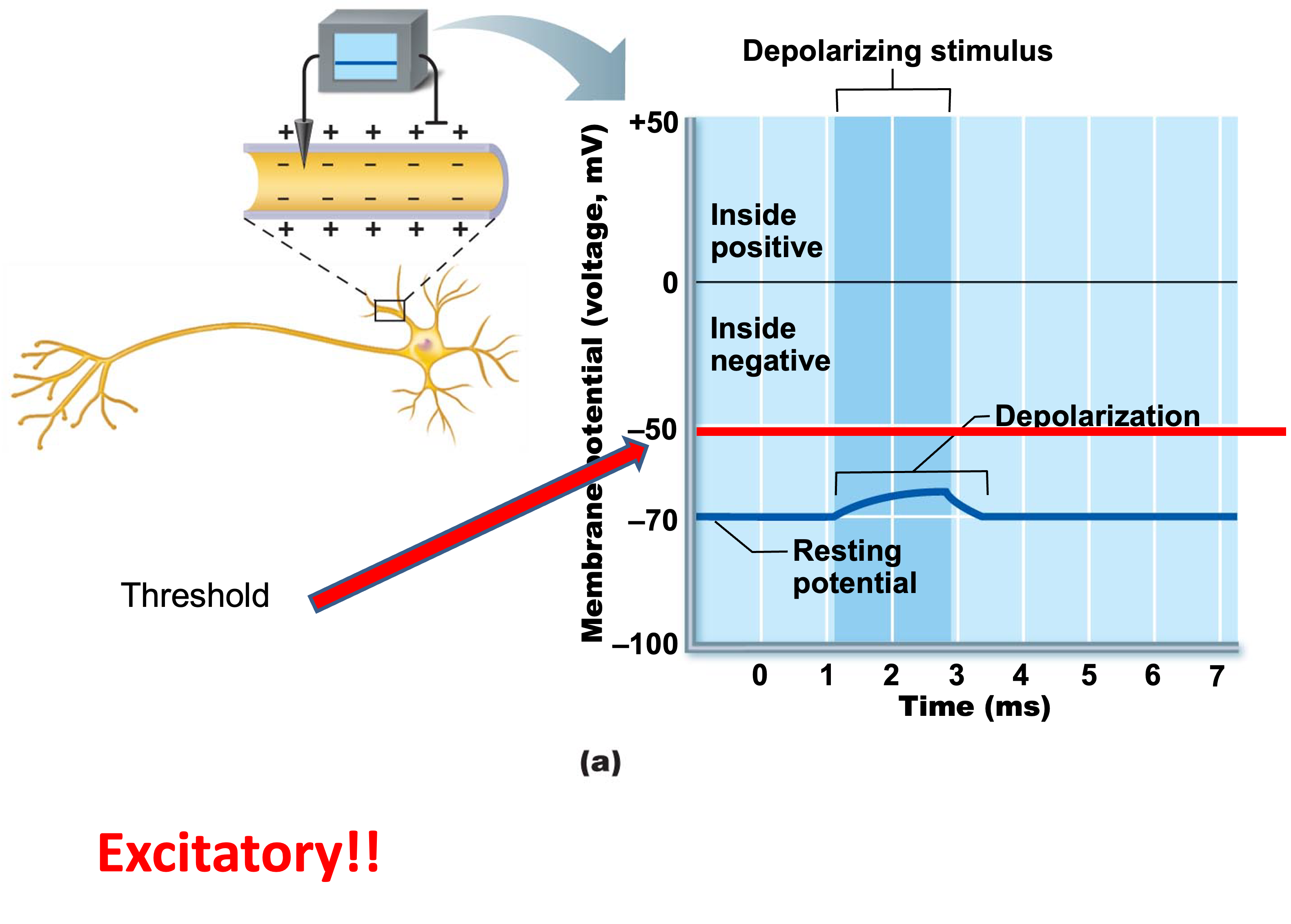
depolarization
decrease in membrane potential
inside of membrane (cytoplasmic side) becomes less negative (i.e. towards and above 0) rises above -70mV
→ INCREASES probability of producing a nerve impulse = EXCITATORY
hyperpolarization
an increase in membrane potential
the inside of the cell to move away from zero relative to the resting membrane potential
→ DECREASES probability of producing a nerve impluse = INHIBITORY

graded potentials
short-lived, localized changes in membrane potential
happen on dendrites
magnitude varies depending on stimulus strength
stronger stimulus → more voltage changes → farther current flows through the axon and to the next neuron
2 types: depolarization/excitatory or hyperpolarization/inhibitory
stimulus opens gated ion channels
current flows but dissipates quickly: only over short distances
SYNAPTIC POTENTIALS
excitatory postsynaptic potentials (EPSP)
membrane change in which neurotransmitter opens Na+ channels,
DEPOLARIZES (less negative on inside of cell) membrane of postsynaptic neuron, as Na+ enters axon
action potential in postsynaptic neuron becomes more likely
inhibitory postsynaptic potential (IPSP)
membrane change in which neurotransmitter opens K+ channels (or Cl- channels)
HYPERPOLARIZES membrane of postsynaptic neuron, as K+ leaves axon
action potential of postsynaptic neuron becomes less likely
summation
EPSPs and IPSPs are added together
→ net excitatory effect leads to greater probability of an action potential does not generate action potentials
occurs at trigger zone: axon hillock
threshold potential
degree of depolarization reaches -55mV → action potential
action potential
main way neurons send signals
principle mean of long-distance neural comunication- do not decay over distance
only in muscle cells and axons of neurons
brief reversal of membrane potential with a change in voltage ~100mV

depolarizing phase of action potentials
depolarizing local currents open voltage-gated Na+ channels causing Na+ to rush into the cell
if neuron reaches threshold of -55 to -50 mV, positive feedback causes opeing of all Na+ channels
repolarizing phase of action potential
Na+ channel gates close
membrane permeability to Na+ declines to resting state
slow voltage-agted K+ channels open → K+ exits the cell restoring internal negativity
hyperpolarization phase of action potential
some K+ channels remain open, allowing excessive K+ efflux, potassium leaves the cytoplasm
→ inside of membrane more negative than resting state → slight dip below resting voltage (-90 mV)
Na+ channels begin to reset (get closer to -70mV
impulse conduction in non-myelinated axons
action potentials are conducted down the length of the axon, each segment must depolarize
salutatory conduction
impulse conduction in myelinated axons
speed increases with myelination: lipids act as an electrical insulator
ions cross membrane only through nodes of ranvier (gaps) by “jumping” from node to node down the axon
also affected by axon diameter: thick acons transmit faster
synaptic transmission
transmission of nerve impulse from one neuron to another
impulse travels down axon of presynaptic neuron to axon terminal
reach synaptic knob → influx of Ca+2 ions
released neurotransmitters from synaptic vesicles by exocytosis
cross the synaptic cleft and react with specific receptors in the membrane of postsynaptic neuron
chemically gated ion channels responds to NTs → synaptic potentials
excitatory NTs and inhibitory NTs
excitatory neurotransmitters
chemicals released at synaptic transmission that increase permeability to Na+ ions
bring membrane closer to threshold → increase likelihood of generating impulses (action potentials)
inhibitory neurotransmitters
chemicals released at a synaptic transmission that move membrane farther from threshold → decrease likelihood of generating impulses
neurotransmitters
at least 100
monoamines, amino acids, peptides
produced in the rough ER or cytoplasm
released by exocytosis when impulse reaches synaptic knob of axon
ex disorders associated with NT imbalances: depression, epilepsy, huntington disease, insomnia, mania, parkinson’s disease, schizophrenia, etc
ex drugs that alter NT levels: cocaine, nicotine, antidepressants, SSRIs
vesicle trafficking
process of membrane recycling
synaptic vesicle becomes part of cell membrane as it releases NT
enodcytosis returns membrane to cytoplasm, forms new vesicles
opiate drugs
potent painkillers derived from poppy plant
→ human body produces opiates called endorphins which relieve pain in acupuncture, childbirth, well-being during exercise
very useful for severe pain, also very addictive
withdrawal symptoms are caused by the drug causing the body to stop producing natural endorphins, so when drug is absent, severe pain is felt
subdural space means
between arachnoid