knowt logo

Biological_Psychology_11_ATAR_Psychology

1. Structure of the Nervous System
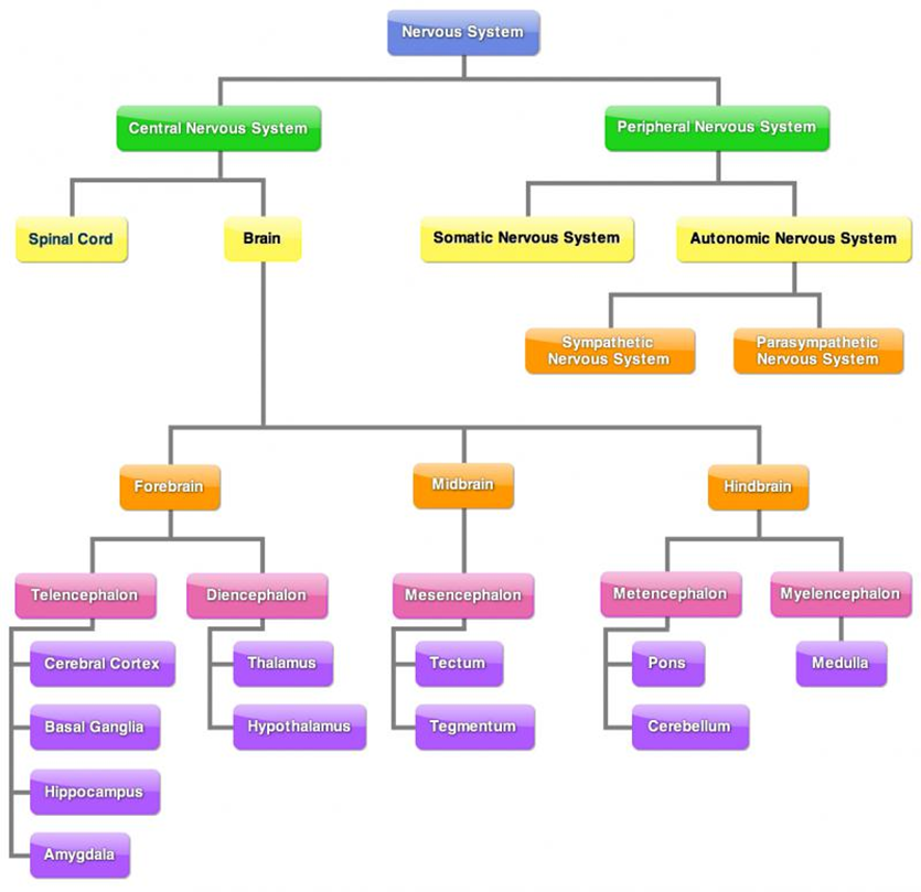
  • Central Nervous System (CNS)

    • Composed of the brain and spinal cord

    • Processes sensory information from PNS and sends motor commands back out

  • Peripheral Nervous System (PNS)

    • Comprises nerves outside the CNS

    • Divided into two main divisions:

      • Somatic Nervous System

        • Controls voluntary muscle movements

        • Carries sensory information to CNS

      • Autonomic Nervous System (ANS)

        • Regulates involuntary body functions

        • Subdivided into:

          • Sympathetic Nervous System

            • Prepares body for fight-or-flight response

          • Parasympathetic Nervous System

            • Calms the body and maintains homeostasis

2. Features of Neurons

  • Definition: Neurons are specialized cells responsible for receiving, transmitting, and processing information.

  • Structure:

    • Dendrites: Receive signals from other neurons.

    • Cell Body (Soma): Contains the nucleus and integrates incoming signals.

    • Axon: Transmits impulses away from the cell body.

    • Axon Terminals: Release neurotransmitters into the synapse.

    • Myelin Sheath: Insulates axon, increasing impulse speed.

  • Types of Neurons:

    • Motor Neurons: Transmit impulses from CNS to muscles and glands.

    • Sensory Neurons: Carry sensory information to CNS.

    • Interneurons: Relay messages between sensory and motor neurons.

3. Neural Transmission

  • Process:

    • Action potentials travel in one direction from dendrites to axon terminals.

    • At axon terminals, neurotransmitters are released into the synaptic cleft.

  • Electro-Chemical Signal:

    • Action potentials = electrical signals within neurons.

    • Neurotransmitters = chemical signals between neurons.

  • Synapse:

    • Junction between pre-synaptic and post-synaptic neurons.

    • Converts electrical impulses into chemical signals and back.

4. Brain Structure and Functions

  • Hindbrain: Controls basic autonomic functions; includes medulla (heart rate, breathing). Controls voluntary muscle movements; includes cerebellum (balance, coordination, swallowing). At base of brain, back of skull.

    • Medulla:

      • At base of brain stem. Front of Cerebellum.

      • Relays information between spinal cord and brain.

      • Regulates vital voluntary bodily functions (coughing, sneezing, blood pressure).

      • Damage = interruptions between brain and spinal cord. Can be fatal, as medulla controls vital organs.

    • Cerebellum:

      • Behind brainstem, beneath occiptal and temporal lobes.

      • Assist in coordinating voluntary movement and balance by relaying motor information to/from cerebral cortex.

      • Involved in balance, judging distance, coordination of fine motor movements.

      • Damage = reduced motor control, difficulty in balance, maintaining equilibrium.

      • Affected by alcohol.

  • Midbrain: Involved in processing information related to hearing, vision, and movement; contains reticular formation (attention and arousal).

    • Reticular formation:

      • Throughout brainstem, from spinal cord to midbrain.

      • Stimulates brain with bombarding of sensory information - keeps cerebral cortex active and alert.

      • Controls physiological awareness and muscle tone by regulating function of ANS (sleep/wake cycle).

      • Damage = disruption to cleep/wake cycle, loss of attention, pain management and balance.

  • Forebrain: Largest brain region; responsible for complex functions including cognition and emotion; includes cerebral cortex and structures like hypothalamus and thalamus.

    • Cerebrum:

      • Largest part of brain, consists of white matter (myelinated axons), cerebral cortex (grey matter; dendrites, unmyelinated axons, cell bodies of neurons).

      • Outer layer = cerebral cortex. Responsible for higher cognitive functions, voluntary movement, emotions + personality.

      • Split into two cerebral hemispheres.

    • Thalamus:

      • Two small egg-shaped structures joined together. One at top of brain, one at top of brainstem.

      • Thalamus receives all information (except smell) on way to cerebral cortex. Information travels up spinal cord and to thalamus.

      • Conducts motor signals and relays information from brainstem to cortex.

      • Coordinates shifts in consciousness.

      • Damage = loss of senses, issues with movement or tremors, attention problems, etc.

    • Hypothalamus:

      • Peanut-sized structure.

      • Located below thalamus.

      • Maintains homeostasis, regulates hormone release (connected to endocrine system); hunger/thirst.

      • Controls ‘body-clock’, assists with sleep/wake cycle.

      • Damage = disruption in body temperature regulation, growth, eating habits, weight control, emotions, sexual behaviour, motivation and sleep cycles.

4.1 Cerebral Cortex
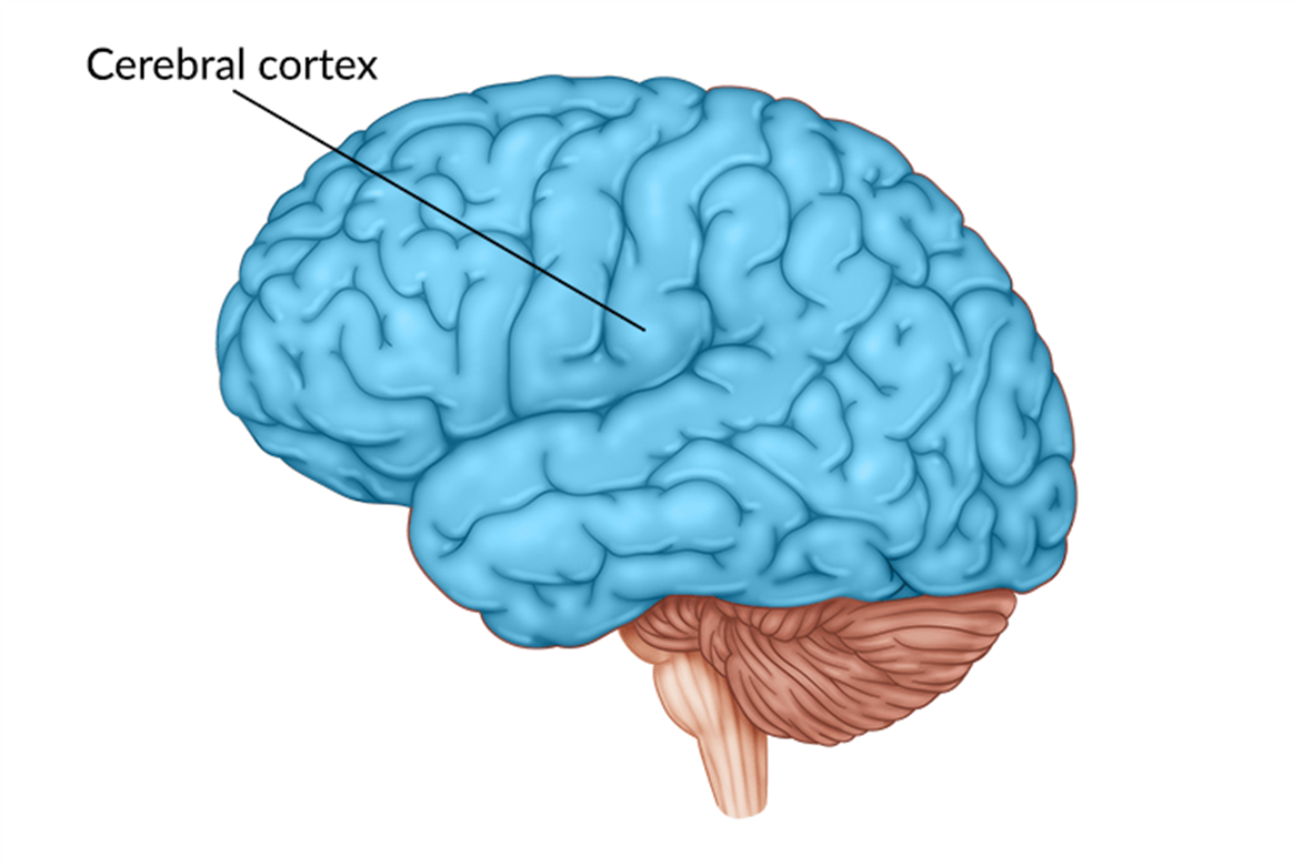
  • At surface of brain.

  • Damage = problems with cognition, sensation, movement, behaviour.

  • Two hemispheres:

    • Left (intellect) = verbal, analytical functions; writing, speaking, understanding. Mathematics, judging time, rhythm.

    • Right (creativity) = information processing, detecting/expressing emotion, non-verbal communication; jokes, irony, spacial/visual skills.

  • Hemispheric specialisation, contralateral control of body.

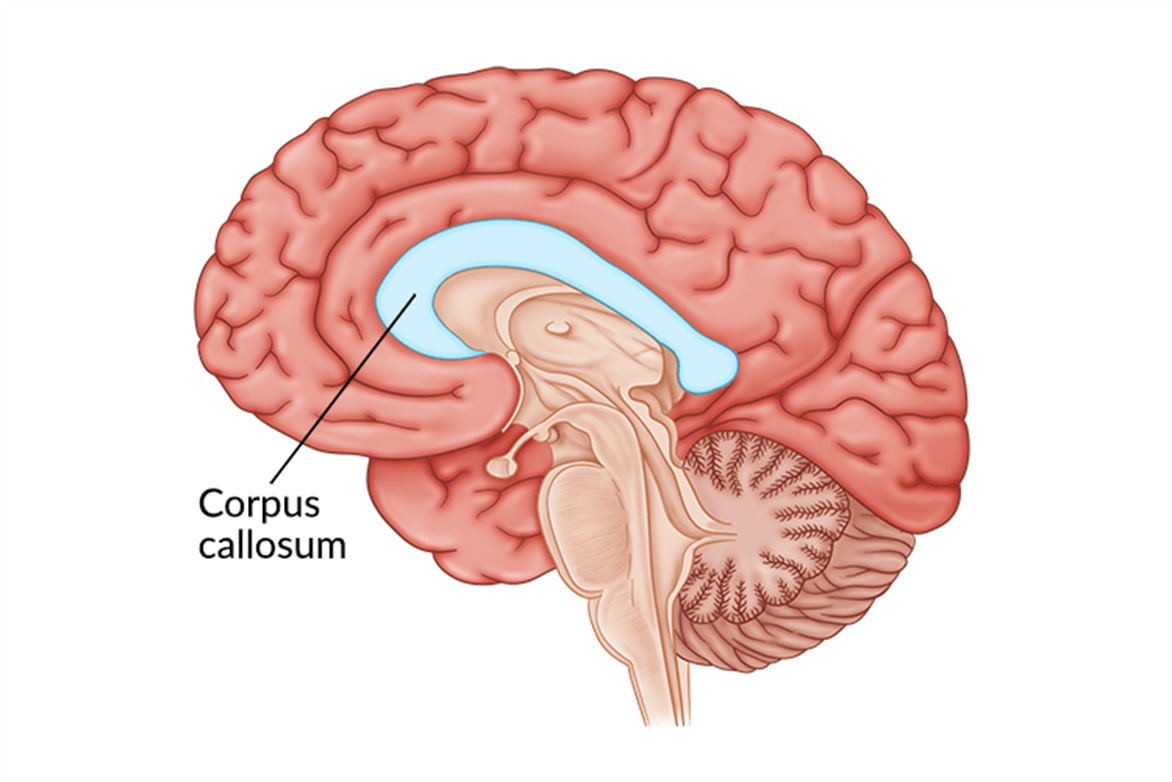
  • Corpus Callosum:

    • Longitudinal fissure of a thick band of nerve fibres connecting hemispheres.

    • Physically connects hemispheres.

    • Transmits information.

  • Divided into four lobes:

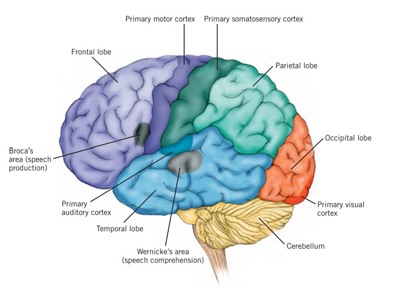
    • Frontal Lobe: reasoning, planning, movement, emotions, and personality.

      • Primary motor cortex:

        • Rear of frontal lobe.

        • Directs skeletal muscles ~ voluntary movement (left/right - contralateral control).

      • Broca’s area:

        • Production of articulate speech.

        • Left frontal lobe, near primary motor cortex.

        • Controls respiration muscles, cheeks, lips, jaws, tongue.

        • Sentence structure, grammar.

        • Related to Wernicke’s area.

        • Damage = Broca’s aphasia, can understand, can’t produce (Broca’s broken speech).

    • Parietal Lobe: Processing sensory information; spatial awareness.

      • Somatic sensations.

      • Spatial awareness.

      • Perception of location, movement of body parts.

      • Integration of sensory language.

      • Sensory cortex:

        • Strip of neurons at front of parietal lobe, next to primary motor cortex.

        • Registers, processes sensations detected by body’s sensory receptors.

        • Contralateral reception.

    • Temporal Lobe: Auditory processing; memory formation; understanding language.

      • Understanding speech.

      • Audio information interpretation.

      • Smell processing.

      • Facial recognition.

      • Long-term memory formation.

      • Primary auditory cortex:

        • Registers, processes auditory information; left is verbal, right is non-verbal.

        • Contribute to memory via link to hippocampus.

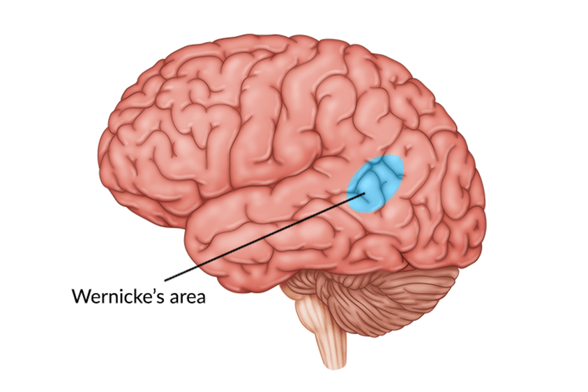
      • Wernicke’s area:

        • Left temporal lobe, near primary cortex.

        • Identifying sounds to comprehend meaning.

        • Accesses words in memory, controlling comprehension of speech and formulation of meaningful sentences.

        • Connections to Broca’s area, left temporal lobes receiving sound. Also speech registered in primary auditory cortex of both hemispheres.

        • Damage = Wernicke’s aphasia; can speak fluently but does not make sense. Difficulty in communication. Can see and pronounce but not understand.

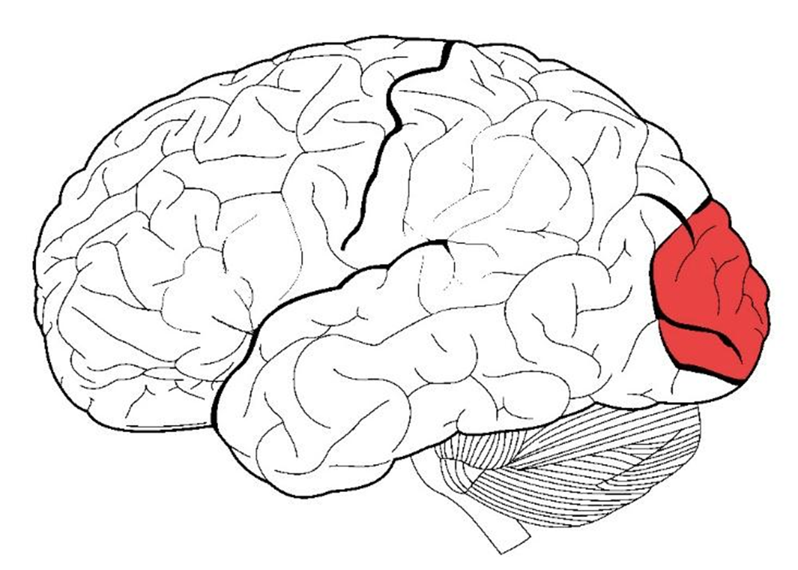
    • Occipital Lobe: Visual processing, facial recognition, perception of distance/depth.

      • Information from each eye is transmitted to primary visual cortex in left occiptal lobe, vice versa.

      • Primary visual cortex:

        • Processes visual information (into whole image or pattern, given meaning) from retinas via optic nerve, then send to other brain lobes.

        • Contains variety of neurons (specialised response neurons; specific to colour, shape, motion, etc.).

4.2 Localisation of Functions

  • Broca’s Area: Speech production.

  • Wernicke’s Area: Language comprehension.

5. Historical Studies

  • Phineas Gage:

    • Case study illustrating the relationship between brain injury and personality changes.

    • Suffered a severe injury when a metal rod penetrated his skull, damaging the frontal lobe.

    • Post-accident changes included:

      • Personality shift: Became impulsive, irritable, and irresponsible.

      • Inability to hold down a job or maintain social relationships.

    • Demonstrated the role of the frontal lobe in regulating personality and social behavior.

  • Roger Sperry:

    • Conducted studies on split-brain patients to explore the functions of cerebral hemispheres.

    • Findings indicated lateralization of brain functions, with each hemisphere specializing in certain tasks.

    • Highlighted how brain function is localized, impacting cognition and behavior.

  • Walter Freeman:

    • Pioneered frontal lobotomies as a treatment for mental illness.

    • His methods raised significant ethical questions regarding neuropsychological treatments.

    • Conducted thousands of lobotomies, often without proper consent, leading to questions about patient rights and treatment efficacy.

6. Contemporary Methods of Study

EEG (Electroencephalogram)

  • How it Works: Measures electrical activity in the brain via electrodes attached to the scalp. It detects voltage fluctuations resulting from ionic current flows within the neurons.

  • Strengths: High temporal resolution allows for the measurement of brain activity in real time, making it effective for tracking dynamic changes in brain activity.

  • Limitations: Low spatial accuracy; does not pinpoint the exact location of brain activity, limiting detailed brain mapping.

  • Uses:

    • Diagnose epilepsy, seizure disorders.

    • Sleep research

CT (Computed Tomography)

  • How it Works: Uses X-rays to create detailed cross-sectional images (slices) of the brain. A computer combines these images to provide a comprehensive view of brain structure.

  • Strengths: Useful for quickly detecting brain injuries, tumors, and bleeding; widely available and fast.

  • Limitations: Exposure to ionizing radiation can pose risks, and less effective in differentiating between types of soft tissues compared to MRI.

  • Uses:

    • Fractures.

    • Diagnose brain tumours, aneurysms.

    • Measure size of brain tumour.

    • Assess brain injury.

MRI (Magnetic Resonance Imaging)

  • How it Works: Utilizes strong magnetic fields and radio waves to generate detailed images of brain structures by aligning hydrogen atoms in water molecules and detecting their signals.

  • Strengths: Provides high-resolution images with detailed structures of the brain; no ionizing radiation exposure.

  • Limitations: More expensive and less accessible than CT; takes longer to conduct, and some patients may find the enclosed space uncomfortable.

  • Uses:

    • Diagnose brain tumours, aneurysms.

    • Measure size of brain tumour.

    • Assess effects of stroke.

    • Assess brain injury.

fMRI (Functional Magnetic Resonance Imaging)

  • How it Works: Measures brain activity by detecting changes in blood flow; areas of the brain that are more active receive more blood, which can be measured using MRI technology.

  • Strengths: Non-invasive method that provides both structural and functional information; excellent spatial resolution allows for precise localization of brain activity.

  • Limitations: Lower temporal resolution than EEG, so it cannot capture rapid brain activity changes with the same effectiveness as EEG.

  • Uses:

    • Activity in brain when performing a task.

    • Plan for tumour removal surgery.

    • Detect brain activity of patients with neurological conditions such as Parkinson’s disease.


Structure of the Nervous System

  1. Central Nervous System (CNS)

    • Composed of brain and spinal cord

    • Processes sensory information from PNS and sends motor commands back out

  2. Peripheral Nervous System (PNS)

    • Comprises nerves outside the CNS

    • Divided into:

      • Somatic Nervous System: Controls voluntary muscle movements

      • Autonomic Nervous System (ANS): Regulates involuntary body functions (divided into Sympathetic and Parasympathetic Systems)

Features of Neurons

  • Definition: Specialized cells for receiving, transmitting, and processing information

  • Structure:

    • Dendrites, Cell Body (Soma), Axon, Axon Terminals, Myelin Sheath

  • Types of Neurons:

    • Motor, Sensory, Interneurons

Neural Transmission

  • Process: Action potentials transmit signals, neurotransmitters released in synapses

  • Synapse: Junction converting electrical impulses to chemical signals

Brain Structure and Functions

  1. Hindbrain: Basic autonomic functions (medulla & cerebellum)

  2. Midbrain: Processes hearing, vision, movement; contains reticular formation

  3. Forebrain: Largest region responsible for complex functions (cerebral cortex, thalamus, hypothalamus)

Cerebral Cortex

  • Two Hemispheres:

    • Left: Analytical, language

    • Right: Creative, non-verbal

  • Lobes:

    • Frontal: Reasoning, movement, Broca's area (speech production)

    • Parietal: Sensory processing, spatial awareness

    • Temporal: Auditory processing, Wernicke's area (language comprehension)

    • Occipital: Visual processing

Historical Studies

  • Phineas Gage: Brain injury affecting personality

  • Roger Sperry: Split-brain studies highlighting lateralization

  • Walter Freeman: Ethical concerns in frontal lobotomies

Contemporary Methods of Study

  • EEG: Measures electrical activity; good temporal resolution

  • CT: Structural imaging using X-rays

  • MRI: High-resolution imaging with no radiation

  • fMRI: Detects changes in blood flow for brain activity

AP

Biological_Psychology_11_ATAR_Psychology

1. Structure of the Nervous System

  • Central Nervous System (CNS)

    • Composed of the brain and spinal cord

    • Processes sensory information from PNS and sends motor commands back out

  • Peripheral Nervous System (PNS)

    • Comprises nerves outside the CNS

    • Divided into two main divisions:

      • Somatic Nervous System

        • Controls voluntary muscle movements

        • Carries sensory information to CNS

      • Autonomic Nervous System (ANS)

        • Regulates involuntary body functions

        • Subdivided into:

          • Sympathetic Nervous System

            • Prepares body for fight-or-flight response

          • Parasympathetic Nervous System

            • Calms the body and maintains homeostasis

2. Features of Neurons

  • Definition: Neurons are specialized cells responsible for receiving, transmitting, and processing information.

  • Structure:

    • Dendrites: Receive signals from other neurons.

    • Cell Body (Soma): Contains the nucleus and integrates incoming signals.

    • Axon: Transmits impulses away from the cell body.

    • Axon Terminals: Release neurotransmitters into the synapse.

    • Myelin Sheath: Insulates axon, increasing impulse speed.

  • Types of Neurons:

    • Motor Neurons: Transmit impulses from CNS to muscles and glands.

    • Sensory Neurons: Carry sensory information to CNS.

    • Interneurons: Relay messages between sensory and motor neurons.

3. Neural Transmission

  • Process:

    • Action potentials travel in one direction from dendrites to axon terminals.

    • At axon terminals, neurotransmitters are released into the synaptic cleft.

  • Electro-Chemical Signal:

    • Action potentials = electrical signals within neurons.

    • Neurotransmitters = chemical signals between neurons.

  • Synapse:

    • Junction between pre-synaptic and post-synaptic neurons.

    • Converts electrical impulses into chemical signals and back.

4. Brain Structure and Functions

  • Hindbrain: Controls basic autonomic functions; includes medulla (heart rate, breathing). Controls voluntary muscle movements; includes cerebellum (balance, coordination, swallowing). At base of brain, back of skull.

    • Medulla:

      • At base of brain stem. Front of Cerebellum.

      • Relays information between spinal cord and brain.

      • Regulates vital voluntary bodily functions (coughing, sneezing, blood pressure).

      • Damage = interruptions between brain and spinal cord. Can be fatal, as medulla controls vital organs.

    • Cerebellum:

      • Behind brainstem, beneath occiptal and temporal lobes.

      • Assist in coordinating voluntary movement and balance by relaying motor information to/from cerebral cortex.

      • Involved in balance, judging distance, coordination of fine motor movements.

      • Damage = reduced motor control, difficulty in balance, maintaining equilibrium.

      • Affected by alcohol.

  • Midbrain: Involved in processing information related to hearing, vision, and movement; contains reticular formation (attention and arousal).

    • Reticular formation:

      • Throughout brainstem, from spinal cord to midbrain.

      • Stimulates brain with bombarding of sensory information - keeps cerebral cortex active and alert.

      • Controls physiological awareness and muscle tone by regulating function of ANS (sleep/wake cycle).

      • Damage = disruption to cleep/wake cycle, loss of attention, pain management and balance.

  • Forebrain: Largest brain region; responsible for complex functions including cognition and emotion; includes cerebral cortex and structures like hypothalamus and thalamus.

    • Cerebrum:

      • Largest part of brain, consists of white matter (myelinated axons), cerebral cortex (grey matter; dendrites, unmyelinated axons, cell bodies of neurons).

      • Outer layer = cerebral cortex. Responsible for higher cognitive functions, voluntary movement, emotions + personality.

      • Split into two cerebral hemispheres.

    • Thalamus:

      • Two small egg-shaped structures joined together. One at top of brain, one at top of brainstem.

      • Thalamus receives all information (except smell) on way to cerebral cortex. Information travels up spinal cord and to thalamus.

      • Conducts motor signals and relays information from brainstem to cortex.

      • Coordinates shifts in consciousness.

      • Damage = loss of senses, issues with movement or tremors, attention problems, etc.

    • Hypothalamus:

      • Peanut-sized structure.

      • Located below thalamus.

      • Maintains homeostasis, regulates hormone release (connected to endocrine system); hunger/thirst.

      • Controls ‘body-clock’, assists with sleep/wake cycle.

      • Damage = disruption in body temperature regulation, growth, eating habits, weight control, emotions, sexual behaviour, motivation and sleep cycles.

4.1 Cerebral Cortex
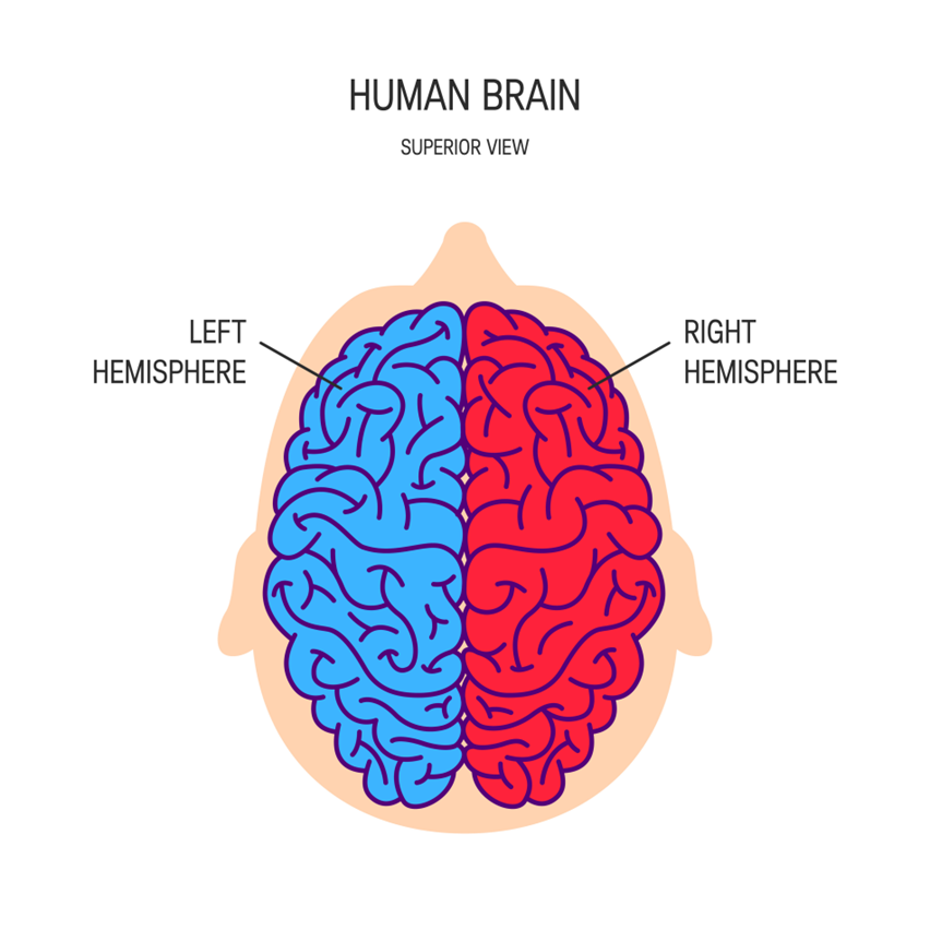
  • At surface of brain.

  • Damage = problems with cognition, sensation, movement, behaviour.

  • Two hemispheres:

    • Left (intellect) = verbal, analytical functions; writing, speaking, understanding. Mathematics, judging time, rhythm.

    • Right (creativity) = information processing, detecting/expressing emotion, non-verbal communication; jokes, irony, spacial/visual skills.

  • Hemispheric specialisation, contralateral control of body.

  • Corpus Callosum:

    • Longitudinal fissure of a thick band of nerve fibres connecting hemispheres.

    • Physically connects hemispheres.

    • Transmits information.

  • Divided into four lobes:

    • Frontal Lobe: reasoning, planning, movement, emotions, and personality.

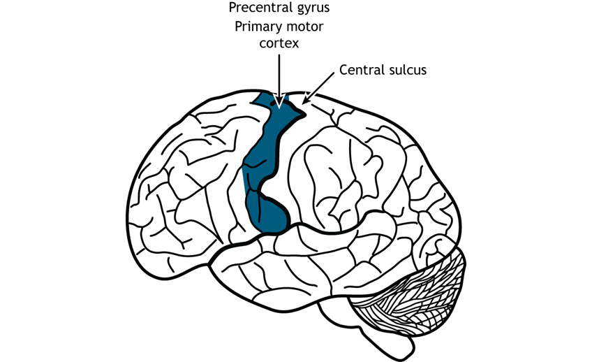
      • Primary motor cortex:

        • Rear of frontal lobe.

        • Directs skeletal muscles ~ voluntary movement (left/right - contralateral control).
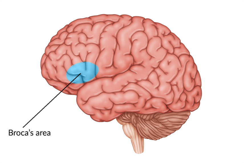
      • Broca’s area:

        • Production of articulate speech.

        • Left frontal lobe, near primary motor cortex.

        • Controls respiration muscles, cheeks, lips, jaws, tongue.

        • Sentence structure, grammar.

        • Related to Wernicke’s area.

        • Damage = Broca’s aphasia, can understand, can’t produce (Broca’s broken speech).
    • Parietal Lobe: Processing sensory information; spatial awareness.

      • Somatic sensations.

      • Spatial awareness.

      • Perception of location, movement of body parts.

      • Integration of sensory language.

      • Sensory cortex:

        • Strip of neurons at front of parietal lobe, next to primary motor cortex.

        • Registers, processes sensations detected by body’s sensory receptors.

        • Contralateral reception.

    • Temporal Lobe: Auditory processing; memory formation; understanding language.

      • Understanding speech.

      • Audio information interpretation.

      • Smell processing.

      • Facial recognition.

      • Long-term memory formation.

      • Primary auditory cortex:

        • Registers, processes auditory information; left is verbal, right is non-verbal.

        • Contribute to memory via link to hippocampus.
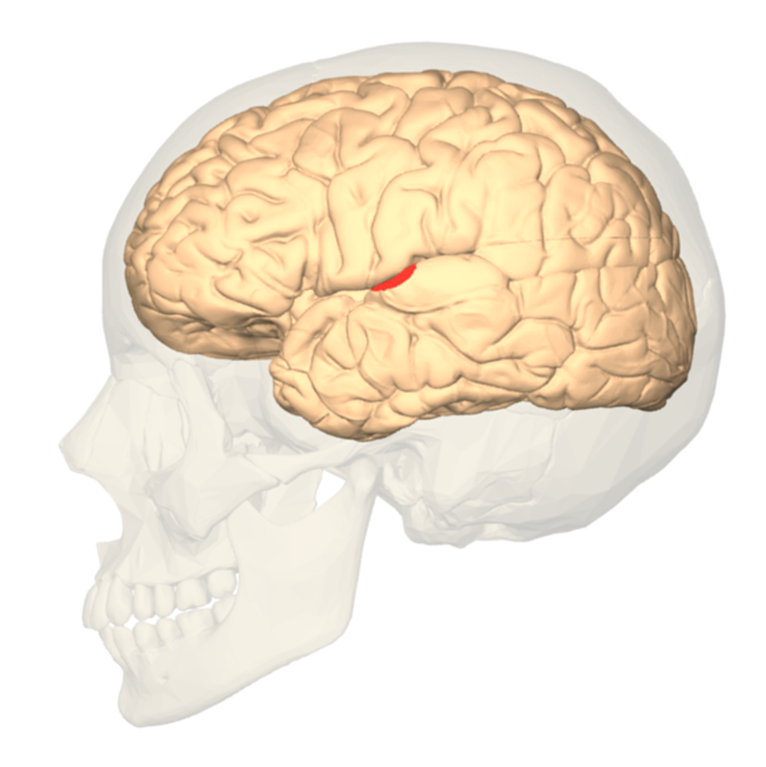
      • Wernicke’s area:

        • Left temporal lobe, near primary cortex.

        • Identifying sounds to comprehend meaning.

        • Accesses words in memory, controlling comprehension of speech and formulation of meaningful sentences.

        • Connections to Broca’s area, left temporal lobes receiving sound. Also speech registered in primary auditory cortex of both hemispheres.

        • Damage = Wernicke’s aphasia; can speak fluently but does not make sense. Difficulty in communication. Can see and pronounce but not understand.
    • Occipital Lobe: Visual processing, facial recognition, perception of distance/depth.

      • Information from each eye is transmitted to primary visual cortex in left occiptal lobe, vice versa.

      • Primary visual cortex:

        • Processes visual information (into whole image or pattern, given meaning) from retinas via optic nerve, then send to other brain lobes.

        • Contains variety of neurons (specialised response neurons; specific to colour, shape, motion, etc.).
4.2 Localisation of Functions

  • Broca’s Area: Speech production.

  • Wernicke’s Area: Language comprehension.

5. Historical Studies

  • Phineas Gage:

    • Case study illustrating the relationship between brain injury and personality changes.

    • Suffered a severe injury when a metal rod penetrated his skull, damaging the frontal lobe.

    • Post-accident changes included:

      • Personality shift: Became impulsive, irritable, and irresponsible.

      • Inability to hold down a job or maintain social relationships.

    • Demonstrated the role of the frontal lobe in regulating personality and social behavior.

  • Roger Sperry:

    • Conducted studies on split-brain patients to explore the functions of cerebral hemispheres.

    • Findings indicated lateralization of brain functions, with each hemisphere specializing in certain tasks.

    • Highlighted how brain function is localized, impacting cognition and behavior.

  • Walter Freeman:

    • Pioneered frontal lobotomies as a treatment for mental illness.

    • His methods raised significant ethical questions regarding neuropsychological treatments.

    • Conducted thousands of lobotomies, often without proper consent, leading to questions about patient rights and treatment efficacy.

6. Contemporary Methods of Study

EEG (Electroencephalogram)

  • How it Works: Measures electrical activity in the brain via electrodes attached to the scalp. It detects voltage fluctuations resulting from ionic current flows within the neurons.

  • Strengths: High temporal resolution allows for the measurement of brain activity in real time, making it effective for tracking dynamic changes in brain activity.

  • Limitations: Low spatial accuracy; does not pinpoint the exact location of brain activity, limiting detailed brain mapping.

  • Uses:

    • Diagnose epilepsy, seizure disorders.

    • Sleep research

CT (Computed Tomography)

  • How it Works: Uses X-rays to create detailed cross-sectional images (slices) of the brain. A computer combines these images to provide a comprehensive view of brain structure.

  • Strengths: Useful for quickly detecting brain injuries, tumors, and bleeding; widely available and fast.

  • Limitations: Exposure to ionizing radiation can pose risks, and less effective in differentiating between types of soft tissues compared to MRI.

  • Uses:

    • Fractures.

    • Diagnose brain tumours, aneurysms.

    • Measure size of brain tumour.

    • Assess brain injury.

MRI (Magnetic Resonance Imaging)

  • How it Works: Utilizes strong magnetic fields and radio waves to generate detailed images of brain structures by aligning hydrogen atoms in water molecules and detecting their signals.

  • Strengths: Provides high-resolution images with detailed structures of the brain; no ionizing radiation exposure.

  • Limitations: More expensive and less accessible than CT; takes longer to conduct, and some patients may find the enclosed space uncomfortable.

  • Uses:

    • Diagnose brain tumours, aneurysms.

    • Measure size of brain tumour.

    • Assess effects of stroke.

    • Assess brain injury.

fMRI (Functional Magnetic Resonance Imaging)

  • How it Works: Measures brain activity by detecting changes in blood flow; areas of the brain that are more active receive more blood, which can be measured using MRI technology.

  • Strengths: Non-invasive method that provides both structural and functional information; excellent spatial resolution allows for precise localization of brain activity.

  • Limitations: Lower temporal resolution than EEG, so it cannot capture rapid brain activity changes with the same effectiveness as EEG.

  • Uses:

    • Activity in brain when performing a task.

    • Plan for tumour removal surgery.

    • Detect brain activity of patients with neurological conditions such as Parkinson’s disease.


Structure of the Nervous System

  1. Central Nervous System (CNS)

    • Composed of brain and spinal cord

    • Processes sensory information from PNS and sends motor commands back out

  2. Peripheral Nervous System (PNS)

    • Comprises nerves outside the CNS

    • Divided into:

      • Somatic Nervous System: Controls voluntary muscle movements

      • Autonomic Nervous System (ANS): Regulates involuntary body functions (divided into Sympathetic and Parasympathetic Systems)

Features of Neurons

  • Definition: Specialized cells for receiving, transmitting, and processing information

  • Structure:

    • Dendrites, Cell Body (Soma), Axon, Axon Terminals, Myelin Sheath

  • Types of Neurons:

    • Motor, Sensory, Interneurons

Neural Transmission

  • Process: Action potentials transmit signals, neurotransmitters released in synapses

  • Synapse: Junction converting electrical impulses to chemical signals

Brain Structure and Functions

  1. Hindbrain: Basic autonomic functions (medulla & cerebellum)

  2. Midbrain: Processes hearing, vision, movement; contains reticular formation

  3. Forebrain: Largest region responsible for complex functions (cerebral cortex, thalamus, hypothalamus)

Cerebral Cortex

  • Two Hemispheres:

    • Left: Analytical, language

    • Right: Creative, non-verbal

  • Lobes:

    • Frontal: Reasoning, movement, Broca's area (speech production)

    • Parietal: Sensory processing, spatial awareness

    • Temporal: Auditory processing, Wernicke's area (language comprehension)

    • Occipital: Visual processing

Historical Studies

  • Phineas Gage: Brain injury affecting personality

  • Roger Sperry: Split-brain studies highlighting lateralization

  • Walter Freeman: Ethical concerns in frontal lobotomies

Contemporary Methods of Study

  • EEG: Measures electrical activity; good temporal resolution

  • CT: Structural imaging using X-rays

  • MRI: High-resolution imaging with no radiation

  • fMRI: Detects changes in blood flow for brain activity

robot