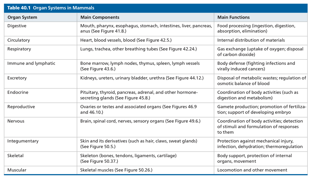
Campbell Unit 7: Animal Form and Function
Chapter 40: Basic Principles of Animal Form and Function
40.1: Animal form and function are correlated at all levels of organization
Anatomy: Biological structure
Physiology: Biological function
A multicellular organization only works if every cell has access to an aqueous environment, either inside or outside the animal’s body
Interstitial Fluid: Fluid that fills spaces between cells
Complex body plans are advantageous for many reasons, especially on land
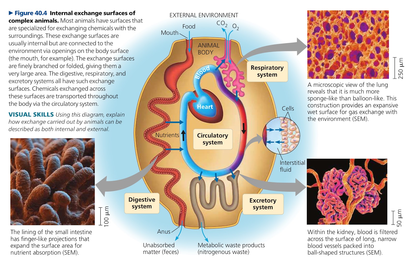
Tissues: Groups of cells with similar appearance and function
Organs: Functional units of tissues
Organ System: Groups of organs that work together
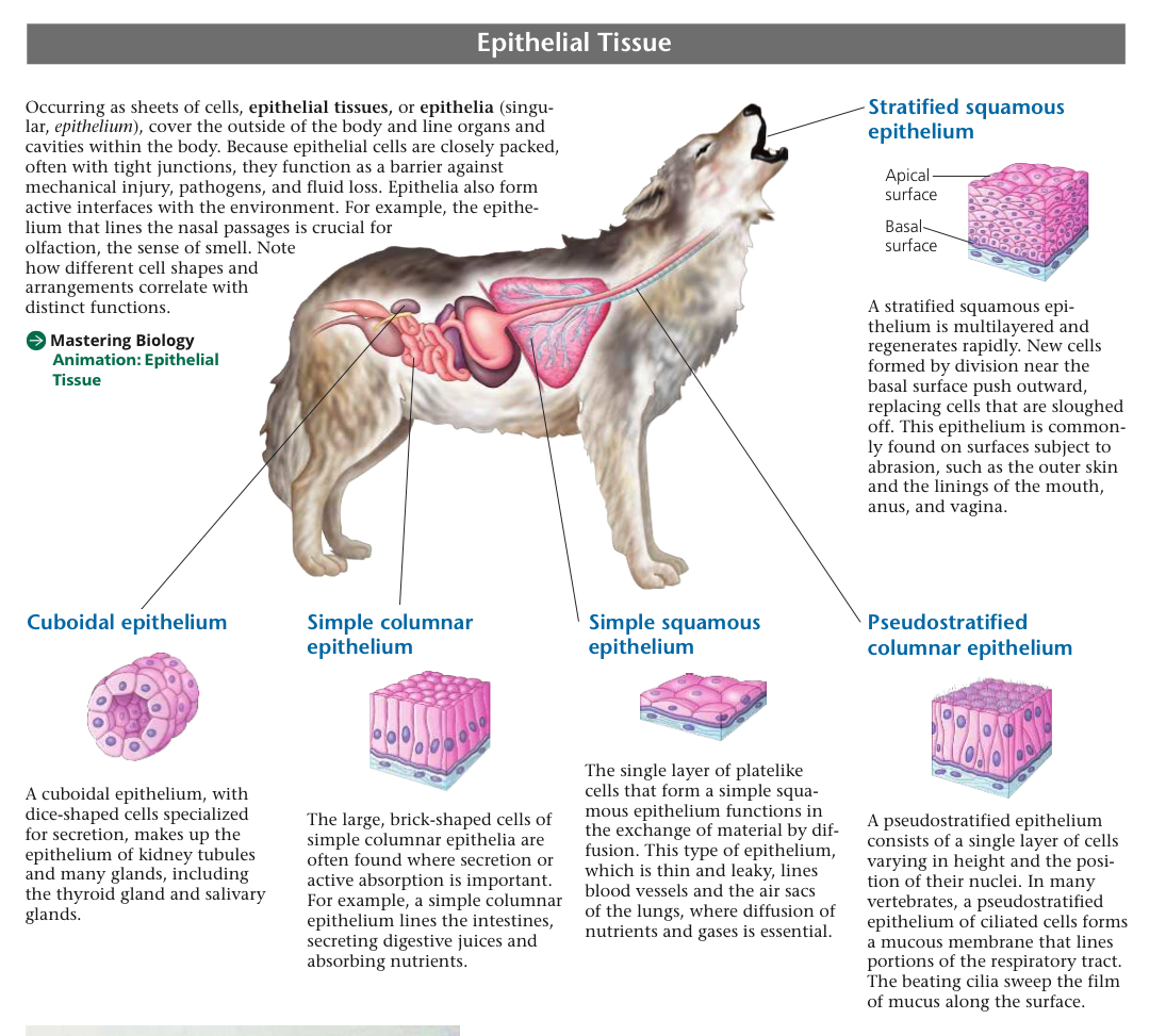
Epithelial Tissues/Epithelia: Sheets of cells that cover the outside of the body, acting as a barrier against mechanical injury, pathogens, and fluid loss
Stratified Squamous Epithelium: Multilayered, regenerates rapidly, for surfaces subject to abrasion
Outer skin, linings of mouth, anus, vagina
Cuboidal Epithelium: Disk shaped cells for secretion
Kidney tubules and many glands, such as thyroid gland and salivary glands
Simple Columnar Epithelium: Large, brick shaped cells for secretion or active absorption
Intestines to secrete digestive juices and absorb nutrients
Simple Squamous Epithelium: Single layer of plate like cells, function in exchange of material via diffusion
Thin and leaky, lines blood vessels and air sacs of lungs
Pseudostratified Columnar Epithelium: Single layer of cells varying in height and position of nuclei
Forms mucous membrane to line portions of respiratory tract
Polarized, so they have an apical surface facing the lumen, with specialized projections, and the basal surface
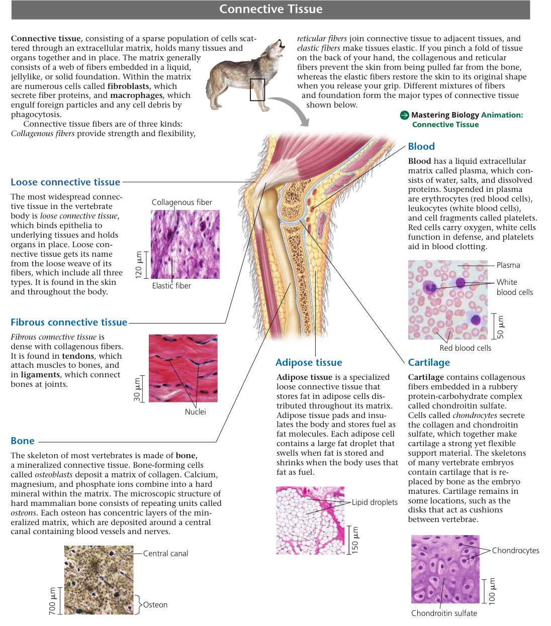
Connective Tissue: Sparse population cells scattered through extracellular matrix (web of fibers in liquid, jelly, or solid), holds tissues and organs together in one place
Fibroblasts: Cells within the matrix, secrete fiber proteins
Macrophages: Engulf foreign particles and debris by phagocytosis
Collagenous Fibers: Provide strength and flexibility
Reticular Fibers: Join connective tissue to adjacent tissues
Elastic Fibers: Make tissue elastic
Loose Connective Tissue: Binds epithelia to underlying tissues and holds organs in place
Most widespread
All three types of fibers, weaved loosely. In skin and throughout body
Fibrous Connective Tissue: Dense with collagenous fibers, found in tendons and ligaments
Tendons: Attach muscles to bones
Ligaments: Connect bones at joints
Bone: Mineralized connective tissue
Osteoblasts: Cells that form bone, deposit a matrix of collagen
Repeating units of osteons
Adipose Tissue: Specialized loose connective tissue that stores fat in its cells
Insulate body, store fuel as fat
Cartilage: Has collagenous fibers embedded in rubbery chondroitin sulfate (protein carbohydrate complex)
Chondrocytes: Cells that secrete collagen and chondroitin sulfate
Muscle Tissue: Tissue responsible for almost all types of body movement
Skeletal Muscle/Striated Muscle: Responsible for voluntary movements
Bundles of long cells, muscle fibers
Sarcomeres: Contractile units, arranged in a way that gives it its striped appearance
Smooth Muscle: Lacks striations, responsible for involuntary body activities
Bladder, digestive tract, arteries
Cardiac Muscle: Forms contractile walls of heart, branched fibers interconnect via intercalated disks, relaying signals from cell to cell for heart contraction
Striated like skeletal muscle
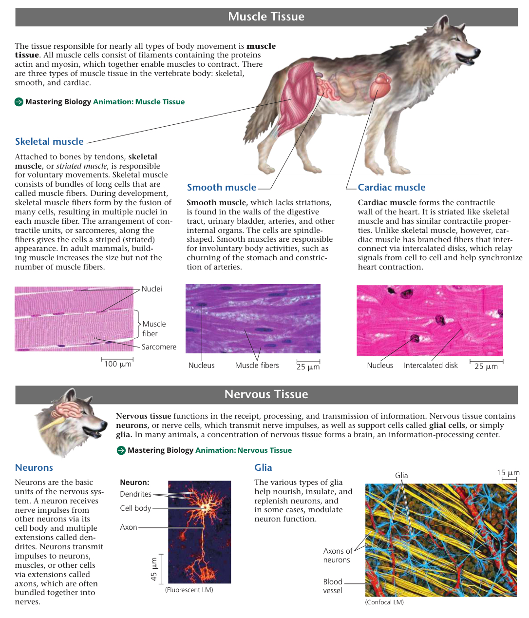
Nervous Tissue: Receiving, processing, and transmission of info
Neurons: Nerve cells, transmit nerve impulses
Glial Cells/Glia: Support cells
Two major systems for controlling responses to stimuli, endocrine and nervous
Endocrine System: Signaling molecules released into bloodstream by endocrine cells
Nervous System: Neurons transmit signals along routes connecting specific locations
Hormones: Signaling molecules broadcast throughout body by endocrine system
Sheets of cells
I’m not sure
Since hormones being released and yeah
40.2: Feedback control maintains the internal environment in many animals
Regulator: Animal that uses internal mechanisms to control change (body temp) during external fluctuation
Conformer: Allows internal condition to change with external changes
Homeostasis: Maintenance of internal balance
Set Point: Value that control system tries to keep values
Stimulus: Fluctuation in variable
Sensor: Detects fluctuations and signals a control center
Response: Triggered by sensor
Negative Feedback: Tries to reduce stimulus to maintain homeostasis
Positive Feedback: Amplifies stimulus, helps finish processes
Circadian Rhythm: Physiological changes that happen ~every 24 hours
Acclimatization: Animal’s physiological adjustment to changes in environment
ex. elk goes to high altitudes and blood pH is raised, so pee becomes more alkaline to return it to normal
40.3: Homeostatic processes for thermoregulation involve form, function, and behavior
Thermoregulation: Process animals maintain their body temperature within a normal range
Endothermic: Warmed mostly by heat generated by metabolism
Ectothermic: Gain most of heat from external sources
Poikilotherm: Animal whose body temp varies
Homeotherm: Animal with relatively consistent body temp
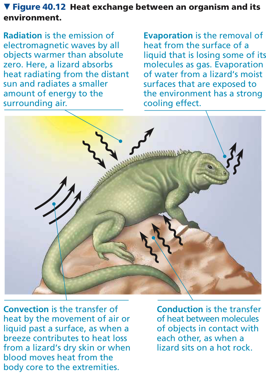
Radiation: Emission of electromagnetic waves by all objects warmer than 0 kelvin
Evaporation: Removal of heat from surface of liquid which is losing some of its molecules as gas
Convection: Transfer of heat by movement of air or liquid past a surface
Conduction: Transfer of heat between objects in contact
Integumentary System: Outer covering of body (skin, hair, nails)
In response to temp changes, many animals alter the amount of blood (so also heat) flowing between their body core and skin
Vasodilation: Widening of superficial (near surface) blood vessels to increase blood flow and heat transfer
Vasoconstriction: Decrease diameter of superficial vessels to reduce blood flow and heat transfer
Countercurrent Exchange: Transfer of heat between fluids flowing in opposite directions
Thermogenesis: Endotherms vary heat production to match changing rates of heat loss
Hypothalamus: Brain region with sensors for thermoregulation, also controls circadian clock
40.4: Energy requirements are related to animal size, activity, and environment
Metabolic Rate: Sum of all the energy an animal uses in a given interval, measured in J, cal, or kcal
Basic Metabolic Rate (BMR): Rate of nongrowing endotherm at rest with an empty stomach not under stress
Standard Metabolic Rate (SMR): Rate of fasting, nonstressed ectotherm at rest
Torpor: State of decreased activity and metabolism
Many birds and small animals do it daily (bats during day, hummingbirds on cold nights)
Hibernation: Long term torpor, to combat winter cold and food scarcity
Chapter 41: Animal Nutrition
41.1: An animal’s diet must supply chemical energy, organic building blocks, and essential nutrients
Nutrition: Process an animal uses to take in and make use of food to satisfy their three needs (chemical energy, organic building blocks, essential nutrients)
Essential Nutrients: Substances that an animal requires but can’t assemble from simple organic molecules
Amino acids and fatty acids, plus certain vitamins and minerals
Too little causes deformities, disease, and death
Essential Amino Acids: The half of amino acids that must be obtained from an animal’s food
Animal has enzymes to produce about half
Plants and microorganisms can produce all 20
Essential Fatty Acids: Fatty acids that animals can’t form the double bonds for that must be acquired by its food
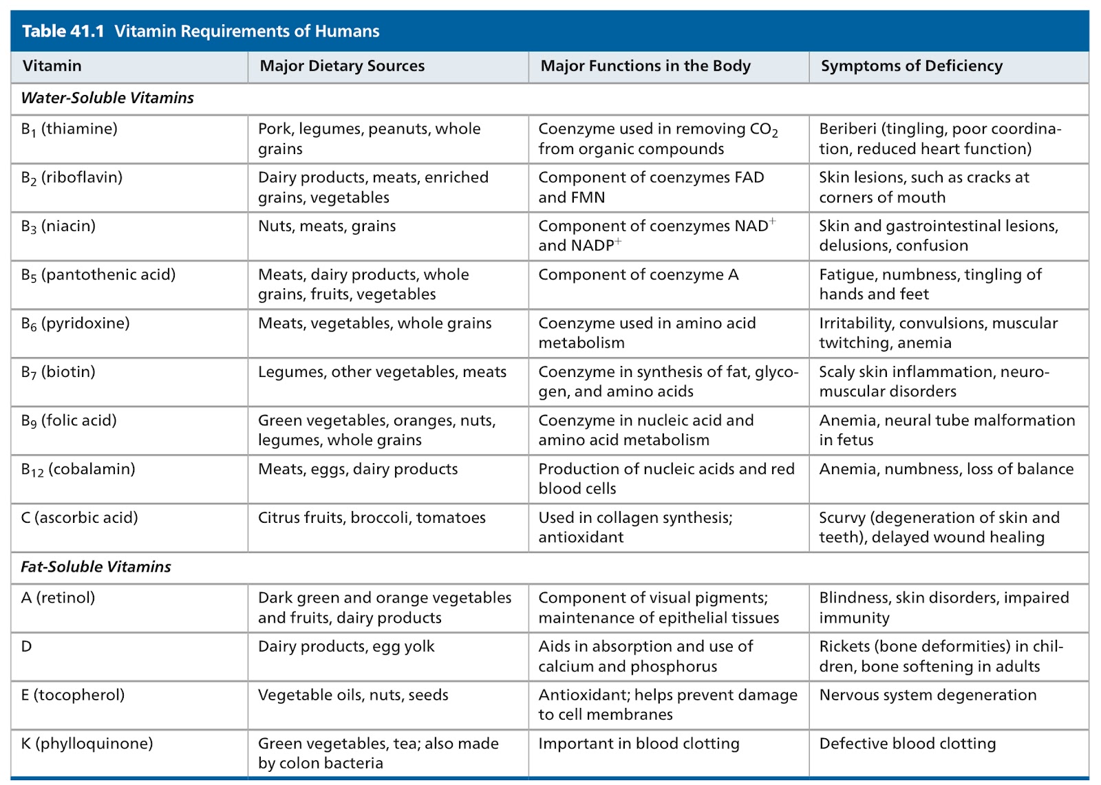
Vitamins: Organic molecules that are required in the diet in very small amounts
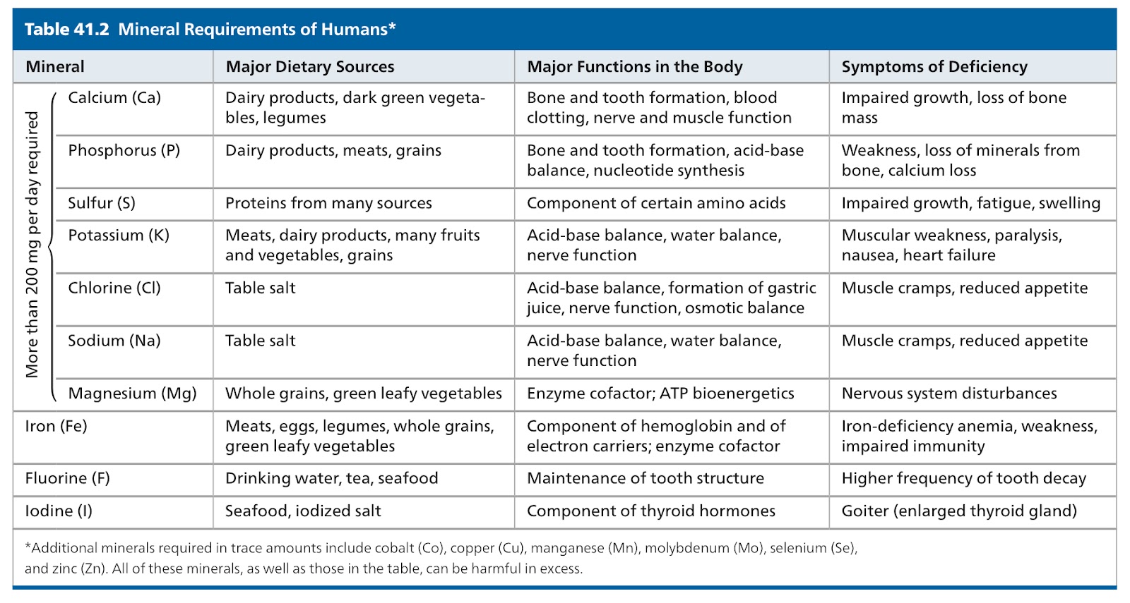
Minerals: Inorganic nutrients required in small amounts
Herbivores: Eat mostly plants and algae
Carnivores: Mostly eat other animals
Omnivores: Consume animals and plants or algae
Too little chemical energy causes malnutrition
We can produce half on our own
I don’t remember this
Looking at its diet and analyzing which nutrients are being consumed and which ones aren’t, or seeing the diseases it contracts
41.2: Food processing involves ingestion, digestion, absorption, and elimination
Ingestion: First stage of food processing, act of eating or feeding
Filter Feeding: Strain small organisms or food particles from surrounding medium
Bulk Feeding: Eat relatively large pieces of food
Using tentacles, pincers, claws, venomous fangs, jaws, and teeth
Substrate Feeding: Animals live in or on their food source
Fluid Feeding: Suck nutrient rich liquid from living host
Digestion: Second stage of food processing, food broken down into small enough molecules for body to absorb
Mechanical Digestion: Chewing or grinding, breaks food into smaller pieces and increases surface area
Chemical Digestion: Cleaves large molecules into smaller components
Enzymatic Hydrolysis: Chemical breakdown by digestive enzymes of fat or a macromolecule
Absorption: Animal’s cells absorb small molecules such as amino acids and simple sugars
Elimination: Undigested material passes out of the digestive system
Intracellular Digestion: Hydrolysis of food in food vacuoles
Cell engulfs food by phagocytosis or pinocytosis. Food vacuoles fuse with lysosomes
Extracellular Digestion: Breakdown of food in compartments continuous with he outside of the animal’s body
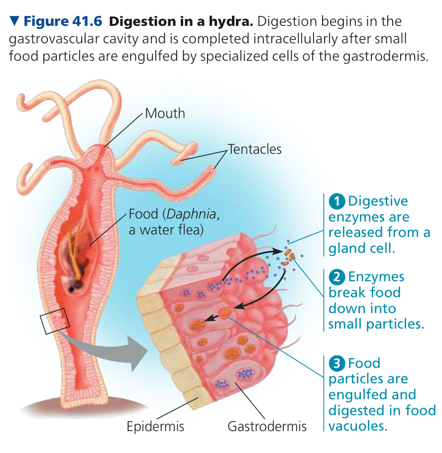
Gastrovascular Cavity: Digestive compartment with single opening in animals with simple body plans, helps in digestion and distribution of nutrients throughout the body
In the hydra, uses tentacles to stuff prey into its mouth and gastrovascular cavity
Specialized gland cells of its gastrodermis secrete digestive enzymes to break the soft tissue of prey into tiny pieces
Gastrodermis: Tissue layer that lines the gastrovascular cavity
Intracellular digestion
Undigested materials eliminated through mouth
Alimentary Canal: Digestive tube with two openings, a mouth and an anus, in animals with complex body plans
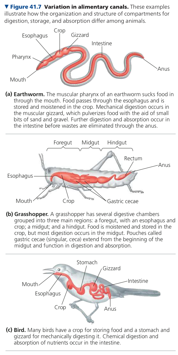
Gastrovascular has one opening, alimentary has two
We start absorbing in absorption, before that it is just floating around but not really doing anything
I’m confused, I have no idea
41.3: Organs specialized for sequential stages of food processing form the mammalian digestive system
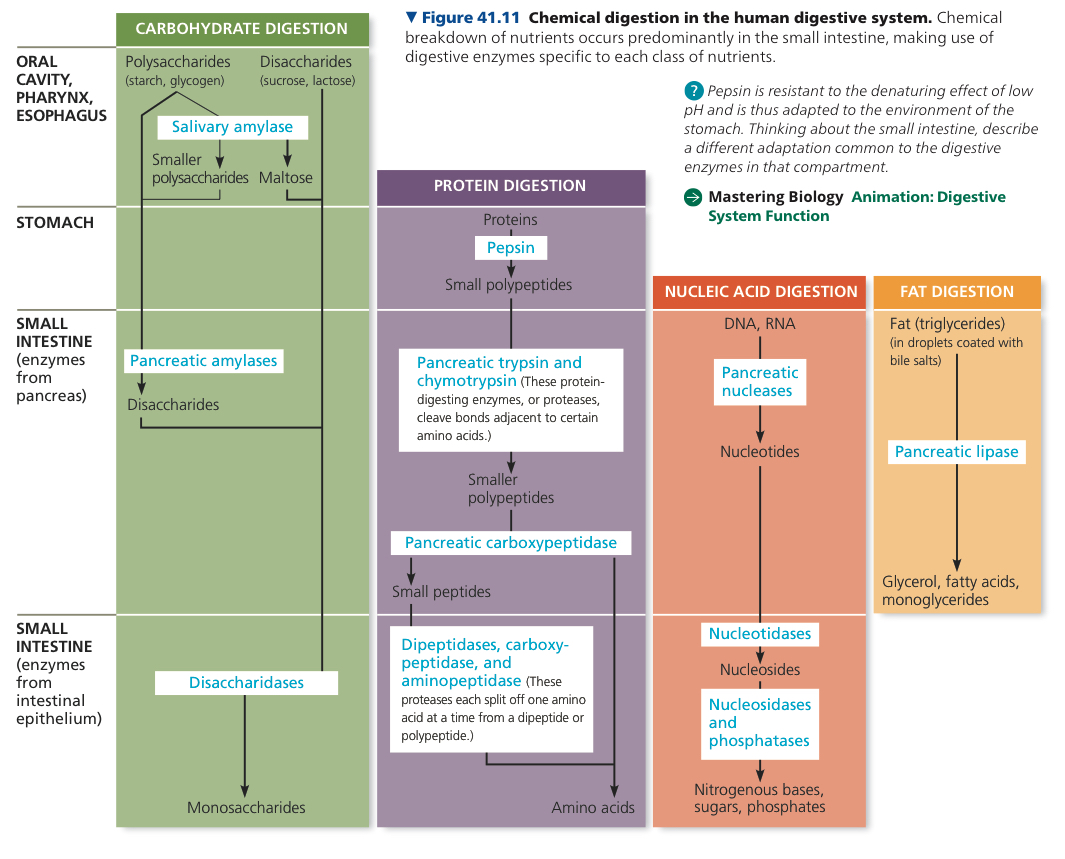
Oral Cavity: Mouth
Salivary Glands: Releases saliva when anticipating food (or it arrives)
Mucus: Viscous mixture of water, salts, cells, and glycoproteins (carbohydrate protein complex)
Contains lots of amylase and glycogen
Amylase: Breaks down starch
Tongue shapes a mixture of saliva and food into a ball called bolus
Pharynx: The throat region which receives the bolus, leading to two passageways, the esophagus and trachea
Esophagus: Muscular tube that connects to the stomach
Trachea/Windpipe: Leads to lungs
Must go into esophagus, going into trachea causes choking
Peristalsis: Pushes food along in the esophagus, alternating waves of smooth muscle contraction and relaxation
Sphincter: Ring like valve of muscle, acts like a drawstring at the end of the esophagus, regulates passage of food into stomach
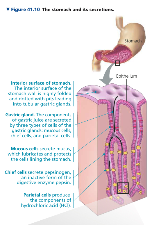
Stomach: Located just below the diaphragm, stores and processes food
Secretes gastric juice and mixes it with food through churning action
Chyme: Mixture of ingested food and gastric juice
Hydrochloric Acid disrupts extracellular matrix that binds cells together in meat and plant material
Pepsin: A protease that attacks exposed bonds weakened by HCl
Protease: Protein digesting enzyme
Two types of cells in gastric glands produce gastric juice components
Parietal Cells use ATP driven pump to expel H+ ions into the lumen while chloride ions diffuse into the lumen, they combine only in the lumen to make HCl
Chief cells release pepsinogen into lumen (inactive form of pepsin), converted into pepsin by HCl which clips off part of the molecule and exposes its active site
Small Intestine: Longest compartment of alimentary canal
Duodenum: First 10 inches of small intestine
Chyme arrive triggers release of secretin hormone, causing pancreas to secrete bicarbonate
Fat is difficult to digest, so it is done by bile salts, which are like emulsifiers that break apart fat and lipids
Bile: Secretion of liver that is stored and concentrated in the gallbladder, largely composed of bile salts
Contents of duodenum moves to remaining regions of small intestine (jejunum and ileum) by peristalsis
Villi: Finger shaped folds in large intestine
Microvilli: Microscopic projections within the villi, a “brush border”
Capillaries and veins carry nutrient rich blood away from villi, converge into hepatic portal vein
Hepatic Portal Vein: Blood vessel that leads directly to the liver
Liver → Heart → other tissues and organs
Allows liver to regulate distribution of nutrients
Allows liver to remove toxic substances before they can circulate
Hydrolysis of a fat lipase in generates fatty acids and a monoglyceride (glycerol + fatty acid), absorbed by epithelial cells and combined into triglycerides
Chylomicrons: Triglycerides coated in phospholipids, cholesterol, and proteins
First enter a lacteal
Lacteal: Vessel at core of each villus
Part of vertebrate lymphatic system, a network of vessels filled with lymph (clear fluid)
Lymph with chylomicrons goes into larger vessels of lymphatic system and then heart
Small intestine also recovers water and ions
Large Intestine: Where alimentary canal ends, including colon, cecum, and rectum
Connected at a T shaped junction with small intestine (arms are colon and cecum)
Colon: Leads to rectum and anus, completes recovery of water (which started in the small intestine)
Cecum: Ferments ingested material, small in humans and has an appendix
Appendix: Finger shaped extension that acts as a reservoir for symbiotic microorganisms
Feces: Wastes of digestive system, becoming increasingly solid as it moves down colon by peristalsis
Rectum: Where feces stored before eliminated
Two sphincters (rings) separate rectum and anus, inner involuntary outer voluntary
41.4: Evolutionary adaptations of vertebrate digestive systems correlate with diet
Microbiome: Collection of microorganisms living in and on the body
Vertebrae digestive systems show evolutionary adaptations
Assortment of teeth correlates with diet
Herbivores have fermentation chambers to digest cellulose
Herbivores have longer alimentary canals than carnivores, since it takes longer to digest veg
It takes longer to digest vegetables, so it helps make sure that it is fully broken down
Don’t wanna go back and check
Maybe because the yogurt itself must also be digested?
41.5: Feedback circuits regulate digestion, energy storage, and appetite
Glucose homeostasis relies on antagonistic effects of hormones insulin and glucagon
Insulin: Decreases blood glucose concentration by triggering uptake of glucose from blood into body cells
Glucagon: Increases blood glucose concentration by releasing glucose into blood from energy stores
In pancreas, pancreatic islets have alpha cells, which make glucagon, and beta cells, which make insulin
Diabetes Mellitus: Caused by deficiency of insulin or decreased response to insulin in target tissues
Ghrelin: Hormone that triggers hunger, secreted by stomach wall
Leptin: Hormone produced by fat, suppresses appetite
Vertebrates store excess calories in glycogen and fat, which can be tapped when it uses more calories than it consumes
Too many calories causes obesity
Appetite issues caused by hormones not working properly (insulin, leptin)
Leptin levels might begin to even out and then they start to gain more
Causes very high glucose levels and liver tries to filter it out but exhausts itself idk
Chapter 42: Circulation and Gas Exchange
42.1: Circulatory systems link exchange surfaces with cells throughout the body
Natural selection has caused two basic adaptations for efficient exchange for all of an animal’s cells
Simple body plan with many or all cells in direct contact with the environment
If not simple body plant, have a circulatory system
Gastrovascular Cavity: Distributes substances throughout the body and in digestion, central in animals with almost all cells in contact with environment
Circulatory system has three components, circulatory fluid, interconnecting vessels, and heart, to connect the aqueous environment to organs that exchange gases, absorb nutrients, and dispose of wastes
Heart: Muscular pump that powers circulation using metabolic energy
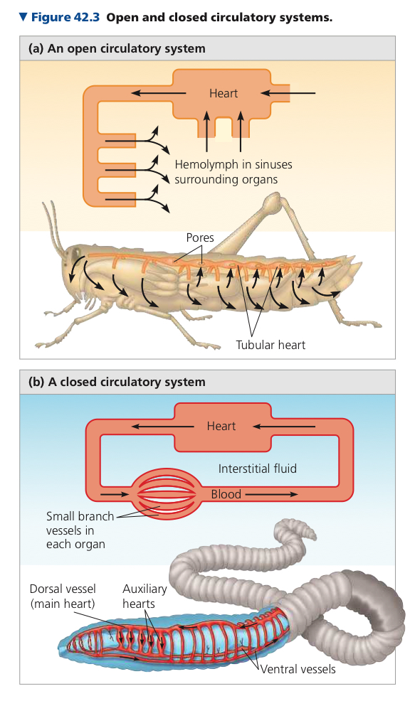
Either opened or closed
Open Circulatory System: Circulatory fluid (hemolymph) is also the interstitial fluid
Contraction of heart pumps hemolymph through circulatory vessels into interconnected sinuses surrounding the organs
In the sinuses, hemolymph and body cells exchange gases
Closed Circulatory System: Circulatory fluid (blood) confined to vessels and distinct from interstitial fluid
1+ hearts pump blood into vessels that branch into smaller ones and infiltrate issues and organs
Cardiovascular System: Heart and blood vessels in vertebrates
3 main types of blood vessels where blood only flows in one direction
Arteries: Blood from heart to organs
Arterioles: What arteries branch into within organs
Capillaries: Microscopic vessels with thin porous walls, get blood from arterioles
Capillary Beds: Networks of capillaries, infiltrate tissues
Converge into venules which converge into veins, which carry blood back to the heart
arteries, away, veins, villain (always comes back)
All hearts have 2+ muscular chambers.
Atria: Receives blood entering the heart
Ventricles: Pumps blood out of the heart
Single Circulation: Blood travels through body in a single cycle then returns to its starting point
Double Circulation: Two circuits of blood flow, with both pumps combined in the heart
Pulmonary Circuit: Right side circuit pumps oxygen poor blood into capillary beds, net movement of O2 into blood, CO2 out
Systemic Circuit: Left side circuit heart pumps oxygen enriched blood to capillary beds in organs
Continues to just circulate
Not sure
It would leak out and the blood being pumped in wouldn’t be oxygen rich
42.2: Coordinated cycles of heart contraction drive double circulation in mammals
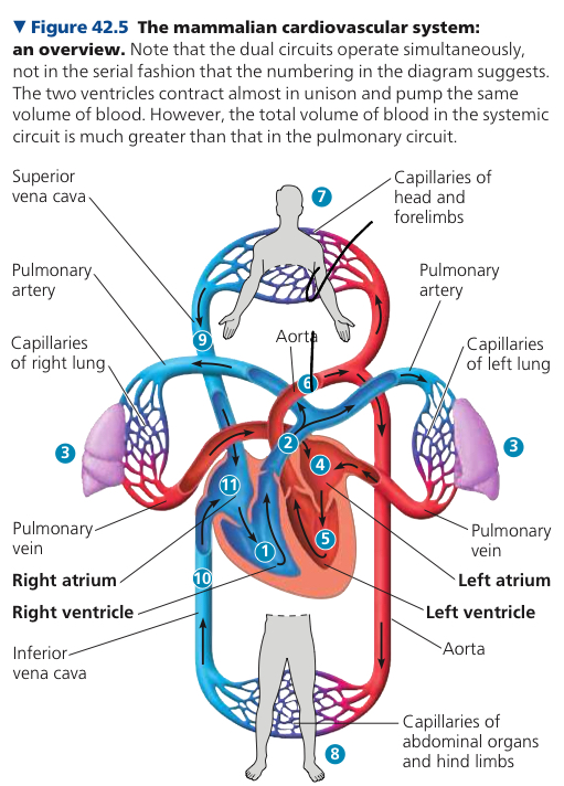
Timely delivery of oxygen (O2) to organs is crucial; brain cells may die if O2 supply is interrupted. Mammalian cardiovascular system meets the body's continuous O2 demand through an organized system
Pulmonary circuit
Right ventricle pumps blood to the lungs via pulmonary arteries.
Oxygen-rich blood returns to the left atrium via pulmonary veins.
Systemic circuit
Left ventricle pumps oxygen-rich blood to body tissues through the aorta.
Branches lead to capillary beds in the head, arms, abdominal organs, and legs.
Capillaries facilitate O2 diffusion to tissues and CO2 absorption.
Veins return oxygen-poor blood to the right atrium via superior and inferior vena cavae.
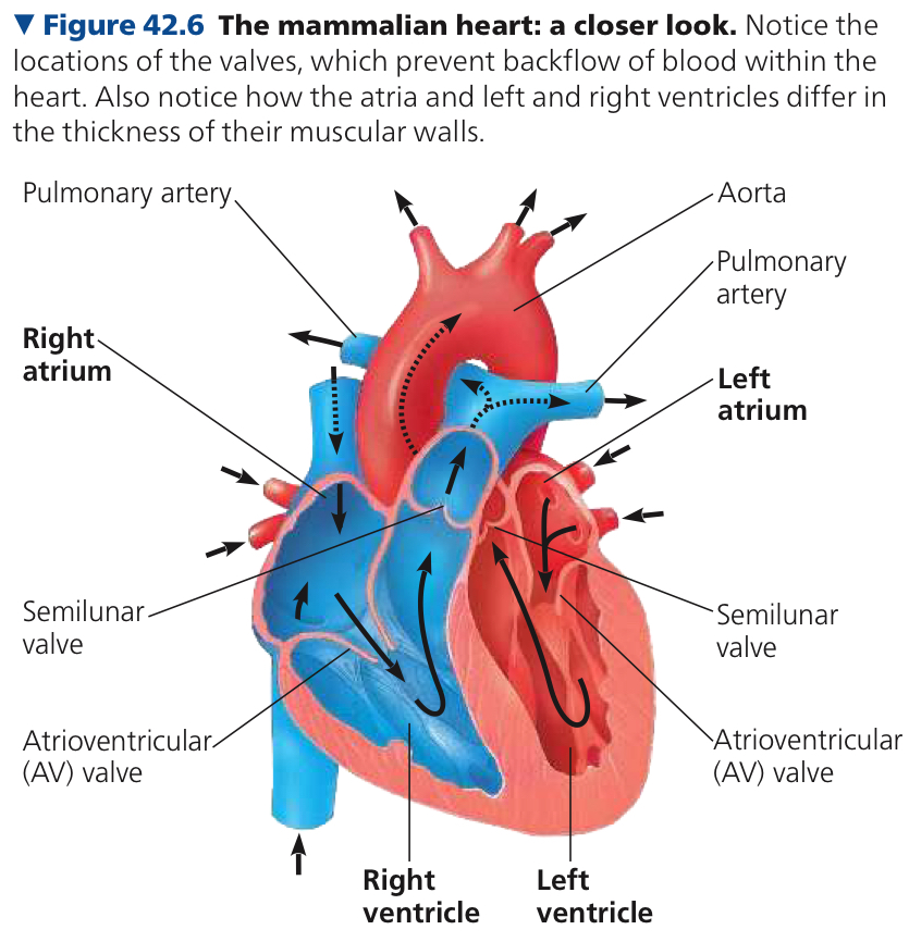
Human heart located behind the sternum, cardiac muscle predominant
Atria serve as blood collection chambers, ventricles pump blood forcefully
Cardiac Cycle: One complete sequence of pumping and filling of the heart
Systole: Contraction phase of the cardiac cycle
Diastole: Relaxation phase of the cardiac cycle
Cardiac Output: Volume pumped per minute; determined by heart rate and stroke volume.
Four valves prevent backflow and keep blood moving in the right direction, 2x AV, 2x semilunar
Antriventricular (AV) Valve: Lies between each atrium and ventricle, anchored by strong fibers that keep them from turning inside out during ventricular systole
Semilunar Valves: Located at the two exits of the heart
Heart murmurs: Abnormal sound made by blood squirting backward through a defective valve
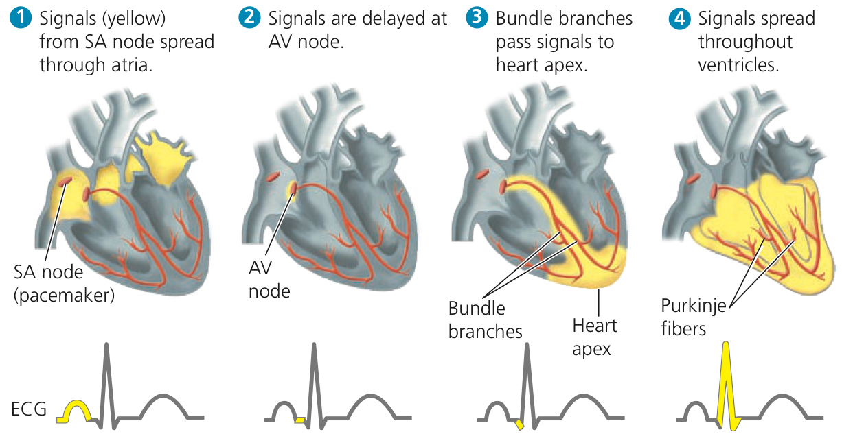
Heartbeat originates in the heart, autorhythmic cells in the right atrium act as pacemaker
Sinoatrial (SA) Node: Cluster of cells in the right atrium; acts as a pacemaker, setting the rate and timing of cardiac muscle contractions.
Impulses spread through atria, delayed at atrioventricular (AV) node for complete atrial contraction
Atrioventricular (AV) Node: Relay point between left and right atria, delays impulses for ~0.1 second before spreading to the heart apex
Bundle branches and Purkinje fibers conduct signals to ventricles
Sympathetic and parasympathetic divisions regulate heart rate; hormones and body temperature also influence pacemaker function.
Sympathetic division accelerates heart rate, parasympathetic division slows it down.
Hormones (e.g., epinephrine) and body temperature also impact pacemaker.
Heart rate increases during activities and fever, decreases during rest
42.3: Patterns of blood pressure and flow reflect the structure and arrangement of blood vessels
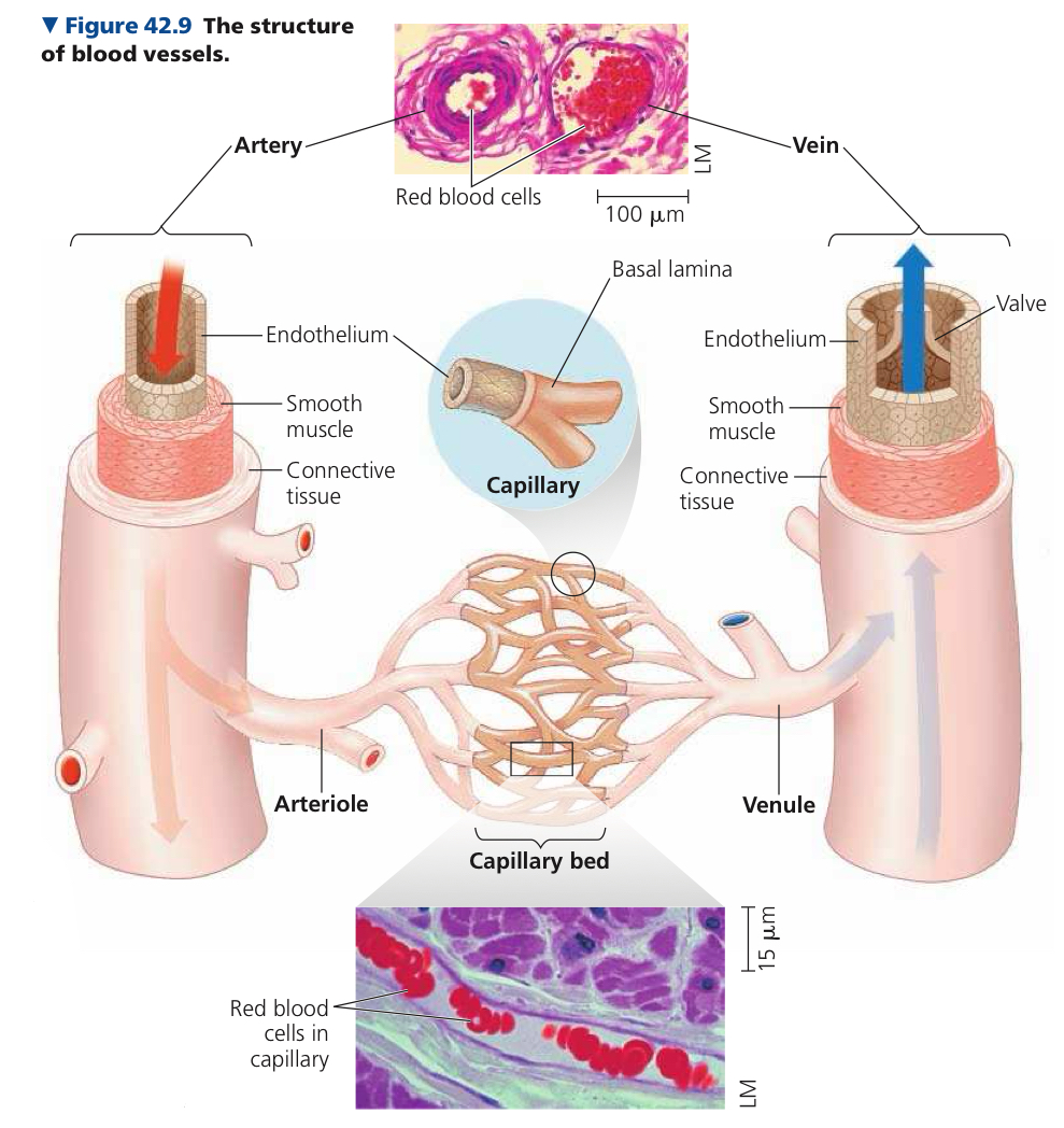
Endothelium: Single layer of flattened epithelial cells, line all blood vessels
Large blood vessel diameter means slow blood flow, small = fast
Systolic Pressure: Arterial blood pressure when the heart contracts during ventricular systole, when it is highest and spikes
Pulse: Rhythmic bulging of artery walls
Diastolic Pressure: Lower blood pressure when ventricles are relaxed
Vasoconstriction: When arterioles narrow after smooth muscles in arteriole walls contract. Increases blood pressure upstream in arteries
Vasodilation: Increase in diameter of arterioles when smooth muscles relax
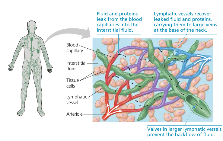
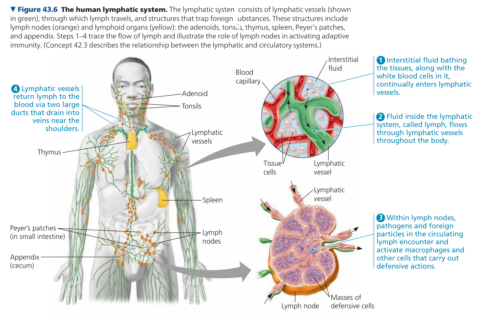
Lymphatic System: Returns lost fluid and proteins in capillaries, which leak into tissues
Lymph: Returned fluid that circulates in the lymphatic system
Lymph Nodes: Lymph filtering organs which help in defense
42.4: Blood components function in exchange, transport, and defense
Plasma: Liquid matrix, holds whole blood
Whole blood consists of cells and cell fragments (platelets) suspended in plasma
Plasma proteins influence blood pH, osmotic pressure, viscosity, lipid transport, immunity, and blood clotting
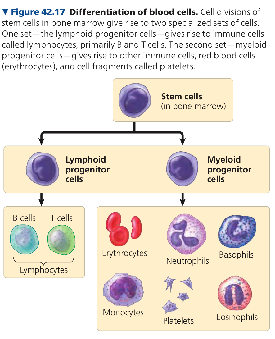
Erythrocytes: Red blood cells, transport O2. Mature ones lack nuclei, small disks. Short lives, ~120 days before being replaced
Hemoglobin: Iron containing proteon that transports O2
Sickle Cell Disease: Abnormal form of hemoglobin plymeries into aggregates, which distort the shape into a curved shape that looks like a sickle
Leukocytes: White blood cells, defend against microorganisms and foreign substances in blood
Platelets, erythrocytes, and leukocytes are all stem cells, which can reproduce indefinitely
Thrombus: Blood clot that forms within a blood vessel and blocks flow of blood
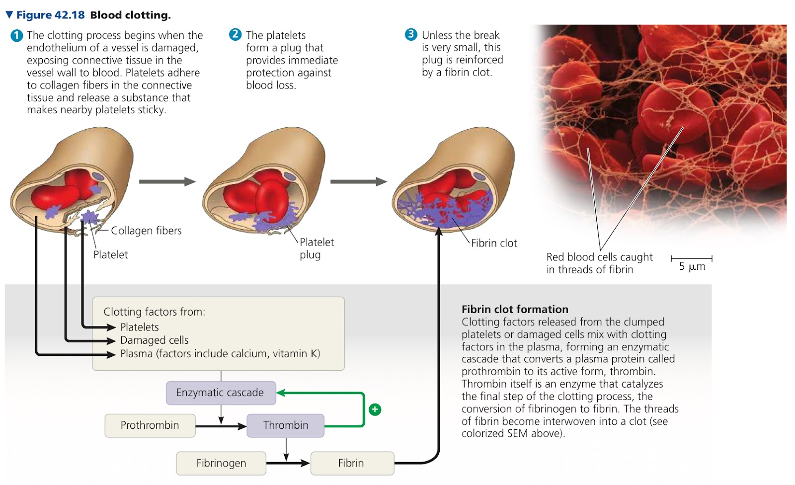
Atherosclerosis: Hardening of the arteries by accumulation of fatty deposits
Heart Attack: Damage or death of cardiac muscle tissue from blockage of one or more coronary arteries
Stroke: Death of nervous tissue in the brain due to lack of O2
Hypertension: High blood pressure
42.5: Gas exchange occurs across specialized respiratory surfaces
Gas Exchange: Gas undergoes net fiddusion from where its partial pressure is higher to where it is lower
Partial Pressure: Pressure exerted by a particular gas in a mixture of gases
Structure and organization of respiratory surfaces differ among animal species
Effectiveness of gas exchange in some gills is increased by ventillation and countercurrent exchange
Ventilation: Movement of the respiratory medium over the respiratory surface to maintain partial pressure gradients necessary for gas exchange
Countercurrent Exchange: Exchange of a substance or heat between two fluids flowing in opposite directions (in fish, blood and water)
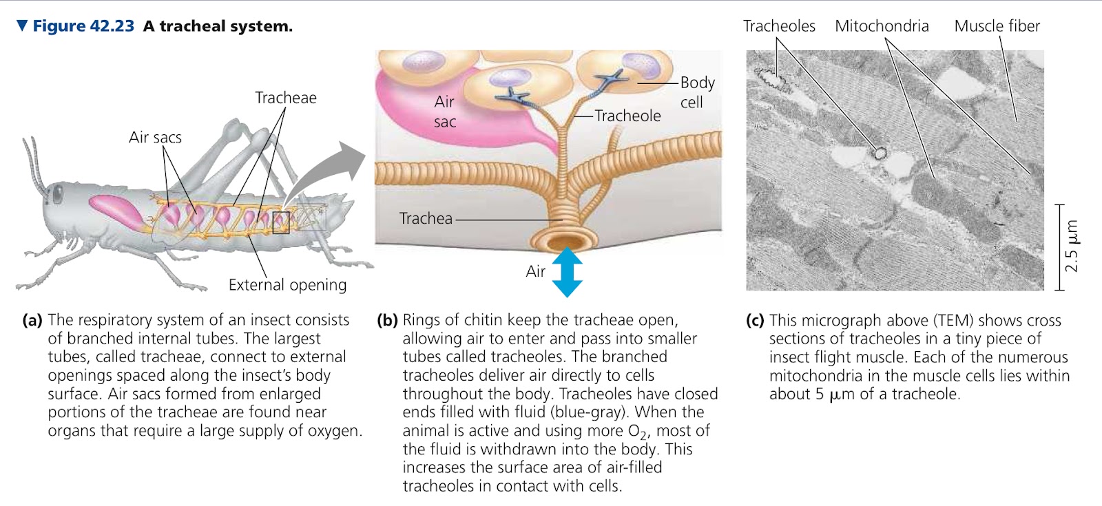
Tracheal System: Branched network of tubes that brings O2 directly to cells in insects
Largest tubes (trachea) open to the outside
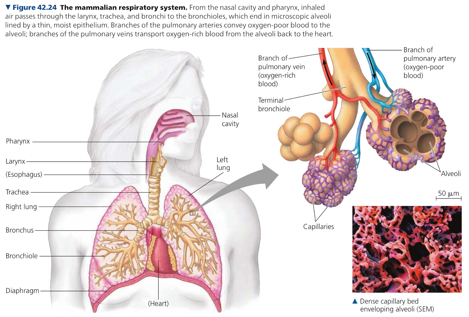
Internal Lungs: Localized respiratory organs in most terrestrial vertebrates
In mammals, air → nostrils → nasal cavity → pharynx → larynx → trachea → bronchi
Pharynx: Where paths for air and food cross
Larynx: Upper part of respiratory tract, moves upward and tips epiglottis over the glottis when food is swallowed
Trachea: Windpipe. Closed by larynx when swallowing, branches into two bronchi
Bronchi: Each lead to one lung, and branch into bronchioles
Bronchioles: Finer and finer tubes
Alveoli: Where gas exchange in mammals occurs
Surfactant: Mixture of phospholipids and proteins, produced by air sacs, coat alveoli and reduces surface tension
42.6: Breathing ventilates the lungs
Breathing: Process that ventilates the lungs, alternating inhalation and exhalation of air
Positive Pressure Breathing: Inflating lungs with forced air flow, how amphibians breathe
Negative Pressure Breathing: Pulling air into the lungs instead of pushing, used by mammals
Rib muscles and diaphragm contract, incoming and outgoing air mix and decrease efficiency
Tidal Volume: Amount of air inhaled and exhaled with each breath, ~500 mL avg in resting humans
Vital Capacity: Tidal volume during max, ~3.4-4.8 L
Residual Volume: Air that remains after a forced exhalation
Inhalation takes energy, exhalation is passive
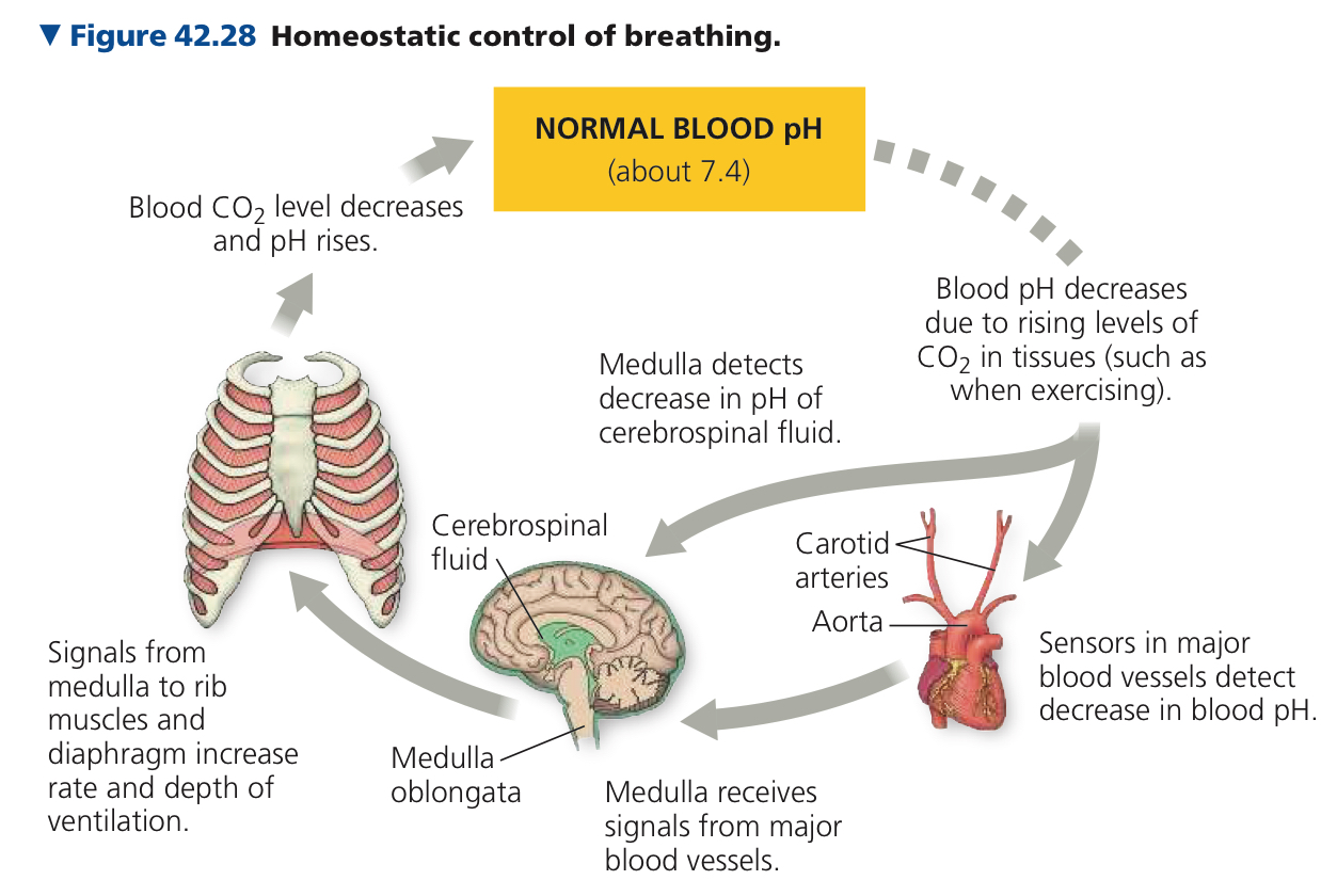
Sensors detect pH of cerebrospinal fluid and adjust accordingly
42.7: Adaptations for gas exchange include pigments that bind and transport gases
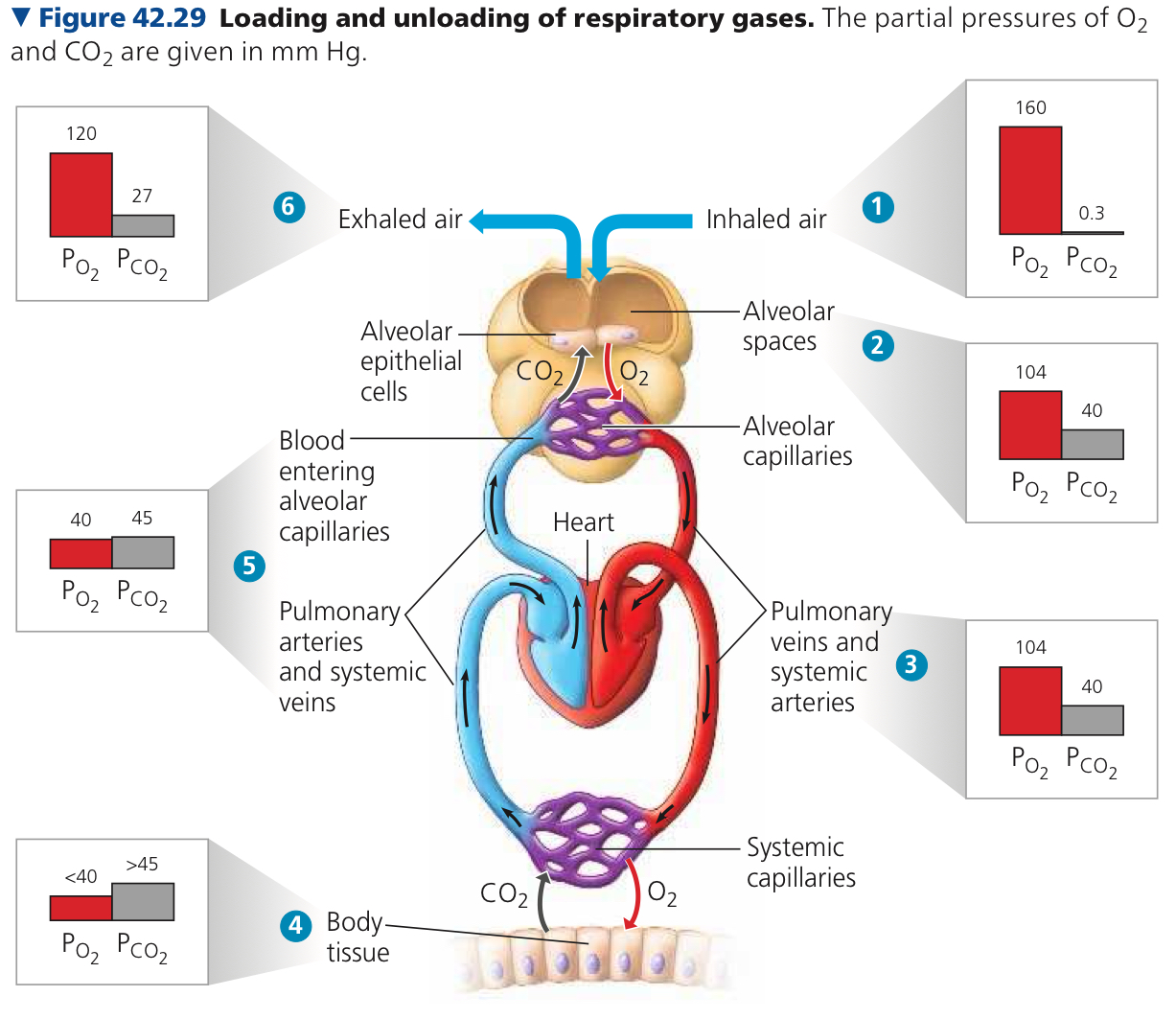
During inhalation, fresh air mixes with air remaining in lungs
Mixture from alveoli has higher Po2 than blood flowing through alveolar capillaries
Net diffusion of O2 from alveoli to blood
Presence of Pco2 in alveoli higher than in capillaries means net diffusion CO2 from blood to air
Po2 and Pco2 match values for air in alveoli. Blood returns to heart and is pumped through systemic circuit
In systemic capillaries, net diffusion of O2 out of blood, CO2 in
Blood is returned to heart and pumped to lungs
Exchange occurs across alveolar capillaries, exhaled air enriched in CO2, depleted of O2
Respiratory Pigments: Proteins, bind to O2 and transport it, circulate with blood or hemolymph
Bohr Shift: Low pH dereases affinity of hemoglobin for O2
Myoglobin: Oxygen storing protein
Chapter 43: The Immune System
43.1: In innate immunity, recognition and responserely on traits common to groups of pathogens
Pathogen: Disease causing agent
Immune System: Lets animal avoid or limit many infections
Molecular Recognition: Specific binding of immune receptors to foreign molecules
Innate Immunity: Set of immune defenses common to all animals
Lysozyme: enzyme that breaks down bacterial cell walls and acts as a chemical barrier against any pathogens ingeste with food
Body secretions make a hostile environment for pathogens and inhibit microbial entry
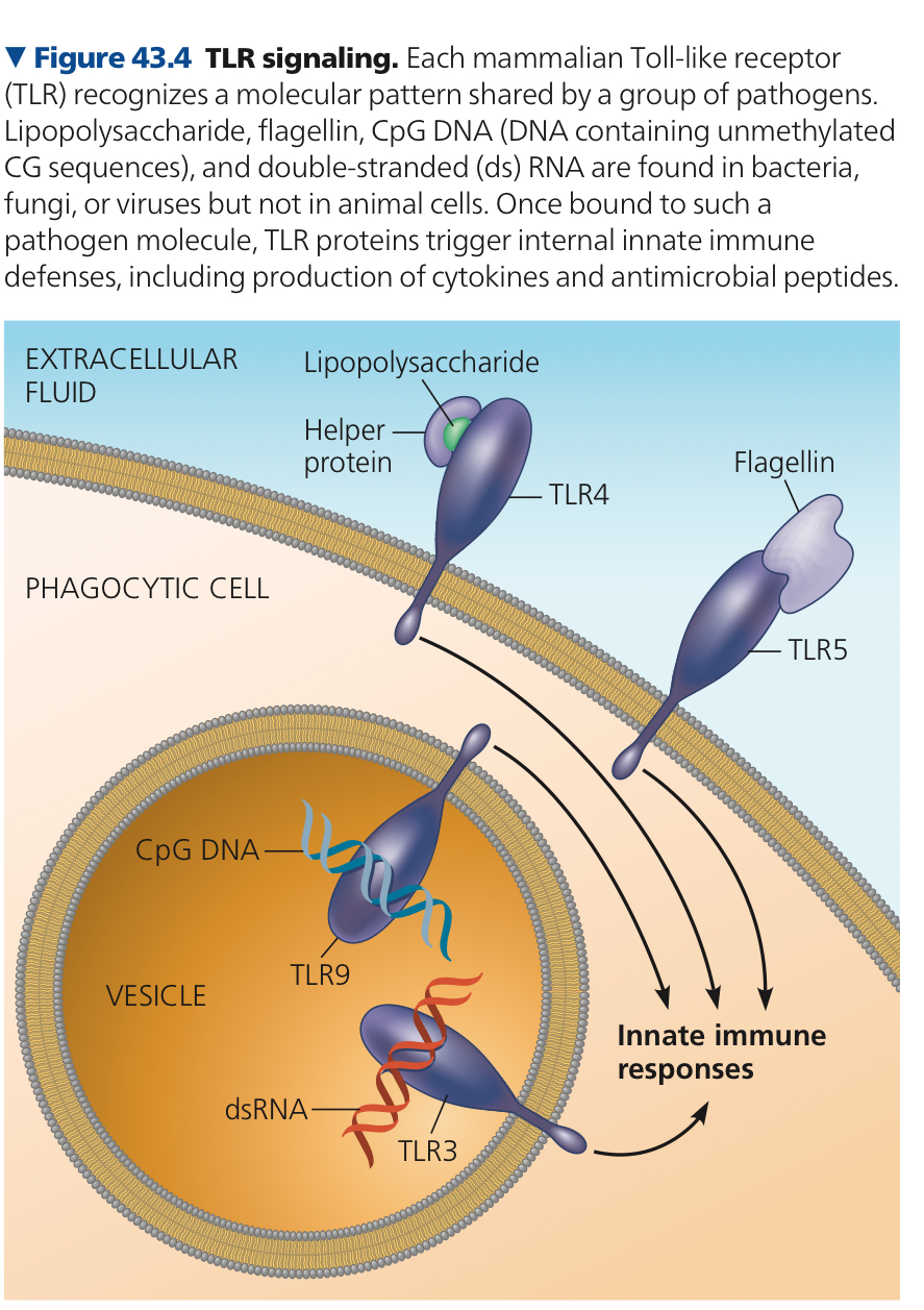
Toll-Like Receptor (TLR): Binds to fragment molecules characcteristic of a set of pathogens
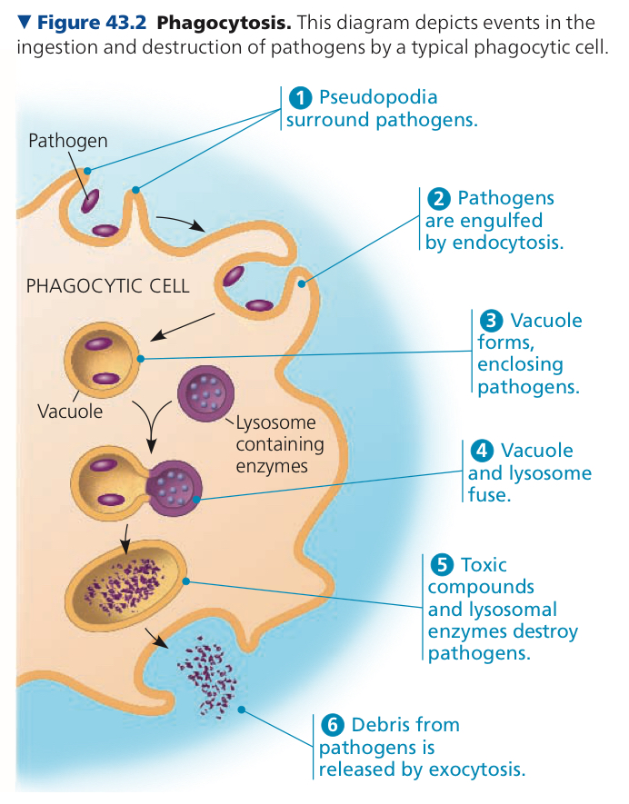
Two main types of phagocytic cells
Neutrophils: Circulate bood, attracted by signals from infected tissues then engulf and destroy infecting pathogens
Macrophages: Larger phagoctic cells that engulf pathogens
Dentritic Cells: Populate tissues that contact the environment, stimulate adaptive immunity against pathogens that they engulf
Eosinophils: Often found beneath an apithelium, defend against multicellular invaders (x. parasites)
Natural Killer Cells: Circulate through the body, detect abnormal surface proteins and release chemicals that lead to cell death
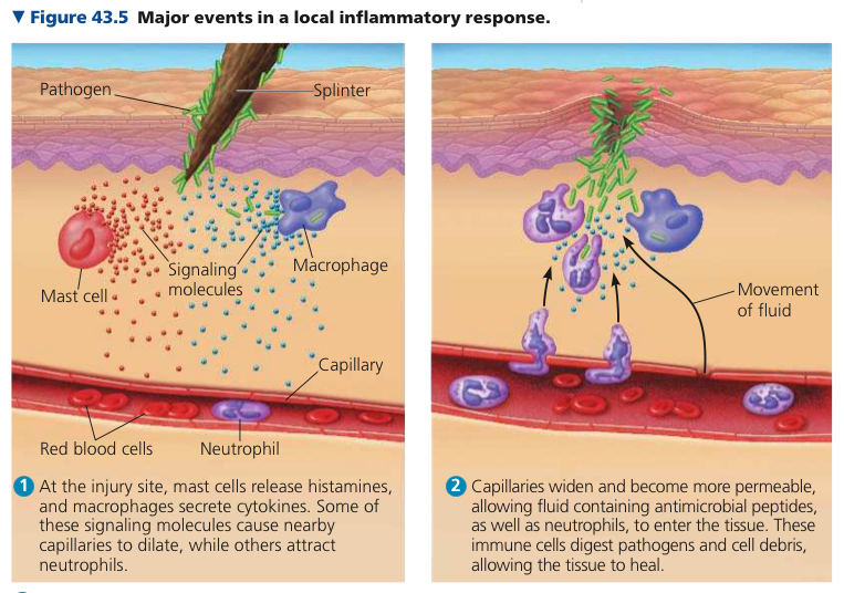
Mast Cells: Found in connective tissue, contribute to inflammatoryresponse
Inflammatory Response: Set of events triggered by signaling molecules released upon injury or infection
Histamine: Signaling molecule at sites of damage, blood vessels dilate
Interferons: Proteins that provide innate defense by interfering with viral infections
Complement System: ~30 proteins in blood plasma which circulate in an inactive state, activated by substances on the surface of many pathogens
It both tries to be used to push the pus out, and also is secreted to prevent more from coming in
Not sure
Ok
43.2: In adaptive immunity, receptors provide pathogen specific recognition
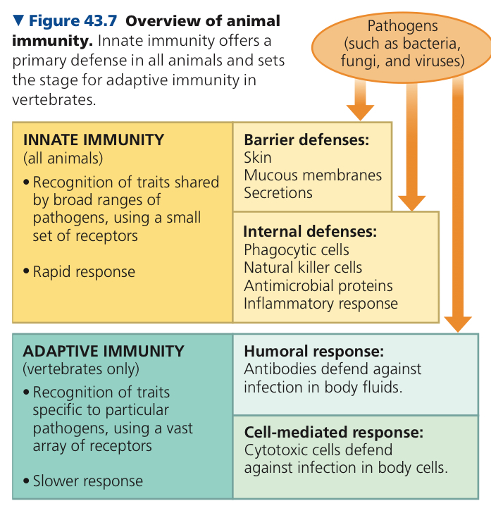
Adaptive Immunity: Set of molecular and cellular defense only among vertebraes
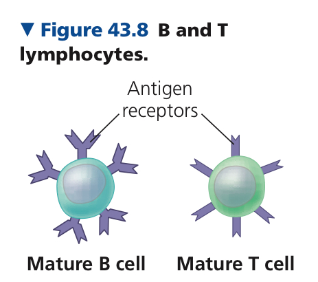
Adaptive Immunity mostly relies on T and B cells, which are lymphocytes
Lymphocyte: Originate from stem cells in bone marrow, white blood cells, 3 types
T Cells: Mature lymphocytes that migrate to the thymus
Thymus: Organ in the thoracic cavity above the heart
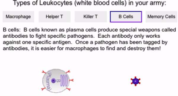
B Cells: Mature lymphocytes that stay in the bone marrow
Natural Killer Cells in innate immunity remain in blood
Antigen: Any substance that elicits either a B or T response
Antigen Receptor: Protein that binds a cell to an antigen
Epitope: Part of antigen that binds to an antigen receptor
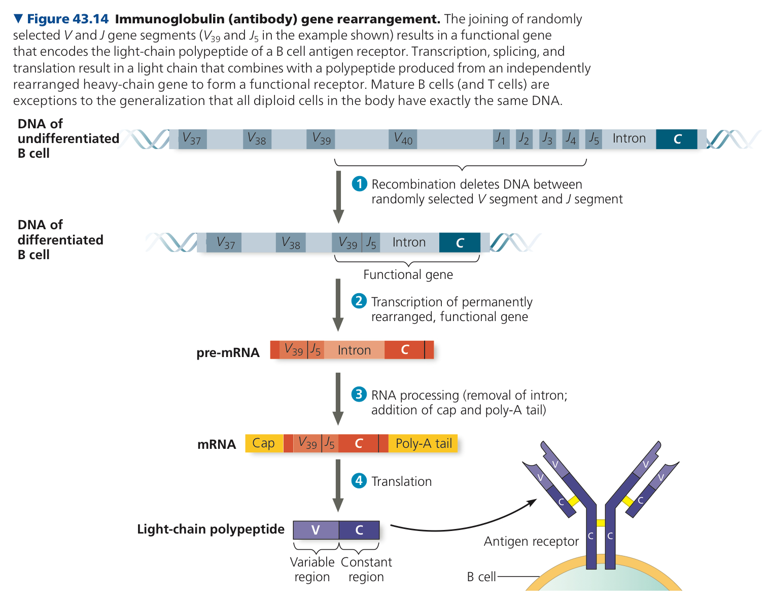
B cell antigen receptors are Y shaped proteins with four polypeptide chains, two heavy chains and two light chains, linked by disulfide bridges
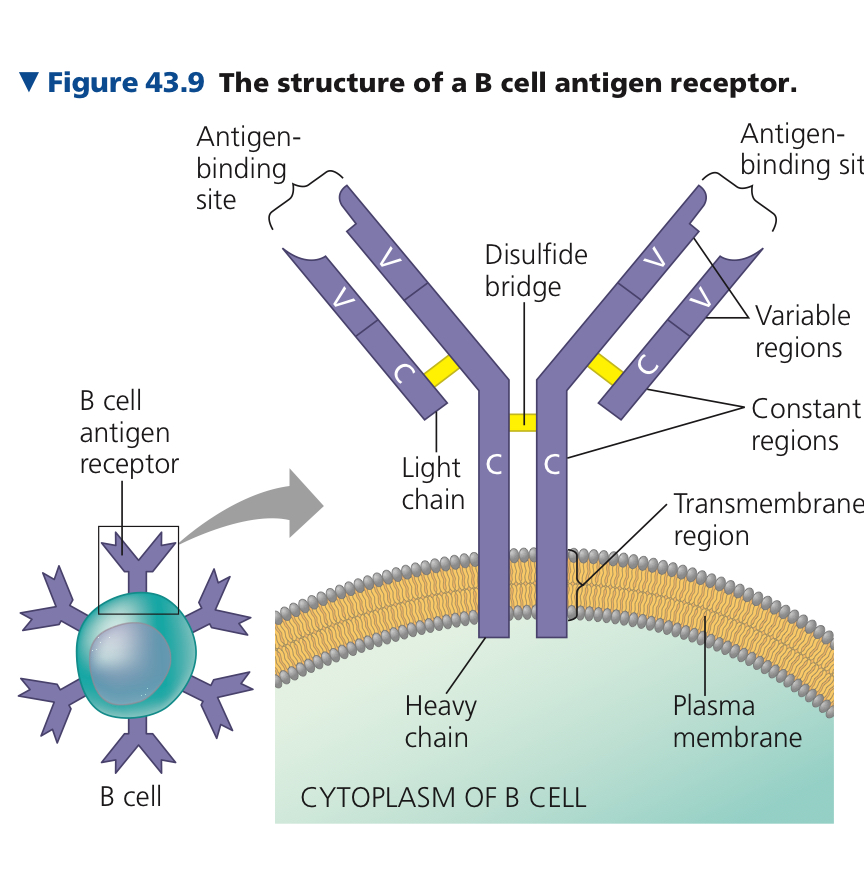
Antibody/Immunoglobulin (Ig): Secreted protein, soluble form of antigen receptor bycells resulting from binding of B antigen receptor to antigen
Major Histocompatibility Complex (MHC): Host protein that displays an antigen fragment on the cell surface
Effector Cells: Clones that take effect immediately against the antigen and any pathogens producing it
Memory Cells: Remaining cells in the clone, give rise to effector cells if the same antigen is encountered again later
Clonal Selection: Encounter with an antigen selects which lymphocyte will divide to produce a clonal population
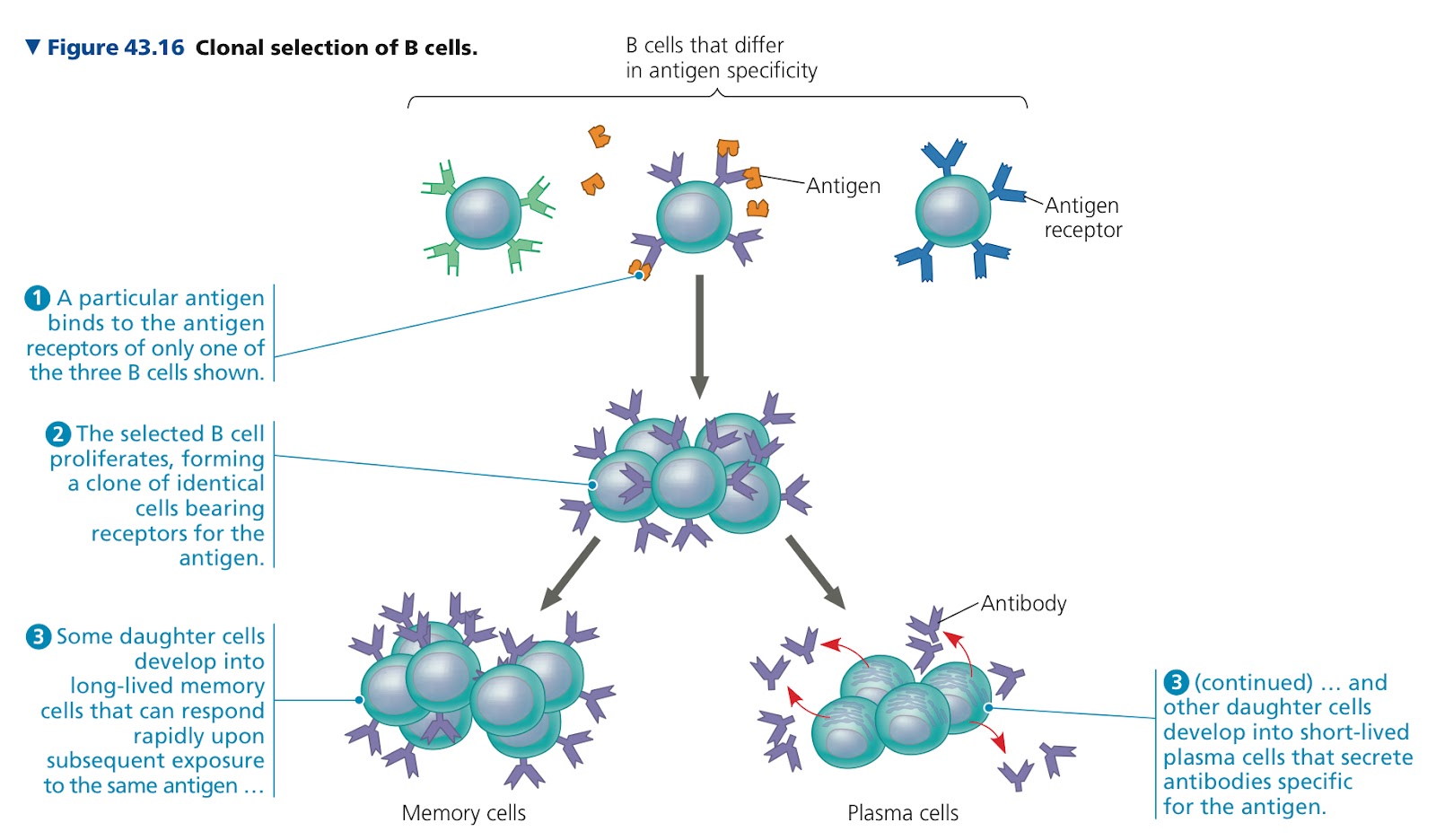
Primary Immune Response: Effector cells formed by clones of lymphocytes after an initial exposure to an antigen
Secondary Immune Response: Response that is faster and of greater magnitude and more prolonged
43.3: Adaptive immunity defends against infection of body fluids and body cells
Humoral Immune Response: Protects blood and lymph by using antibodies to neutralize or eliminate toxins and pathogens in body fluids
Cell Mediated Immune Response: Specialized T cells destroy infected host cells
Both this and humoral can include primary and secondary immune response with memory cells enabling the secondary response
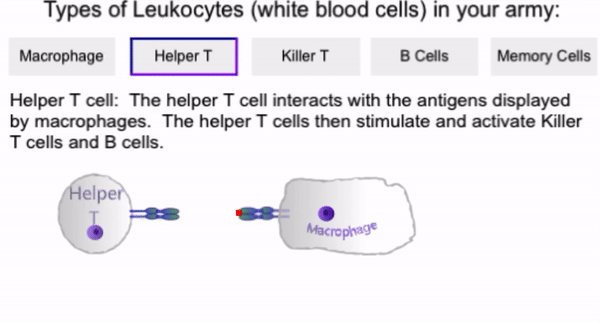
Helper T Cells: Activates humoral and cell mediated immune responses
Two conditions
Foreign molecule that can bind specifically to the antigen receptor of the helper T cell must be present
Antigen must be displayed on the surface of an antigen presenting cell
Antigen presenting cell can be dendritic, macrophage, or B cell
B cells only present the antigen to which it specially binds
Single activated B cell gives rise to thousands of identical plasma cells which stop expressing antigen receptors and begin producing and secreting antibodies
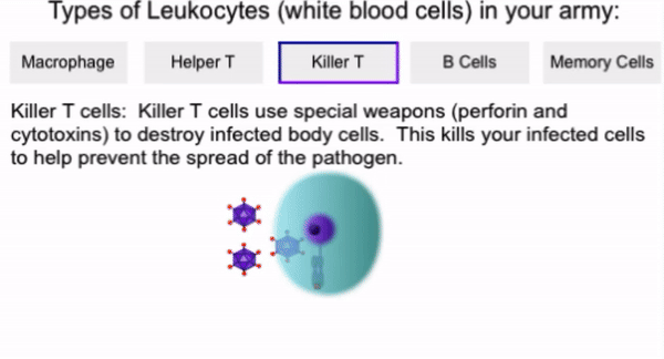
Cytotoxic T Cells: Use toxic proteins to kill cells infected by viruses or other intracellular pathogens before they fully mature
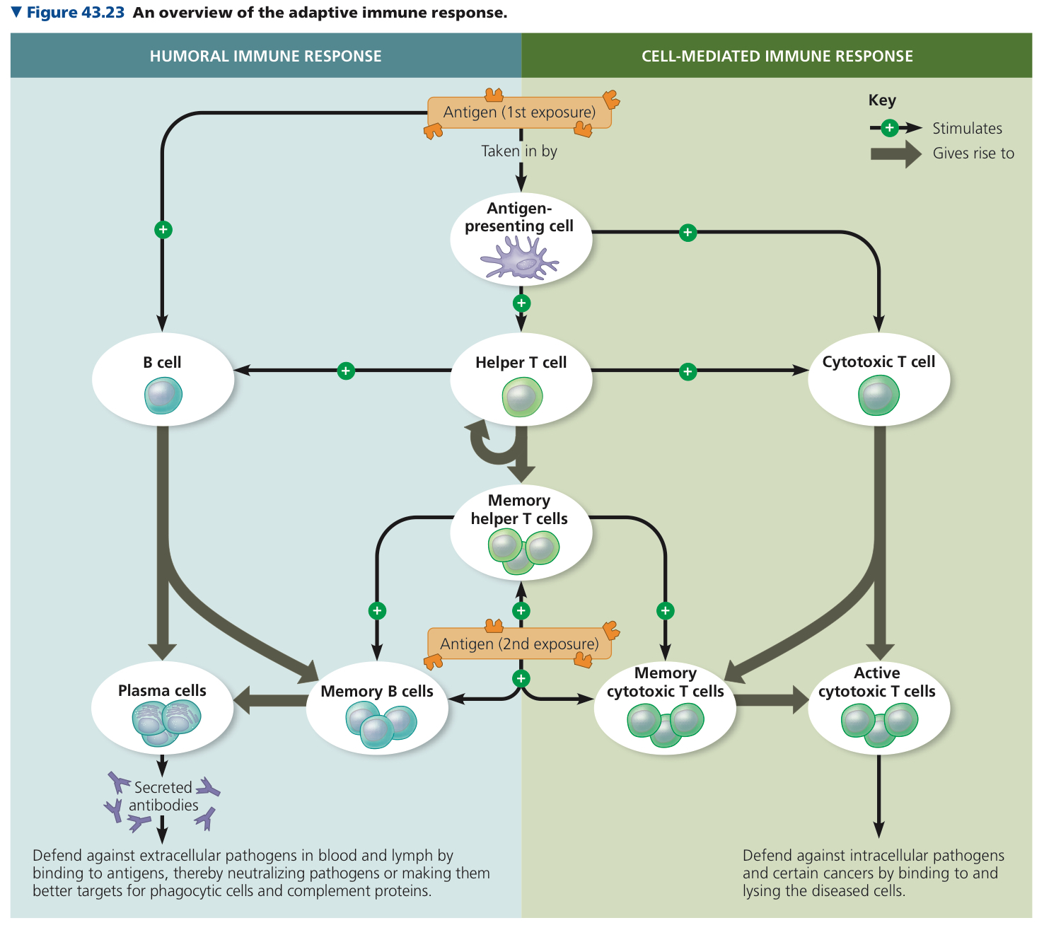
Immunization: Use of antigens artificially introduced into the body to generate an adaptive response from the body and memory cell formation
Active Immunity: Defenses that arise when a pathogen infection or immunization prompts an immune response
Passive Immunity: Antibodies in the recipient are produced by another individual
ex. Pregnant female gets antibodies so the fetus does too
Monoclonal Antibodies: Identical and specific for the same epitope (spot) on an antigen
43.4: Disruptions in immune system function can elicit or exacerbate disease
Allergens: Antigens with exaggerated responses
Autoimmune Disease: Immune system is active against particular molecules of the body
Human Immunodeficiency Virus (HIV): Attacks adaptive immune response and infects helper T cells
Acquired Immunodeficiency Syndrome (AIDS): Impairment in immune responses that leaves the body susceptible to infections and cancers that would be beatable for a healthy immune system
Chapter 44: Osmoregulation and Excretion
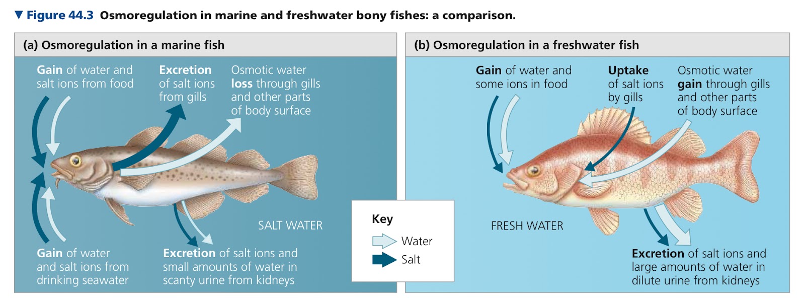
44.1: Osmoregulation balances the uptake and loss of water and solutes
Osmoregulation: Process by which animals control solute concentrations and balance water gain and loss
Excretion: Process for ridding the body of metabolic waste
Osmolarity: Number of moles of solute per liter of solution
Hyperosmotic: Higher concentration of solutes
Hypoosmotic: Lower concentration of solutes
Two ways for animals to maintain water balance
Osmoconformer: To be isoosmotic with its surroundings, marine animals
Osmoregulator: To control internal osmolarity independent of that of the external environment
Anhydrobiosis: Animals enter a dormant state when their habitats dry up
44.2: An animal’s nitrogenous wastes reflect its phylogeny and habitat
Ammonia: Toxic metabolite produced by dismantling of nitrogenous molecules, can only be excreted in large volumes of dilute solutions
Urea: Product of energy consuming metabolic cycle that combines ammonia with carbon dioxide in the liver
Higher energy cost
Uric Acid: Relatively nontoxic, doesn’t readily dissolve, more energetically expensive than urea
Used by insects, land snails,and reptiles
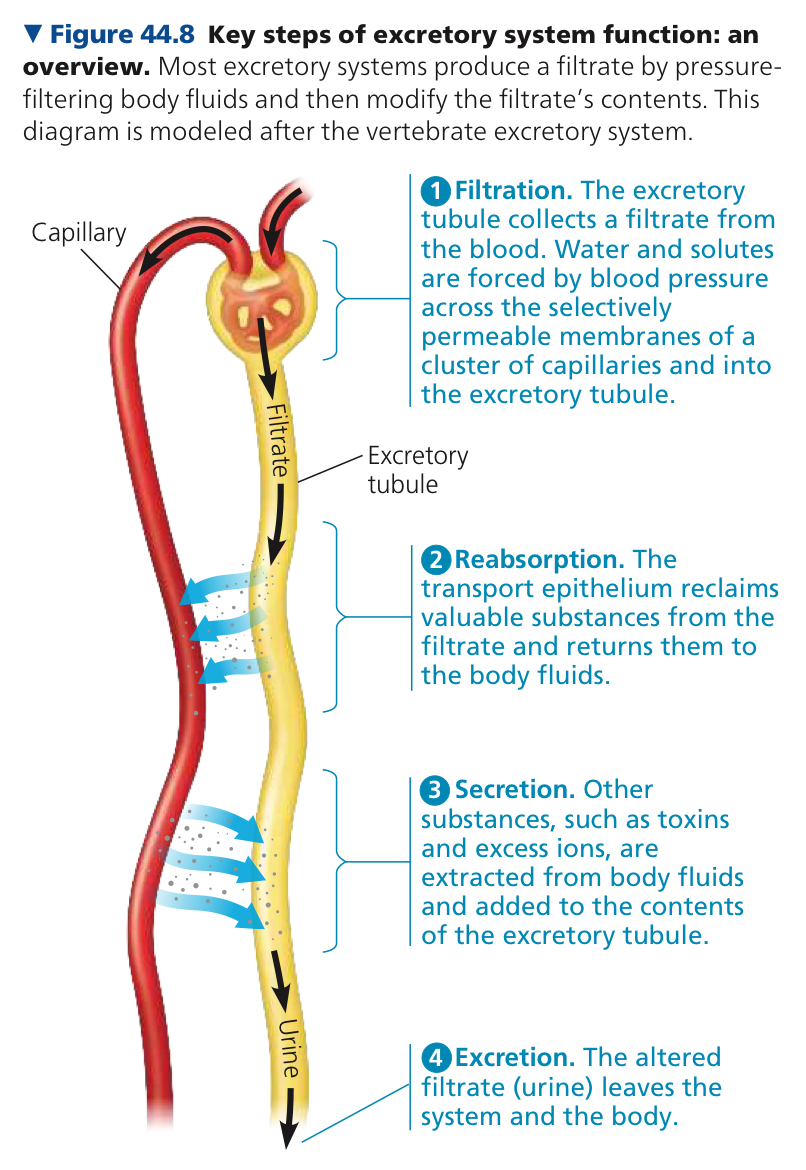
44.3: Diverse excretory systems are variations on a tubular theme
Filtration: Excretory tubule collects a filtrate from the blood, and water and solutes are forced by blood pressure across the membranes of a cluster of capillaries and into the excretory tubule
Filtrate: Water and small solutes, such as salts, sugars, amino acids, and nitrogenous wastes, which can cross the membrane
Converted into waste fluid by specific transport of materials
Reabsorption: Transport epilithium finds useful molecules and water from filtrate and returns them to the body fluid
Secretion: Other waste substances are extracted from body fluids and added to the contents of the excretory tubule
Excretion: Altered filtrate leaves the body as urine
Protonephridia: Network of dead end tubules that branch throughout the body
Metanephridia: Exretory organs that collect fluid directly from the coelom in annelids
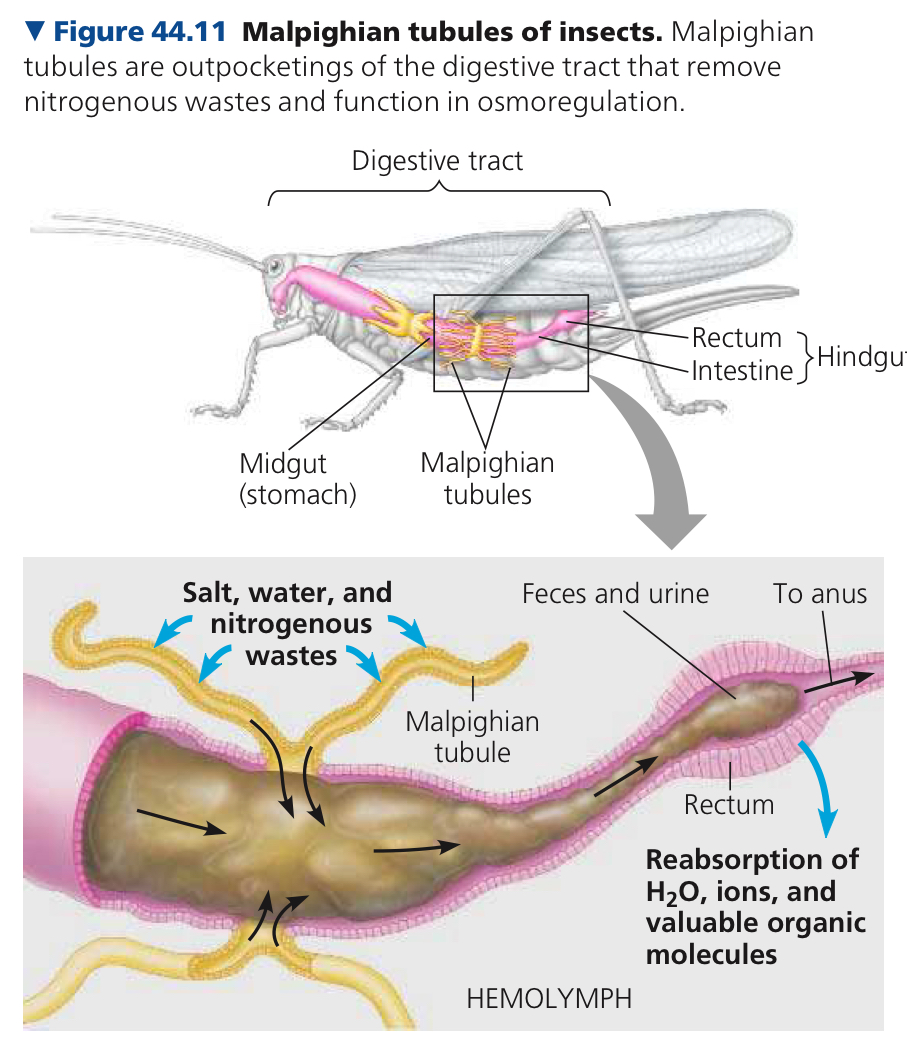
Malpighian Tubules: Remove nitrogenous wastes and function in osmoregulation
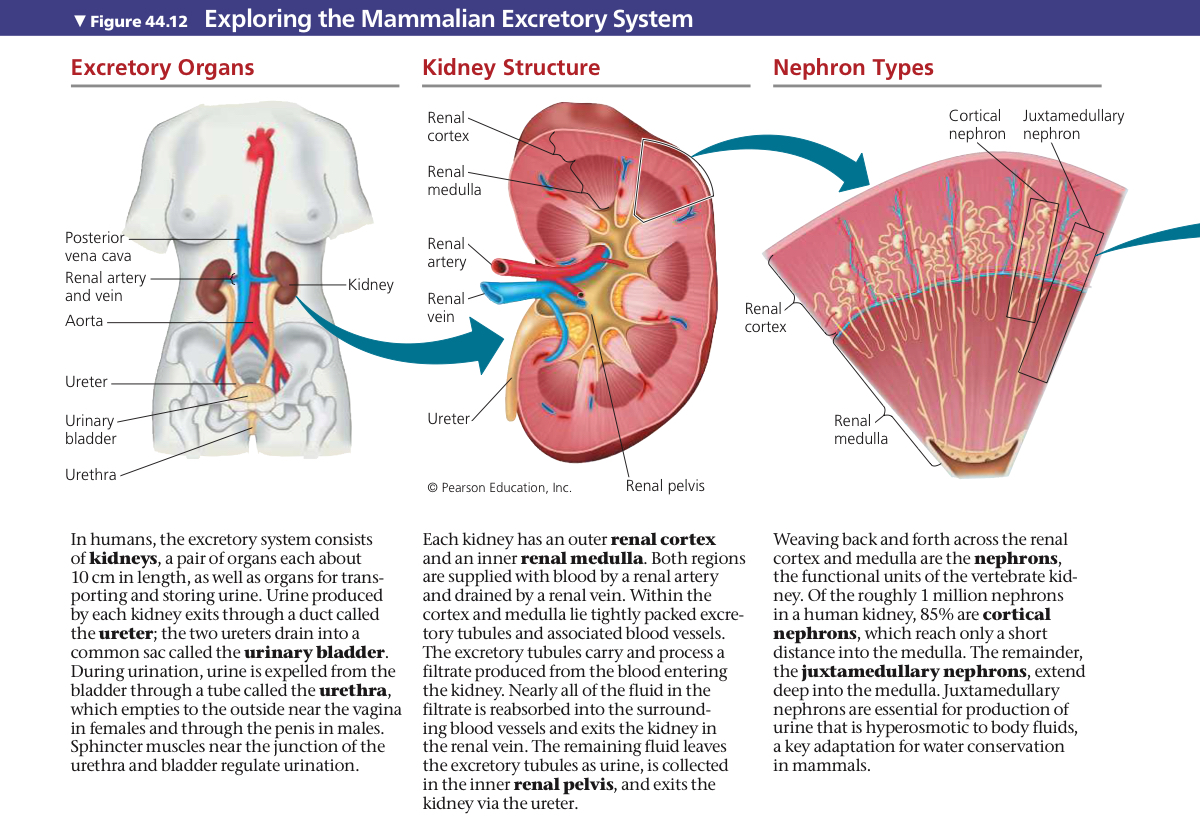
Kidney: Functions in both osmoregulation and excretion for transporting and storing urine
Urine produced by kidney → ureter (duct) → drain into urinary bladder → urethra
Outer renal cortex and inner renal medulla, both supplied with blood by renal arteries, drained by a renal vein. Excretory tubules carry and process a filtrate produced from blood entering the kidney
Neurphrons: Functional units of the vertebrae kidney
Cortical Nephrons: Only reach a short distance into the medulla, 85% of nephrons
Juxtamedullar Nephrons: Other 15%, extend deep into the medulla
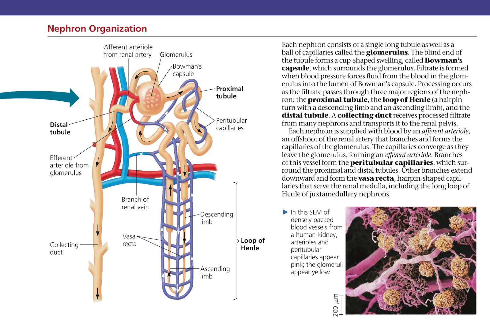
Glomerulus: Ball of capillaries, + a single long tubule = a nephron
Bowman’s Capsule: Cup-shaped swelling which surrounds the glomerous, blind end of the tubule
Processing occurs as the filtrate passes through three major regions of the nephron
Proximal Tubule: First nephron segment after the glomerulus where reabsorption commences
Loop of Henle: Hairpin turn with descending limb and ascending limb
Distal Tubule: Short nephron segment, interposed between the macula densa and collecting duct
Collecting Duct: Receives processed filtrate from nephrons and transports it to the renal pelvis
Peritubular Capillaries: Surround the proximal and distal tubules, branches of the efferent arteriole
Vasa Recta: Branches that extend downward form it, hairpin shaped capillaries that serve the renal medulla
44.4: The nephron is organized for stepwise processing of blood filtrate
Proximal tubule. Reabsorption in proximal tubule is used for recapture of valuable nutrients from initial filtrate.
Descending limb of the loop of Henle. When leaving the proximal tubule, filtrate enters the loop of Henle. In the first portion of the loop (descending limb), water channels formed by aquaporins make transport freely permeable to water and the filtrate loses water and increases solute concentration
Ascending limb of the loop of Henle. The membrane is impermeable to water. There are two specialized regions, a thin segment near the loop tip and a thick one adjacent to the distal tubule.
In the thick part, NaCl is moved out of the filtrate and into the interstitial fluid, and the filtrate is diluted. Loop of henle recovers water (descending) and salt(ascending) from the filtrate
Distal tubule. Regulates K+ and NaCl concentration in filtrate
Collecting duct. Processes filtrate into urine which is carried into the renal pelvis.
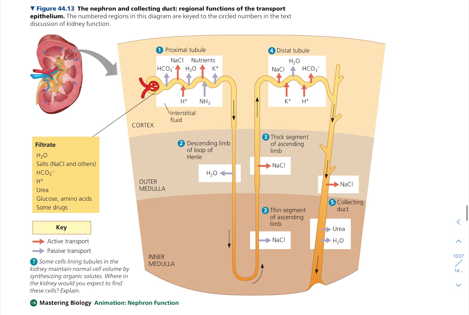
Countercurrent Multiple Systems: Systems that expend energy to create concentration gradients.
In loop of Henle maintains gradient of salt concentration in kidney interior
44.5: Hormonal circuits link kidney function, water balance, and blood pressure
Antidiuretic Hormone (ADH): Activate membrane receptors on the surface of collecting duct cells and reduce urine volume
Renin-Angiotensin-Aldosterone System (RAAS): Endocrine circuit, regulates kidney function, increases water and Na+ absorption when blood volume drops
Juxtaglomerular Apparatus (JGA): Specialized tissue consisting of cells of and around afferent ateriole, used in RAAS
Atrial Natriuretic Peptide (ANP): Opposes the RAAS, inhibits the release of renin from JGA and inhibits NaCl absorption by collecting ducts, reduces aldosterone release from adrenal glands if blood volue and pressure increase
Chapter 45: Hormones and the Endocrine System
45.1: Hormones and other signaling molecules bind to target receptors, triggering specific response pathways
Hormone: Secreted molecule that circulates throughout the body and stimulates specific cells
Two basic systems for communication and regulation in the animal body
Endocrine System: Controls chemical signaling by hormones
Nervous System: Network of specialized cells, neurons, thatt transmit signals along dedicated pathways
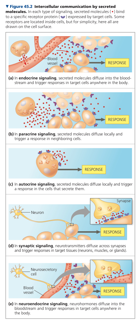
In endocrine signaling, hormones secreted by endocrine cells reach the target cells via the bloodstream
Local Regulators: Molecules that act over short distances and reach their target cell through diffusion
Paracrine Signaling: Target cells are near the secreting cell
Autocrine Signaling: Secreting cells are the target cells
Prostaglandins: Local regulator, modified fatty acid, that is produced throughout the body and have diverse functions
In immune system, promote inflammation and sensation of pain in response to injury
Nitric Oxide (NO): Local regulator, gas, synthesized when level of blood oxygen falls
Neurotransmitters: Diffuse a very short distance and bind to receptors on target cells, molecules secreted by neurons, synaptic signaling
Neurohormones: Diffuse from nerve cell endings into the bloodstream, secreted by neurosecretory cells
Pheromones: Chemicals released into the external environment
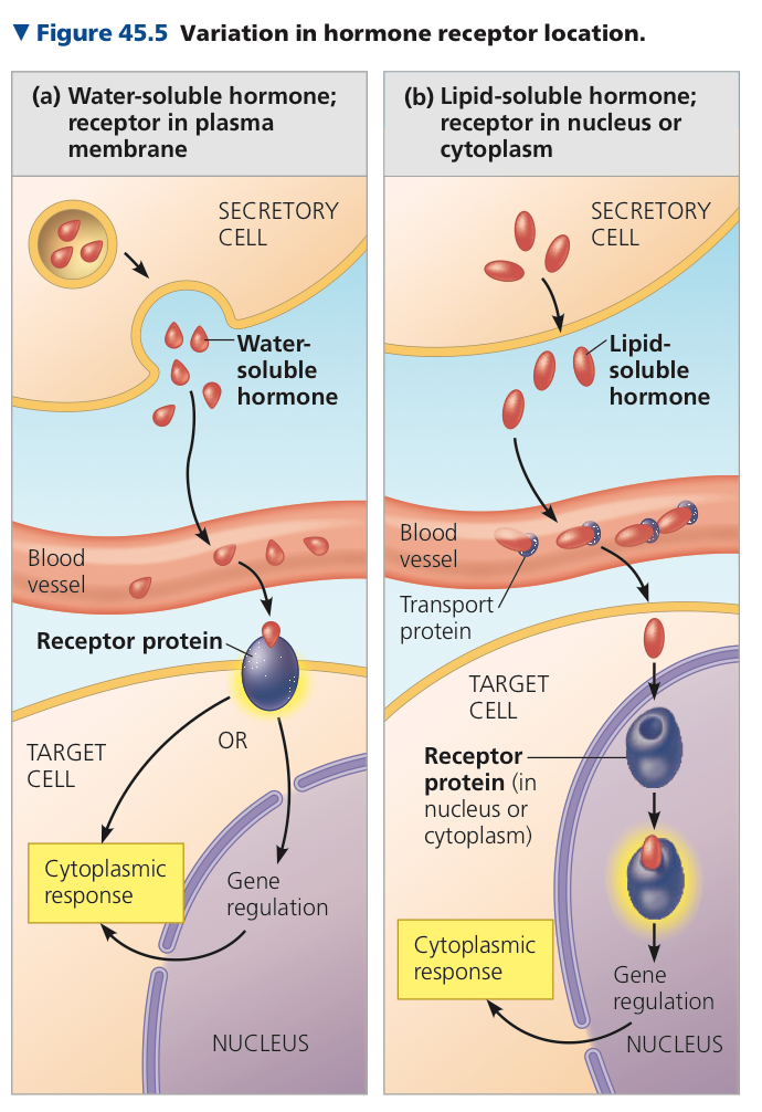
Binding of water soluble hormone to cell surface receptor protein triggers a response—either the activation of an enzyme, a change in the uptake/secretion of specific molecules, or rearrangement of the cytoskeleton
Signal Transduction: Chain of events that converts the extracellular chemical signal to an intracellular response
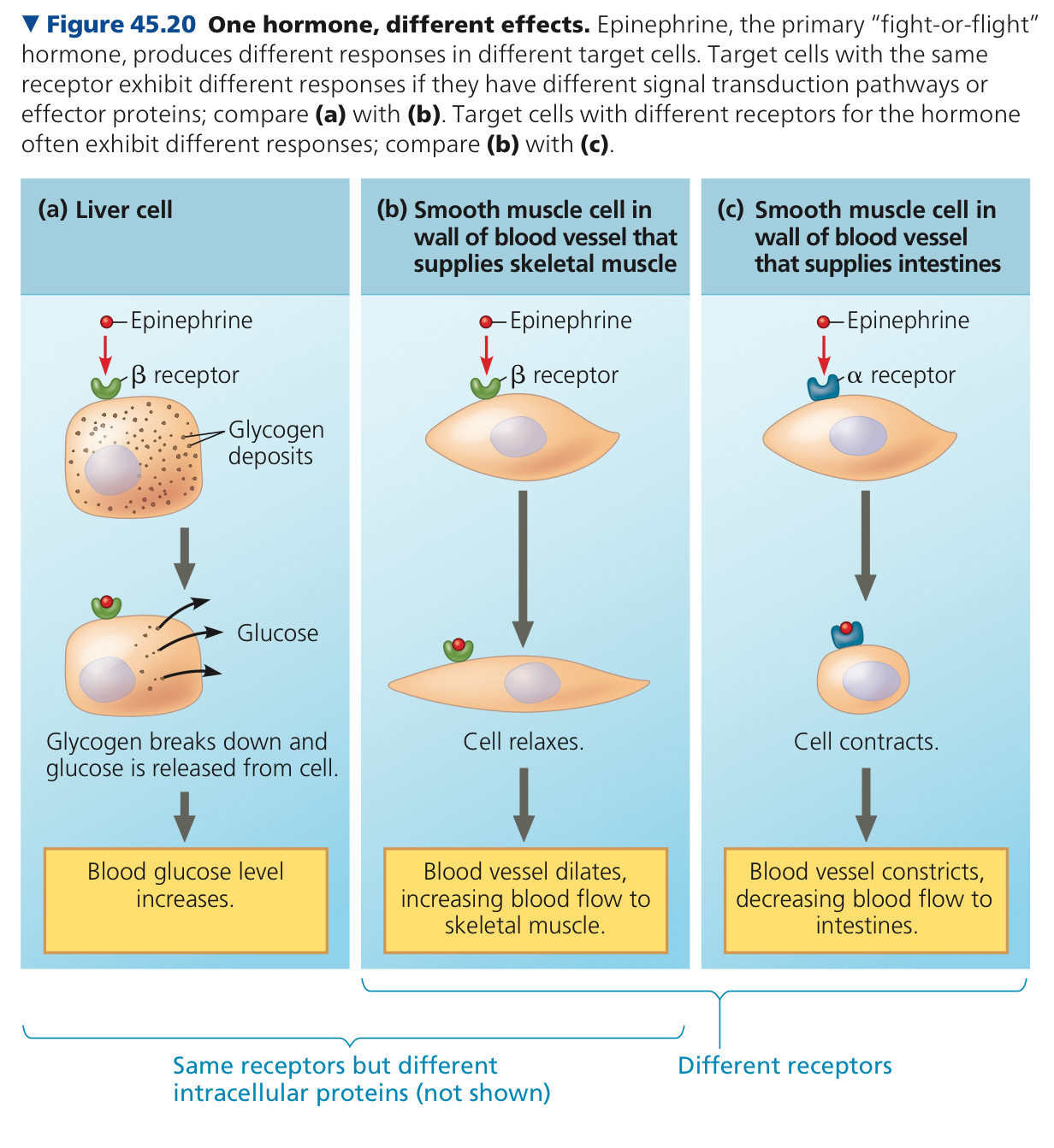
Epinephrine/Adrenaline: Hormone that regulates many organs
In lipid soluble hormone it activates the receptor which directly triggers the cell’s response, usually a change in gene expression
Endocrine Glands: Groups of endocrine cells, secrete hormones into surrounding fluid
Exocrine Glands: Have ducts that carry secreted substances onto body surfaces or cavities
They trigger different responses and there are two receptor proteins used in lipids
Exocrine, since it goes into the environment
45.2: Feedback regulation and coordination with the nervous system are common in hormone pathways
Simple Endocrine Pathway: Endocrine cells respond directly to internal or external stimulus by secreting a particular hormone, which travels in the bloodstream to the target cells and receptors
Simple Neuroendocrine Pathway: Stimulus received by sensory neuron and not endocrine tissue, which stimulates a neurosecretory cell, which secretes a neurohormone that diffuses in the bloodstream and travels to target cells
ex. Oxytocin
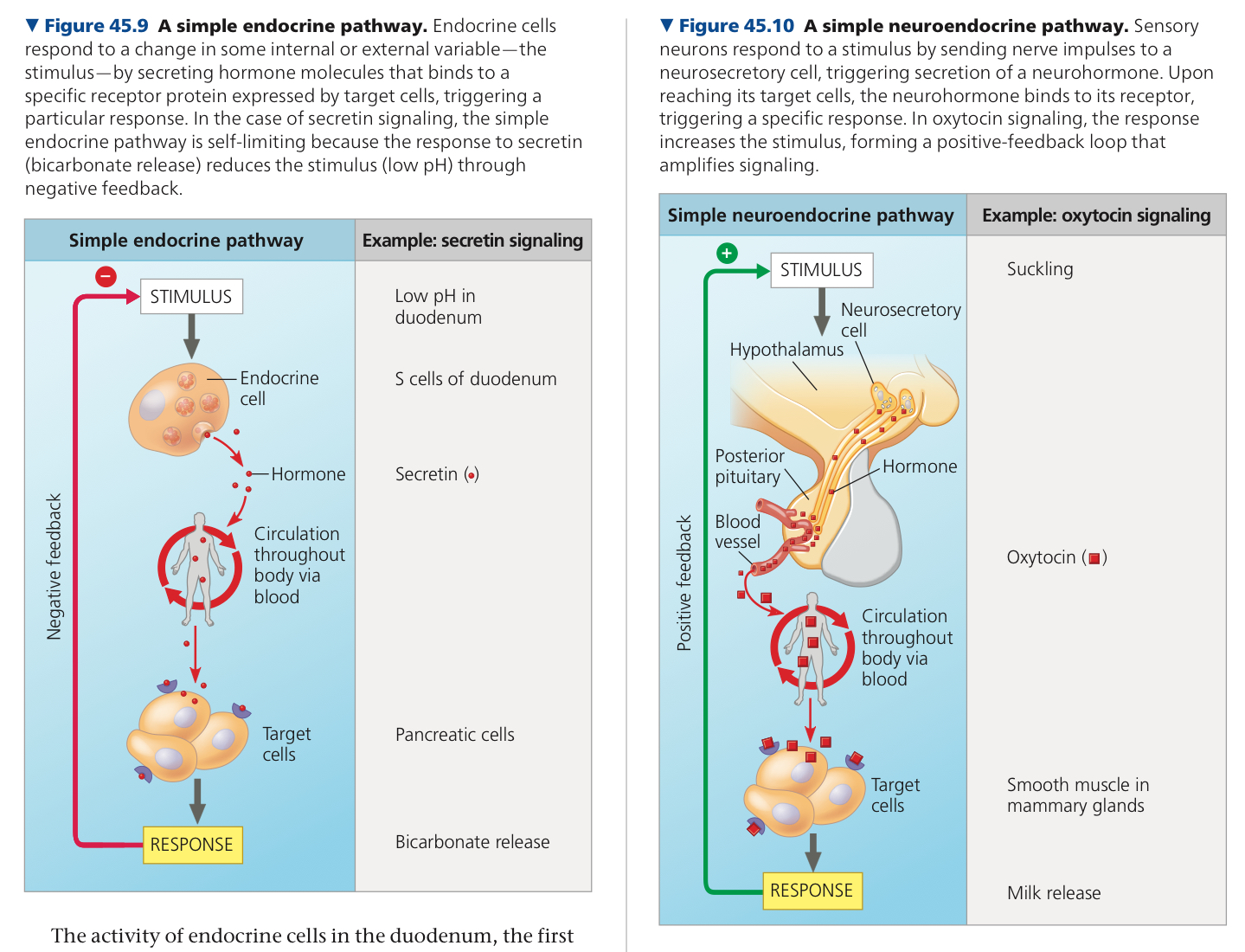
Negative Feedback: Response reduced initial stimulus
Positive Feedback: Reinforces a stimulus to drive a process to completion
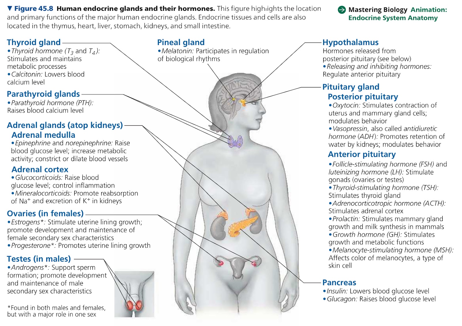
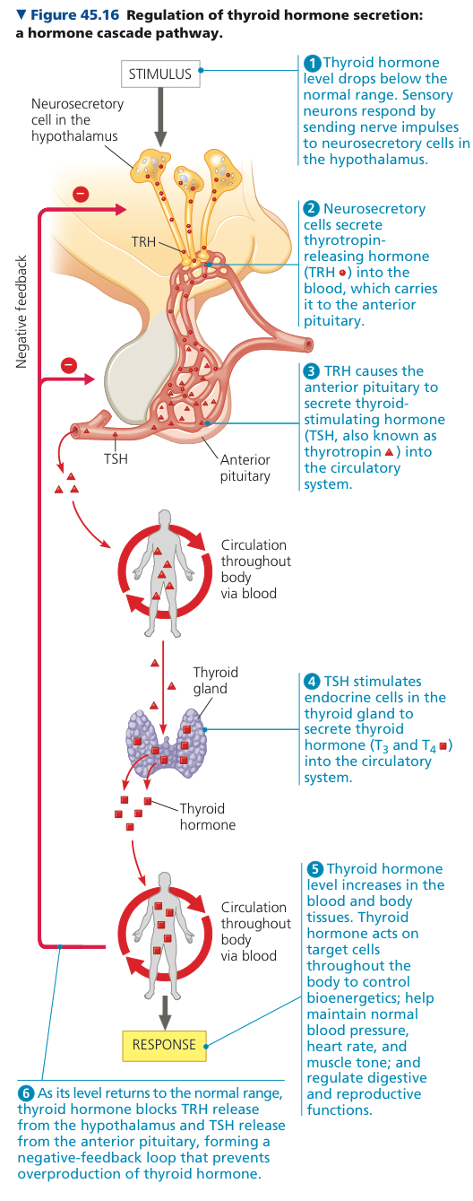
Hypothalamus: Coordination of endocrine signaling relies on this part of the brain, which recieves information from nerves and initiates neuroendocrine signaling
Pituitary Gland: Gland located at the base of the hypothalamus
Posterior Pituitary: Extension of hypothalamus, hypothalamic axons that reach here secrete neurohormones from the hypothalamus
Anterior Pituitary: Endocrine gland that synthesizes and secretes hormones in response to hormones from the hypothalamus
Antidiuretic Hormone (ADH): Regulates kidney function, increases water retention in kidneys
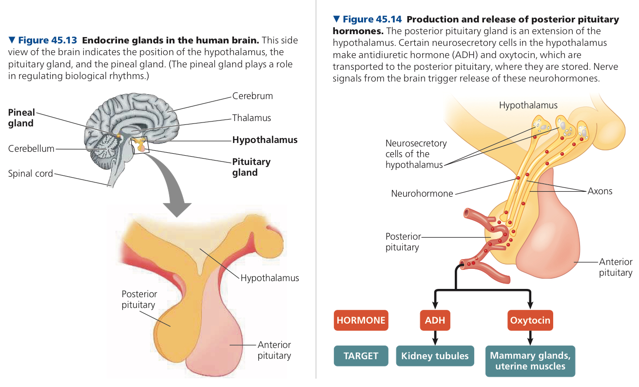
Prolactin: Stimulates milk production
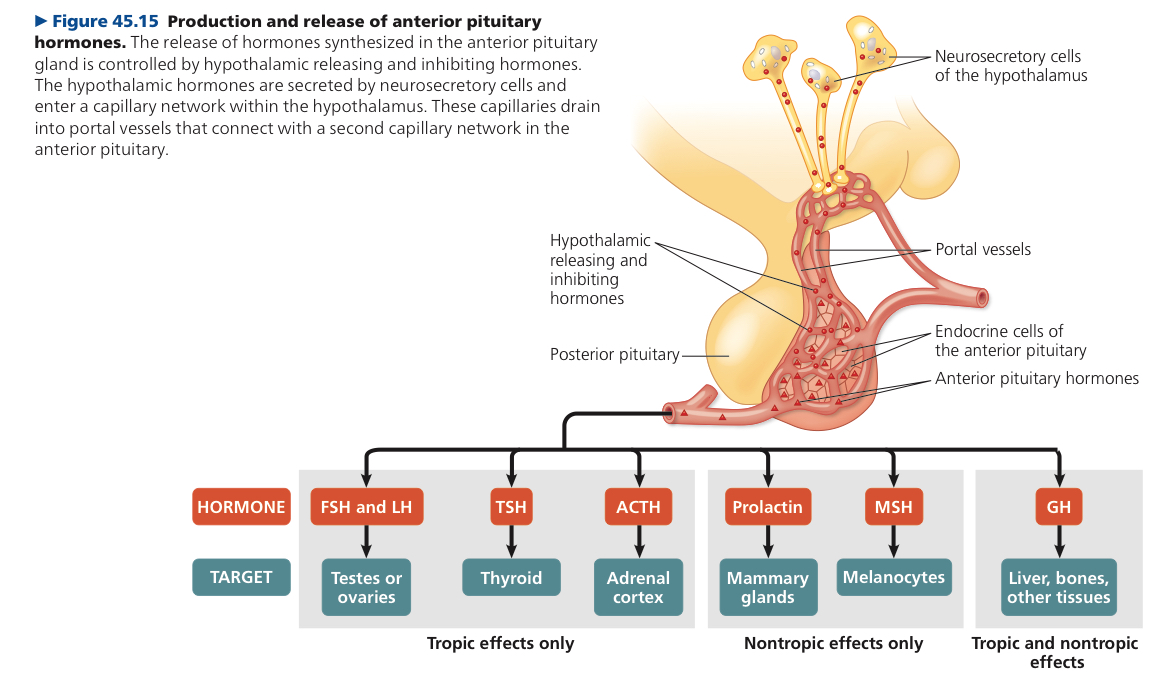
Thyroid Hormone: Regulates bioenergetics, helps maintain normal blood pressure, heart rate, muscle tone, digestive and reproductive stuff
Thyroid Gland: Organ in the neck with two lobes on the ventral surface of the trachea
Growth Hormone: Stimulates growth
Help produce milk for offspring which helps them bond ig?
Posterior is an extension, Anterior responds
45.3: Endocrine glands respond to diverse stimuli regulating homeostasis, development, and behavior
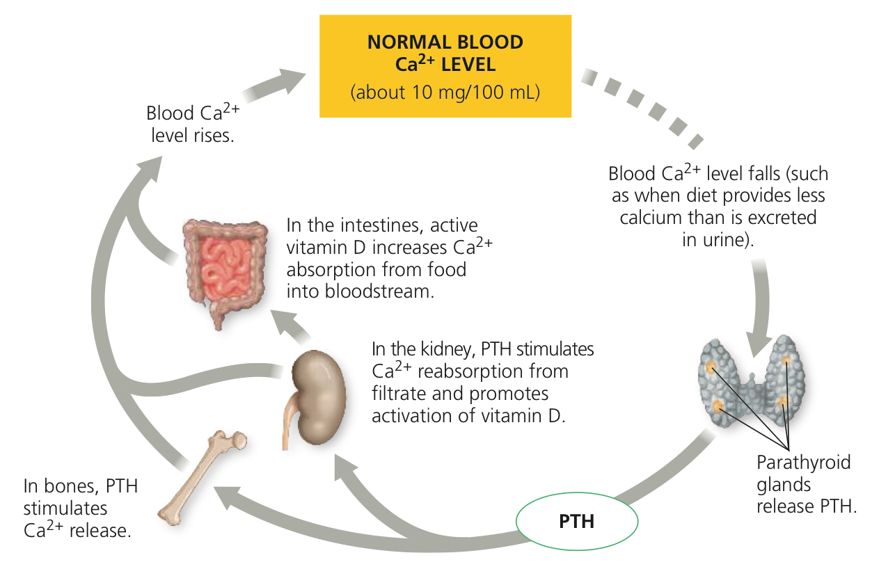
Parathyroid Glands: Set of four small structures embedded in posterior surface of thyroid, regulate Ca2+ levels
Parathyroid Hormone (PTH): Hormone released by parathyroid glands when blood Ca2+ levels fall below 10 mg
Calcitonin: Hormone that inhibits bone breakdown and enhances Ca2+ excretion
Adrenal Gland: Made of adrenal cortex and adrenal medulla
Norepinephrone/Noradrenaline: Neurotransmitter, catecholamine, “fight or flight” along with adrenaline
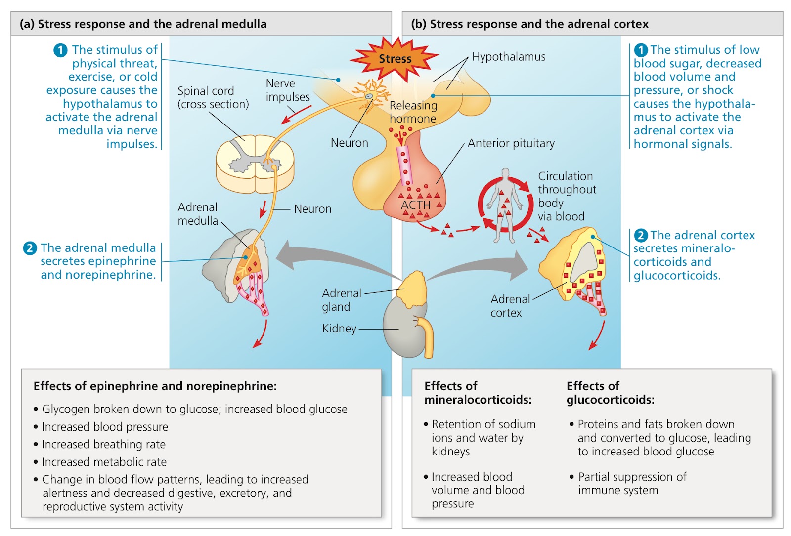
In liver cells, adrenaline binds to a B type receptor in the plasma membrane, which activates protein kinase A which regulates enzymes of glycogen metabolism, and glucose is released into blood
In smooth muscle cells lining blood vessels supplying skeletal muscle, the same kinase is activated by the same receptor that inactivates a muscle specific enzyme which results in smooth muscle relaxation
In smooth muscle cells lining blood vessels of the intestines, adrenaline binds to an a type receptor, triggering a signaling pathway that involvs enzymes other than kinase A which causes smooth muscle contraction
Glucocorticoids: Make more glucose available as fuel, promotes glucose synthesis from noncarbohydrate sources such as proteins
Mineralocorticoids: Act principally in maintaining salt and water balance
Testes synthesize androgens, main one being testosterone
Estrogens (most important is estradiol) in females for development of reproductive system
Progesterone: Involved in preparing and maintaining tissues of uterus to support development of embryo
Melatonin: Regulates functions related to light and seasons
Pineal Gland: Produces melatonin, a small mass of tissue near the center of the brain
Melanocyte Stimulating Hormone (MSH): Secreted by anterior pituitary, controls hunger, metabolism, and skin coloration
Chapter 46: Animal Reproduction
46.1: Both asexual and sexual reproduction occur in the animal kingdom
Asexual Reproduction: New individuals generated without the fusion of the egg and sperm
Fission: Splitting and seperation of a parent organism into two individuals of ~equal size
Fragmentation: Breaking of parent body into several pieces
Regeneration: Regrowth of lost body parts
ex. worms that split into several fragments, each regenerating into a complete worm (ew gross)
Parthenogenesis: Egg develops without being fertilized
Sexual Reproduction: Fusion of haploid gametes forms ygote
Egg: Large and nonmotile, the female gamete
Sperm: Smaller and more motile, the male gamete
Hermaphroditism: Each individual has both male and female reproductive systems
Any two organisms can mate
Ovulation: Production and release of mature eggs, midpoint of each cycle
Sexual reproduction has a “twofold cost”, which means asexually, a female can produce 2x as many offspring as the female that reproduces sexually. Not sure what sexual reproduction’s benefit is that counteracts its twofold cost. Possibly genetic variation is higher.
Asexual reproduction can produce twice as many offspring and they would be all females
Not too sure
No, since it is still sexual reproduction and hence won’t result in a clone
Not sure
46.2: Fertilization depends on mechanisms that bring together sperm and eggs of the same species
Fertilization: Union of sperm and egg, can be internal or external
External fertilization, female releases eggs into environment and male finds and fertilizes them
Must have moist environment
Spawning happens in certain species, where animals clustered in the same area release their gametes at the same time
Internal fertilization, sperm is deposited in or near the female reproductive tract
Allows sperm to reach an egg even when external environment is dry
Requires sophisticated and compatible reproductive systems, plus cooperative behavior
Pheromones are used no matter what type of fertilization it is
Gonads: Organs that produce gametes, found in many but not all animals
Spermathecae: Sacs in which sperm is kept alive and stored (sometimes a year or more), in many insect species, and females fertilize their own eggs only in response to the right stimuli
Cloaca: Digestive, excretory, and reproductive systems’ common opening, in many nonmammalian vertebrates, prob present in ancestors of all vertebrates
Since the fertilization is immediate and there is no risk of the eggs being eaten or something
Spawning in external environments and going all at once, sophisticated reproductive systems in internal
I’ll go back later or smth
46.3: Reproductive organs produce and transport gametes
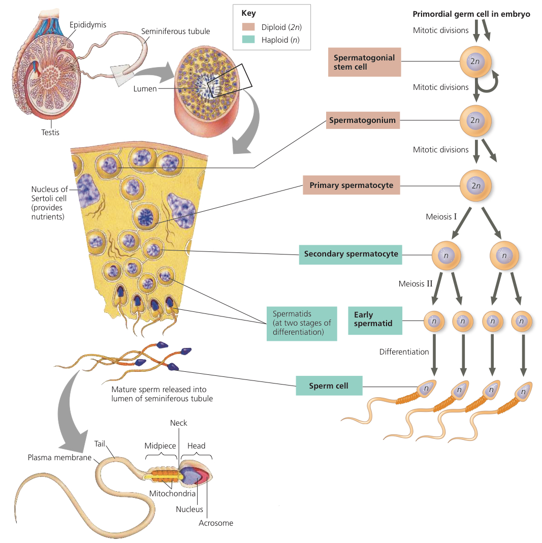
Testes: Male gonads, must be cooler than the rest of the body to produce sperm
Seminiferous Tubules: Where testes produce sperm, highly coiled tubes
Scrotum: Fold of the body wall, keeps the testes (2 degrees celcius) cooler than the body temperature
Seminiferous tubules → coiled duct of epididymis (takes about 3 weeks for sperm to travel through the duct, and in the process they become matured and more motile)
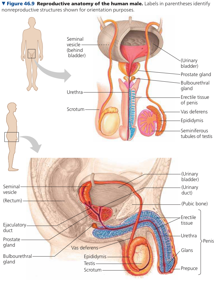
Ejaculation: Sperm propelled from each epididymis through the vans deferens
Vans Deferens: Extends around and behind the urinary bladder and joins a duct from the seminal vesicle to form an ejaculatory duct, which open into the urethra
Urethra: Outlet tube for excretory system and reproductive system
Semen: Fluid that is ejaculated, secretions produced by three accessory glands + semen
Seminal Vesicles: 2 contribute ~60% the volume of semen
Prostate Gland: Secretes products directly into urethra, has anticoagulant enzymes and citrate, a sperm nutrient
Bulbuorethral Glands: Pair of small glands along the urethra below the prostate, secrete clear mucus that neutralizes acidic urine in the urethra
Penis: Urethra + three cylinders of spongey erectile tissue, which fill with blood from the arteries when aroused
Glans: Head of the penis with a sensitive and thin outer layer, covered by the prepuce (foreskin)
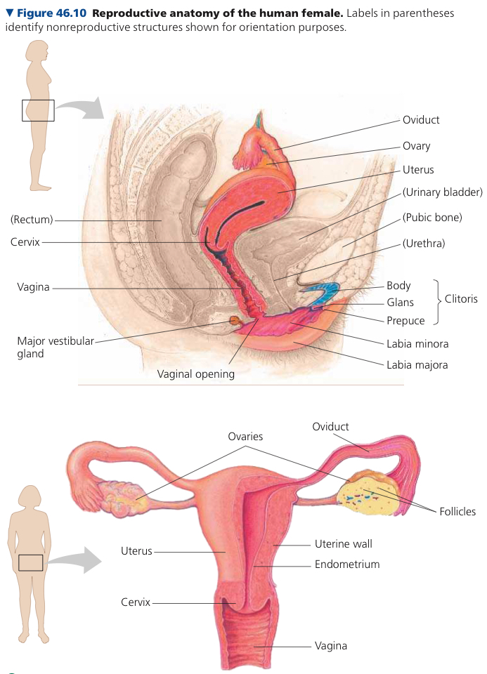
Female has clitoris and two sets of labia
Female gonads are ovaries. The outer layer of each one is packed with follicles, each with an oocyte (partially developed egg) surrounded by support cells
Oviduct/Fallopian Tube: From uterus to a funnel like opening at each ovary
Cilia in the oviduct pushes the egg down to the uterus in wave like motions
Uterus/Womb: Thick, muscular organ that can expand during pregnancy
Endometrium: Lining of the uterus, richly supplied with blood vessels
Cervix: Neck of the uterus, opens into the vagina
Vagina: Muscular but elastic chamber that is the site for insertion of penis and deposition of sperm during copulation, as well as the birth canal where a baby is born
Vulva: External female vagina
Labia Majora: Enclose and protect the rest of the vulva, thick, fatty ridges
Labia Minora: Slender skin folds bordering the cavity of the vaginal and urethra openings
Clitoris: Erectile tissue supporting a round glans covered by the prepuse
Mammary Glands: Present in both sexes, produce milk only in females
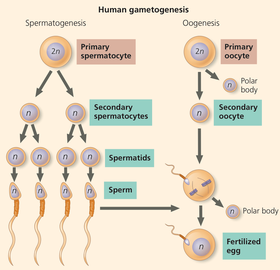
Gametogenesis: Production of gametes
Spermatogenesis: Production of sperm
Spermatogonia: In mature testes, stem cells divide mitotically to form them, and they generate spermatocytes by mitosis
Acrosome: Specialized vesicle with enzymes to help the sperm penetrate an egg
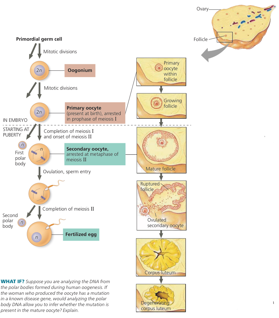
Oogenesis: Production of eggs
Oogonia: Divide by mitosis to form the cells that begin meiosis
Primary Oocytes: Cells that begin meiosis but are stopped at prophase I before birth, reside within small follicles
Secondary Oocytes: Arrested in metaphase II, released at ovulation
Corpus Lutem: Develops from ruptured follicle left behind after ovulation, secretes estradiol and progesterone which maintains the uterine lining during pregnancy
46.4: The interplay of tropic and sex hormones regulates reproduction in mammals
Hypothalamus secretes gonadotropin releasing hormone (GnRH) which directs secretion of follicle stimulating hormone (FSH) and luteinizing hormone (LH), tropic hormones that regulate activity of endocrine cells or glands
Support gametogenesis
Gonads produce and secrete three major types of steroid sex hormones: Adrogens (mostly testosterone), estrogens (mostly estradiol), and progesterone. Concentrations vary between the genders
In spermatogytis, FSH stimulates sertoli cells to nourish developing sperm, LH causes Leydig cells to produce testosterone and other androgens
Leydig cells also secrete small quantities of other hormones and local regulators like oxytocin, renin, angiotensin, corticotropin releasing factor, growth factors, and prostaglandins
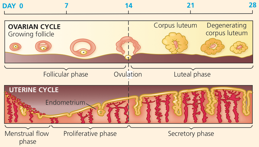
Ovarian Cycle: Cyclic events in the ovary, once per cycle a follicle matures and an oocyte is released
Uterine Cycle: Changes in the uterus
Menstrual Cycle: Endometrium thickens and develops a rich blood supply before being shed through the cervix and vagina if pregnancy doesn’t occur
Menstruation: Cyclic shedding of blood rich endometrium from the uterus in a flow through the cervix to the vagina
28 days average but can be 20 to 40
In females, hypothalamus releases GnRH and anterior pituitary secretes FSH and LH, stimulate follicle growth aided by LH which start to make estadiol
Follicular phase days 0-14, estradiol concentration slowly rises, follicles grow and oocytes mature and LH level rises and peaks at day 13
Proliferative phase days 6-14
Luteal phase days 15-28, remaining follicular tissue forms corpus luteum which secretes progesterone and estradiol, which exert negative feedback on the hypothalamus and pituitary and reduces LH and FSH secretion
Secretory phase days 15-28
Endometriosis: Some cells of uterine lining migrate to abdominal location that is ectopic (abnormal)
Menopause: After about 500 cycles, ovaries lose responsiveness to FSH and LH so less estradiol production
Estrous Cycle: In animals without menstrual cycles, cyclic changes in uterus control sexual receptivity. They only have intercourse around their cycle
Four phase of sexual response cycle
Excitement, vagina and penis are prepared for coitus (intercourse). Vagina is lubricated
Plateau, outer third of vagina is vasocongested and inner two third slightly expands to receive sperm. Breathing quickens and heart rate rises
Orgasm when rhythmic contractions of reproductive structures
In males, two stages.
First is emission when the semen is forced into the urethra
Expulsion/ejaculation is when urethra contracts and semen is expelled
In females, uterus and outer vagina contract, inner two thirds doesn’t
46.5: In placental mammals, an embryo develops fully within the mother’s uterus
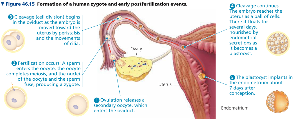
Conception: Fertilization in humans
Zygote behind cleavage cell divisions 24 hours after fertilization, after 4 days produces a blastocyst
Blastocyst: Sphere of cells surrounding a central cavity
Few days later embryo implants into the endometrium of the uterus
Pregnancy/Gestation: Carrying 1+ embryos in the uterus
Averages 266 days from fertilization
In first trimester, implated embryo secretes hormones to signal its presence and regulate the mother’s reproductive system
During first 2-4 weeks embryo gets nutrients directly from the endometrium
Trophoblast: Outer layer of the blastocyst
During first 2-4 weeks grows outward and mingles with the endometrium, eventually helping form the placenta
Organogenesis: Development of body organs, mostly in first trimester
Heart begins beating by weeka 4
Can be detected at 8-10, at 8 all major structures are present in their basic forms, the embryo is a fetus. Well diffrentiated but only 5 cm long
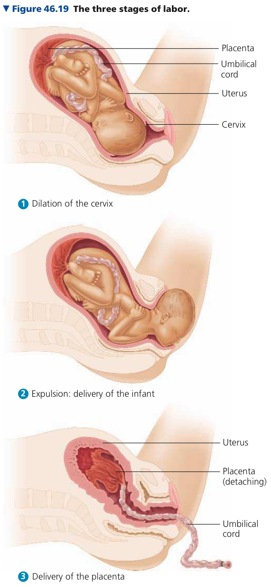
Labor has three stages
Thinning and opening up of the cervix
Expulsion/delivery of the baby
Delivery of the placenta
Contraception: Deliberate prevention of pregnancy
Birth Control Pills: Hormonal contraceptives with pregnancy rates <1%
Sterilization
Tubal Ligation: Sealing shut/tying off a section of each oviduct to prevent eggs from traveling into the uterus
Vasectomy: Cutting and typing off each vans deferens so sperm can’t enter the urethra
Abortion: Termination of pregnancy
In Vitro Fertilization (IVF): Combining oocytes and sperm in the laboratory
Chapter 47: Animal Development
47.1: Fertilization and cleavage initiate embryonic development
Model Organisms: Species chosen for ease that they can be studied
Sea Urchin Fertilization
When a sperm head contacts the egg surface, an acrosomal reaction is triggered
Hydrolytic enzymes are discharged from the acrosome (specialized vesicle at the top of the sperm) which partially digest the jelly coat and allow the sperm to penetrate the jelly coat
Sperm nucleus enters egg cytoplasm, and ion channels open in the egg’s plasma membrane, where sodium ions diffuse in and cause depolarization
Additional sperm can no longer fuse with it, preventing polysperny (>1 sperm nuclei into the egg)
Ca2+ activates the egg
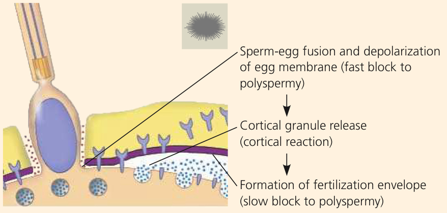
Sperm must travel through a layer of follicle cells before reaching the zona pellucida (extracellular matrix of egg)
Cleavage: Series of rapid cell divisions during early development
Solves the problem that a sing;e nucleus in a newly fertiized egg has too little DNA to produe the amount of mRNA to meet the cell’s need for new proteins
Blastomeres: Smaller cells that make up cytoplasm of the large fertilized egg
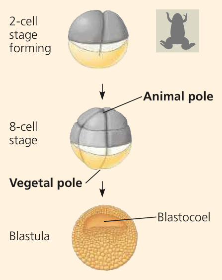
Blastula: Hollow ball of cells surrounding the blastocoel, from first to
Bastocoel: Fluid filled cavity
Yolk: Stored nutrients concentrated towards the vegetal pole, away from the animal pole
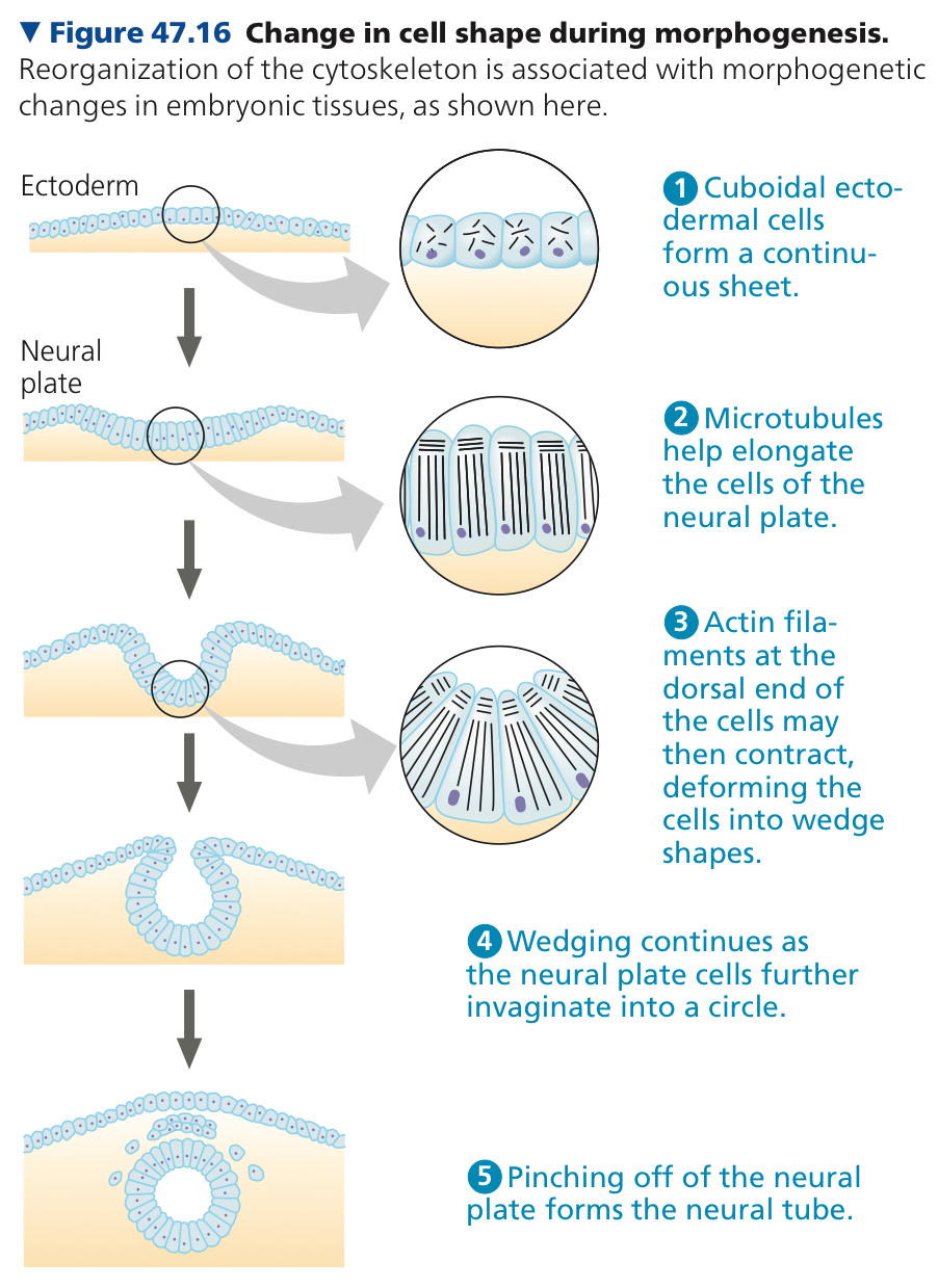
47.2: Morphogenesis in animals involves specific changes in cell shape, position, and survival
Morphogenesis: Processes by which the animal body takes shape, occurs over the last two stages of embryonic development
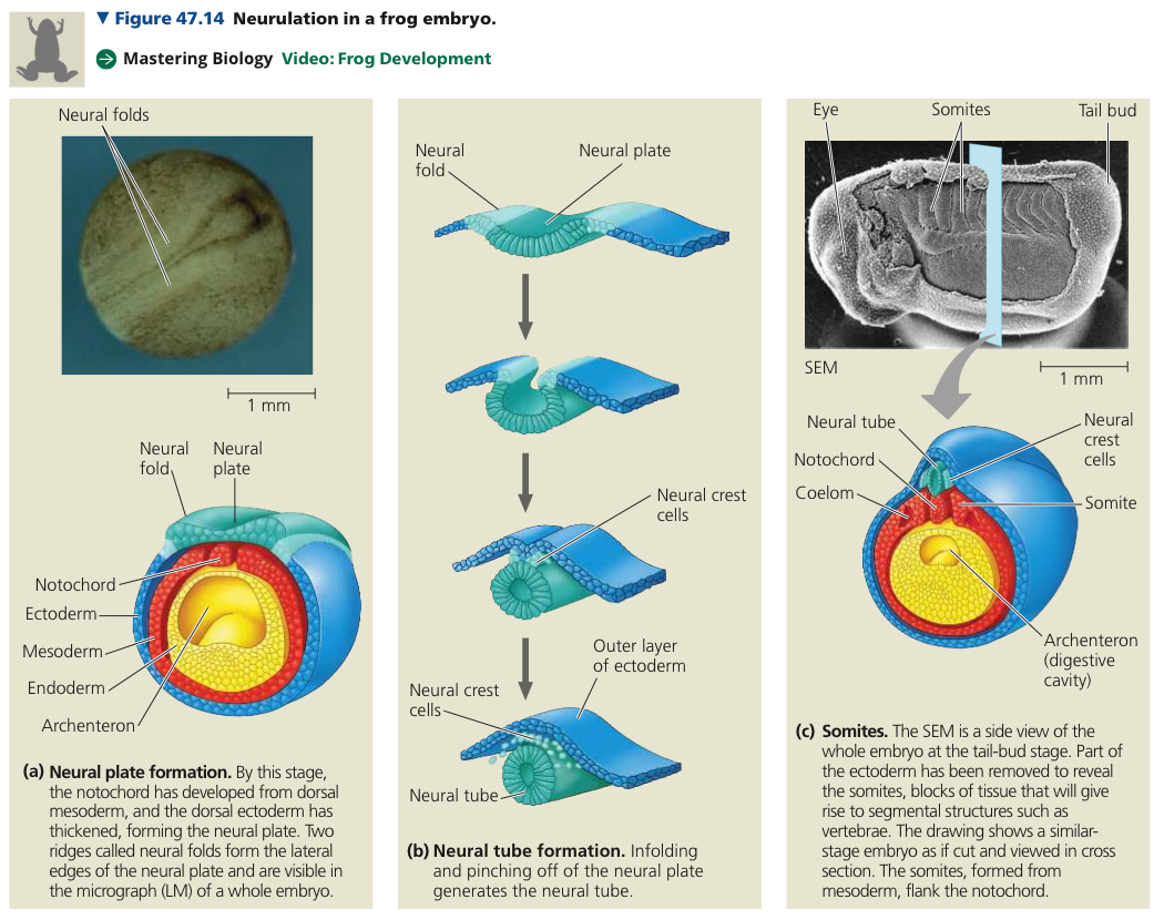
During gastrulation, a set of cells near the surface moves to an interior location, establishing cell layers and a primitive digestive tube
Further transformation happens in oranogenesis
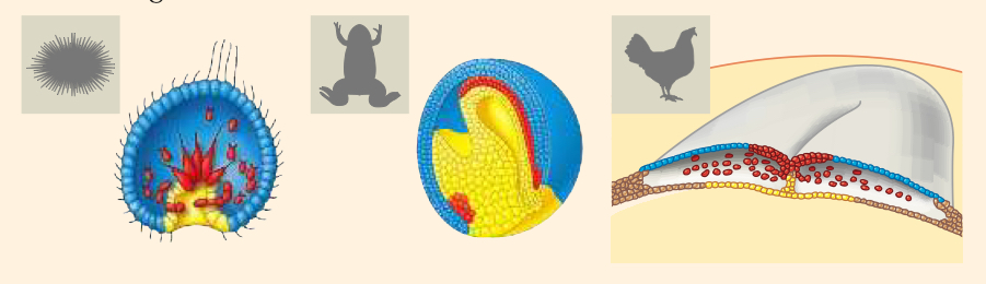
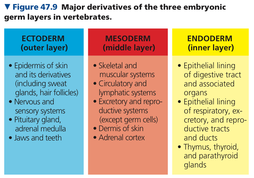
Germ Layers: Cell layers produced collectively
In late gastrula, ectoderm forms the outer layer, endoderm forms the lining of the digestive tract
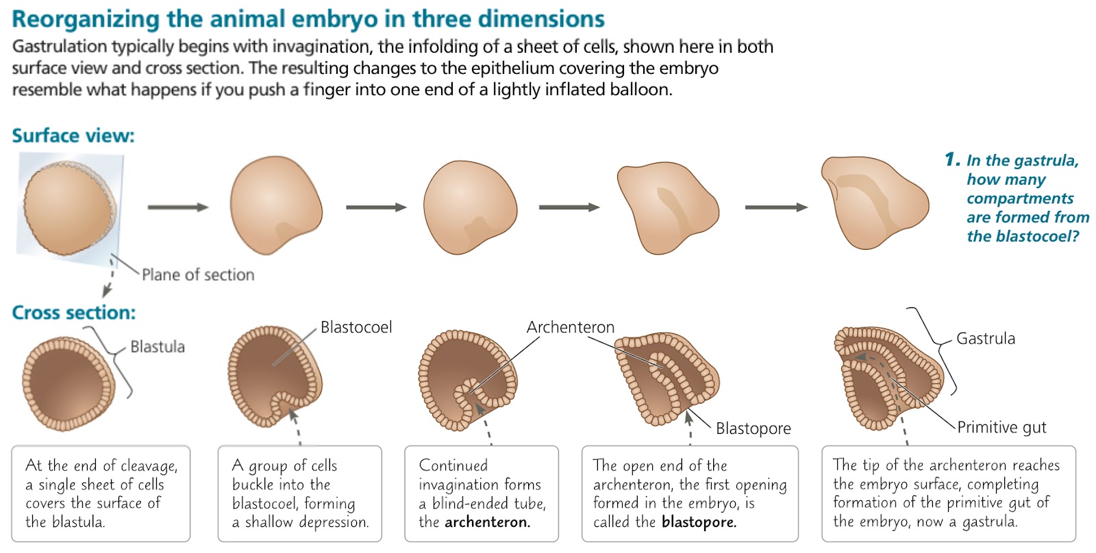
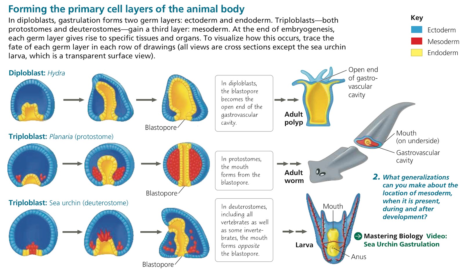
Mesoderm: Third germ layer that forms between the ectoderm and endoderm
Human eggs are pretty small. Development in human embryos is as follows
Embryo is a blastocyst, with a group of cells, the inner cell mass, clustered at one end of the cavity, which develop into the embryo proper
Trophoblast (outer epithelium of the blastocyst) initiates implantation of the embryo
Trophoblast continues to expand into the endometrium and four new extraembryonic membranes appear
Amnion layer
Chorion layer
Yolk sac layer
Allantois layer
Embryonic germ layers have formed. Extraembryonic mesoderm and four distinct extraembryonic membranes now surround the embryo
Amniotes: Mammals and reptiles including birds
Organs of animal body develop from specific portions of embryonic germ layers
Organogenesis in vertebrates have many early events
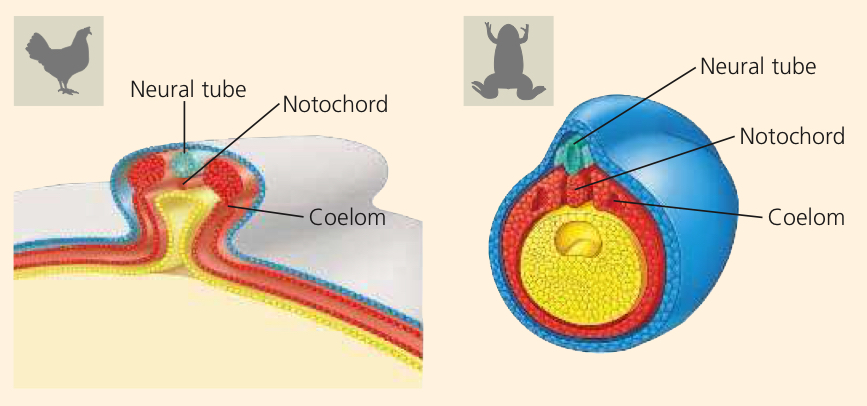
Notochord: Rod that extends along the dorsal side of chordate embryo, formed by cells of dorsal mesoderm
Neural Tube: Developed from infolding of ectodermal neural plate
Induction: Process in which a group of cells or tissues influences development of another group through close range interactions
2 sets of cells that develop near the vertebrate neural tube undergo long range migration
Neural Crest: Set of cells, develops along borders where neural tube pinches off from the ectoderm
Somites: Blocks, groups of mesodermal cells lateral to the notochord seperate into them
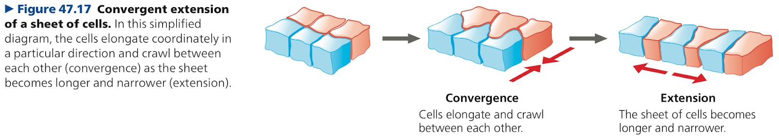
Convergent Extension: Rearrangement that causes a sheet of cells to become narrower while it becomes longer, occurs in gastrulation
47.3: Cytoplasmic determinants and inductive signals regulate cell fate
Determination: Process by which a cell or group of cells becomes committed to a particular fate
Diffrentiation: Resulting specialization in structure and function of determination
Fate Maps: Diagrams showing structures arising from each region of an embryo
Totipotent: Blastomeres that can develop into all the different cell types of a species
Pattern Formation: Process governing arrangement of organs and tissues in their characteristic places
Positional Information: Molecular cues that control pattern formation
In the form of molecules secreted by certain cells such as the dorsal lip of the blastopore in the amphibian gastrula, apical extodermal ridge, and zone of polarizing activity in the vertebrate limb bud
Apical Ectodermal Ridge (AER): Thickened area of ectoderm at the tip of the bud
Zone of Polarizing Activity (ZPA): Specialized block of mesodermal tissye
Chapter 48: Neurons, Synapses, and Signaling
48.1: Neuron structure and organization reflect function in information transfer
Neuron: Nerve cell that can recieve and transmit information
Cell Body: Where most of the neuron’s organelles are located
Dendrites: Stud the cell body, highly branched extensions, recieve signals from other neurons
Axon: Extension that transmits signals to other cells, longer than dendrites, one per neuron
Synapse: Junction between two cells
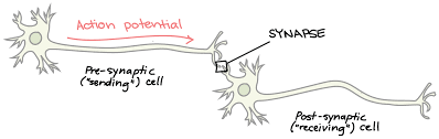
Neurotransmitter: Pass information from transmitting neuron to the recieving cell
Sensory Neuron: Transmit info about external stimuli and internal conditions
Interneurons: Form local circuits connecting neurons in the brain or ganglia, integrate sensory input
Motor Neurons: Transmit signals to muscle cells and cause them to contract
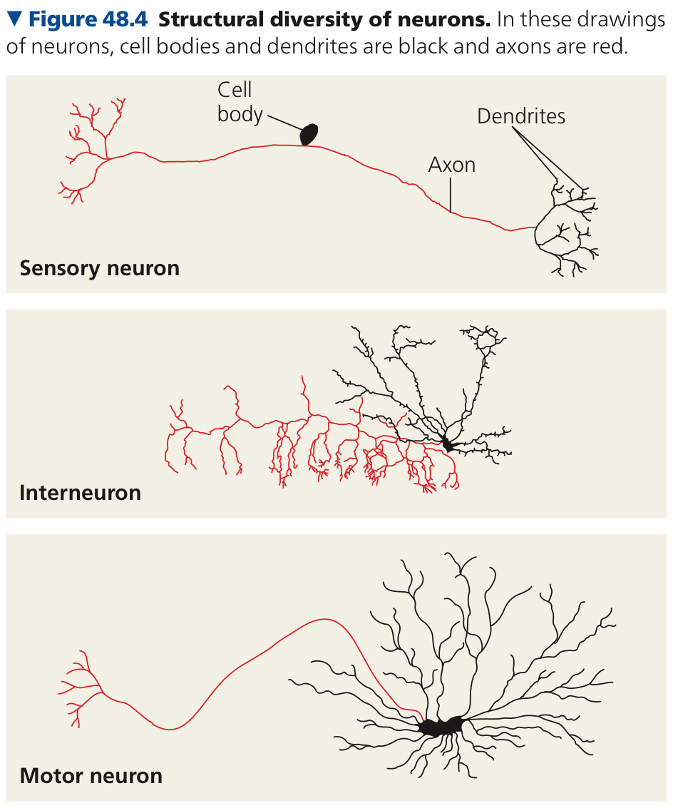
Nerves: Bundles of neurons grouped together
Central Nervous System (CNS): Organized system of neurons that carry out sorting, processing, and integration
May include a brain or ganglia (simpler clusters)
Peripheral Nervous System (PNS): Neurons that carry information in and out of the CNS
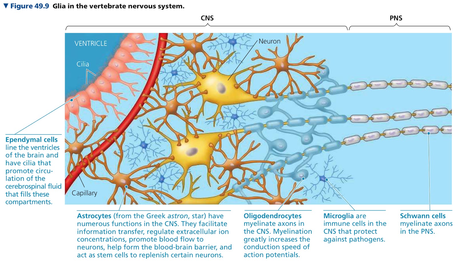
Glia: Supporting cells required in both PNS and CNS
Axons are longer and transmit signals to other cells instead of other neurons
Sensory neurons notice that you’ve been called so they tell the interneurons to tell the brain which tell the motor neurons to turn your head
It can reach more places?
48.2: Ion pumps and ion channels establish the resting potential of a neuron
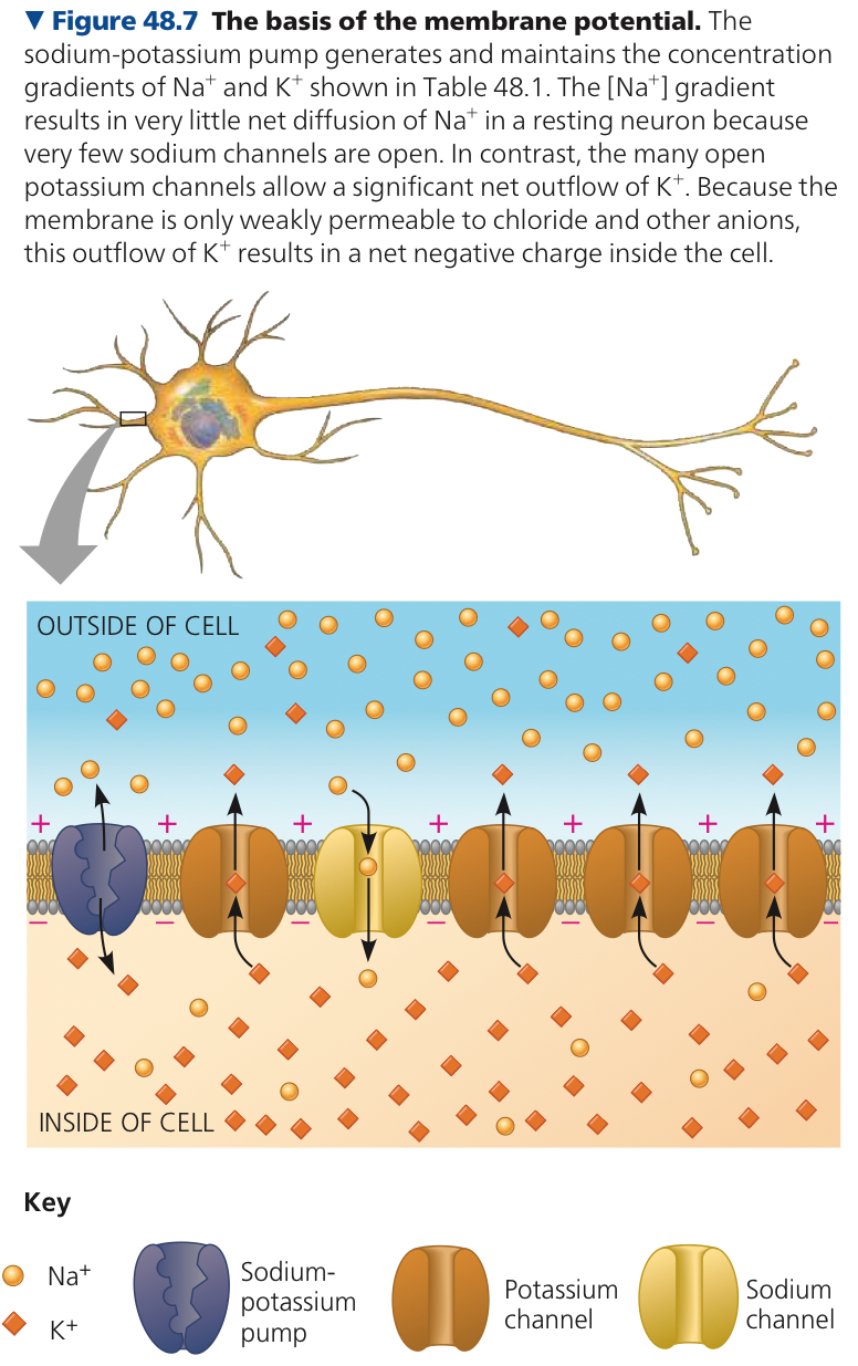
Membrane Potential: Charge difference across the plasma membrane
Resting Potential: Membrane potential of a resting neuron (one that’s not sending a signal)
Sodium Potassium Pump: Transports 3 Na+ out of the cell for every 2 K+ that are pumped in, net positive charge
Very slow so the difference in membrane potential is pretty small
Ion Channels: Pores formed by clusters of specialied proteins that span the membrane
Ions move rapidly through them so the resulting current makes a membrane potential, and is either net positive or negative
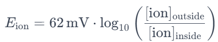
Equilibrium Potential (Eion): Magnitude of membrane voltage at equilibrium for an ion
Nernst Equation: Formula for Eion of a membrane permeable to a single type of ion
Active transport?
More permeable since there is more of the positive ions coming in
48.3: Action potentials are the signals conducted by axons
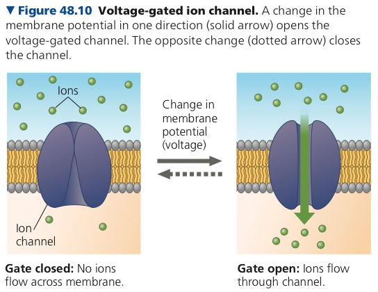
Gated Ion Channels: Ion channels in neurons that open or close in response to stimuli
Alters membrane’s permeability to particular ions
Voltage Gated Ion Channel: Channel that opens or closes in response to a shift in the voltage across the plasma membrane of the neuron
Hyperpolarization: Change in membrane potential that makes the inside of the membrane more negative
Depolarization: Change in membrane potential that makes the inside of the membrane less negative
Graded Potential: Shift in membrane potential in response to hyperpolarization or depolarization, induce small electrical current that dissipates as it flows along the membrane
Action Potential: If depolarization shifts membrane potential a lot, there is a massive change in membrane voltage
Threshold: If depolarization increases membrane potential to this level, voltage gated sodium channels open and there is further depolarization
How do voltage gated channels shape action potential?
At resting potential, most voltage gated sodium channels are closed, some potassium channels are open
When stimulus depolarizes the membrane, some gated sodium channels open and more Na+ diffuses into the cell
Positive feedback rapidly brings to membrane close to the equilibrium potential of Na, aka the rising phase
Two events prevent membrane potential from reaching the equilibrium in the falling phase, which brings the membrane potential back to equilibrium potential of K
Voltage gated sodium channels inactivate soon after opening
Most voltage gated potassium channels open
In the undershoot, membrane is more permeable to K+ than at rest and membrane potential is closer to equillibrium potential of K+ than at resting potential, but it eventually returns to resting potential
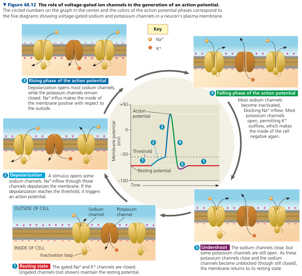
Sodium ions don’t flow once inactivation occurs even though channels are “open”, and this happens during falling phase and early undershoot
Refractory Period: “Downtime” when second action potential can’t be initiated because sodium channels are inactive
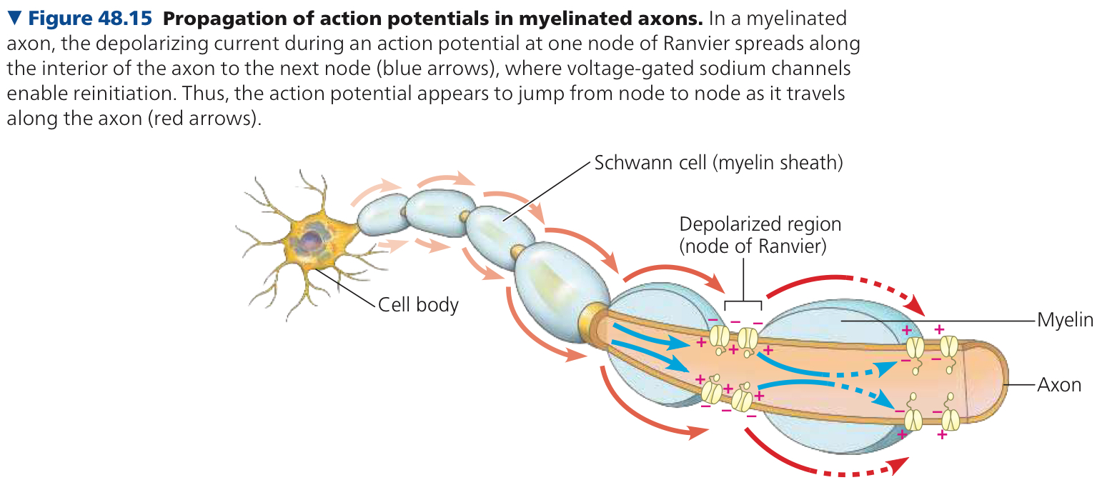
Myelin Sheath: Electrical insulation that surrounds vertebrae axons
Produced by glia
Ogilodendrocytes: Glia in the CNS
Schwann Cells: Glia in the PNS
Nodes of Ranvier: Gaps in the myelin sheath
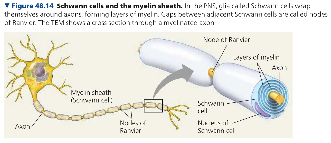
Saltatory Conduction: Action potential skips from node to node
48.4: Neurons communicate with other cells at synapses
Electrical Synapses: Contain gap junctions that allow electrical current to flow between neurons
Chemical Synapses: Rely on the release of a chemical neurotransmitter by the presynaptic neuron to transfer information
Presynaptic Neuron: Synthesizes neurotransmitter at each synaptic terminal and packages it in synaptic vesicles (Membrane enclosed compartments)
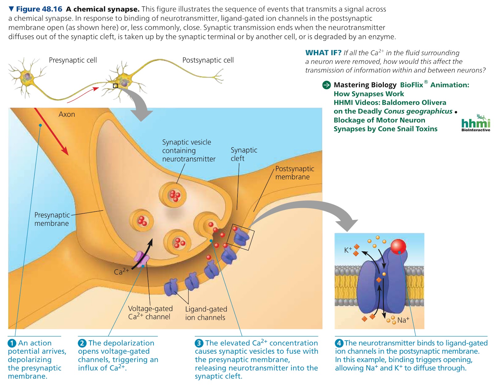
Synaptic Cleft: Gap that seperates presynaptic neuron and postsynaptic cell
Ligand Gated Ion Channel/Ionotropic Receptor: Clustered in mebrane of postsynaptic cell, directly opposite the synaptic terminal
Binding of the neurotransmitter to a particular part of the receptor opens the channel
Postsynaptic Potential: Graded potential in the postsynaptic cell
Excititory Postsynaptic Potential (EPSP): When ligand gated ion channels are permeable to both K+ and Na+ causing membrane potential to polarize to a midway equillibrium value
Inhibitory Postsynaptic Potential (IPSP): When ligand gated ion channels are selectively permeable only for K+ or Cl- so the postsynaptic membrane hyperpolarizes
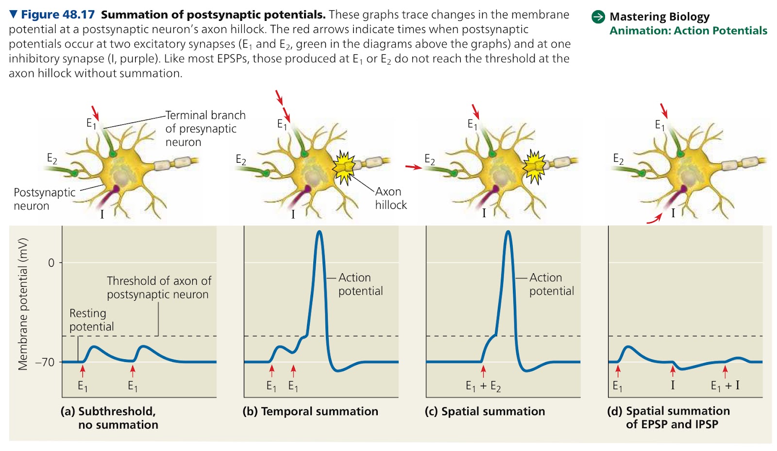
Summation: Individual postsynaptic potentials combine to produce a bigger postsynaptic potential
Temporal Summation: Effects of impulses received at the same place can add up if the impulses are received in close temporal succession
ex. two ESPS happen at a synapse in rapid succession, and the second ESPS arises before the postsynaptic membrane potential returns to its resting value
Spatial Summation: Stimuli are applied at the same time, but in different areas, with a cumulative effect upon membrane potential
ex. synapses are active on the same postsynaptic neuron, and the ESPS add together
When neurotransmitter is not part of an ion channel, it binds to a metabotropic receptor and activates a signal transduction pathway in the postsynaptic cell
Metabotropic Receptors: G protein coupled receptors
Neuropeptides: Relatively short chains of amino acids which act as neurotransmitters that operate via metabotropic receptors
Endorphins: Neuropeptides that act as natural anagesics and decrease pain perception
Chapter 49: Nervous Systems
49.1: Nervous systems consist of circuits of neurons and supporting cells
Nerves: Axons of multiple neurons bundled together
Cephalization: Cluster of sensory neurons and interneurons at the front end of the bodyin bilaterally symmetrical animals
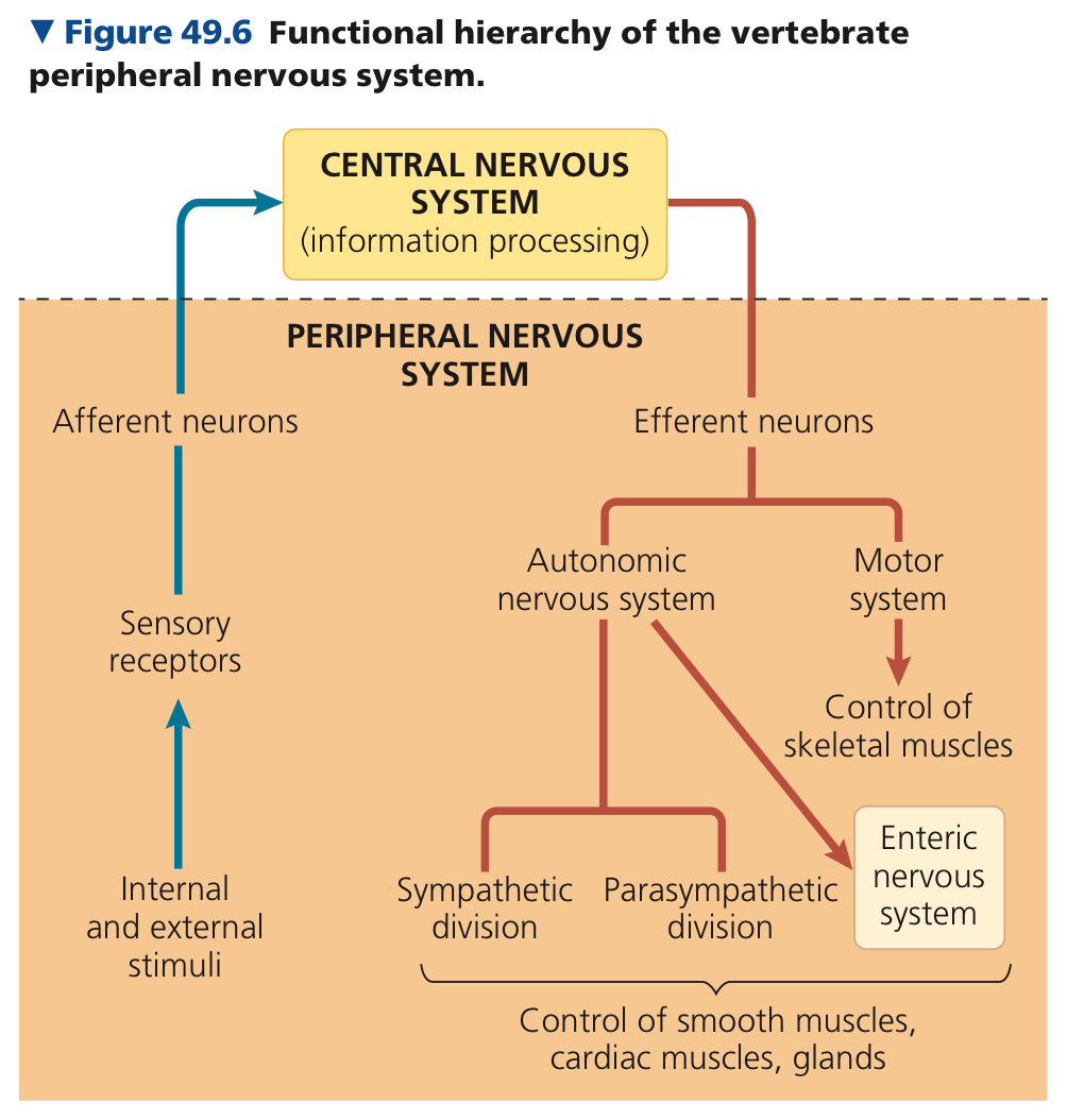
Central Nervous System (CNS): Neurons that carry out integration
Peripheral Nervous System (PNS): Neurons that carry information into and out of the CNS
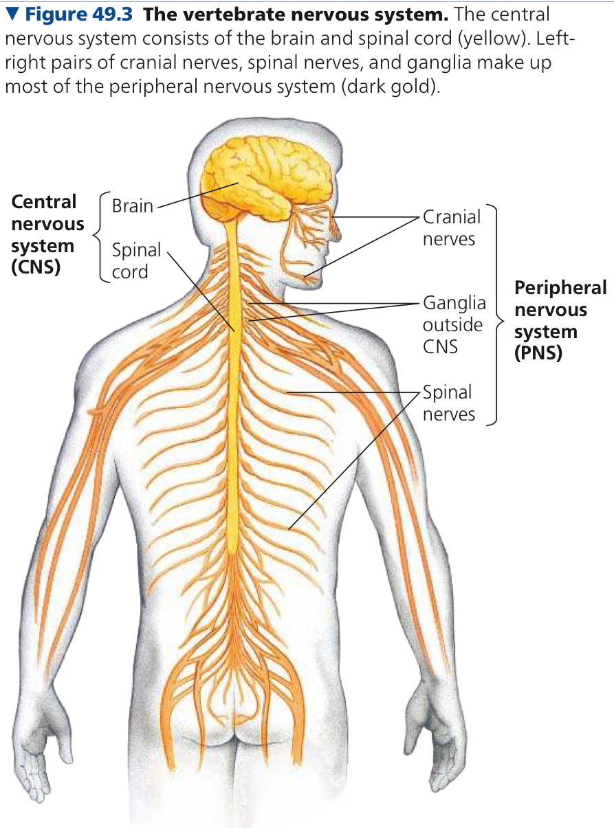
Ganglia: Segmentally arranged clusters of neurons that act as relay points in transmitting information
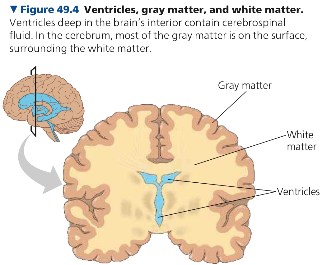
Brain and spinal cord have gray and white matter
Gray Matter: Neuron cell bodies
White Matter: Bundled axons, the outer layer of the spinal cord and in the interior of the brain
Reflexes: Body’s automatic responses to certain stimuli
PNS has two components, the motor and autonomic system
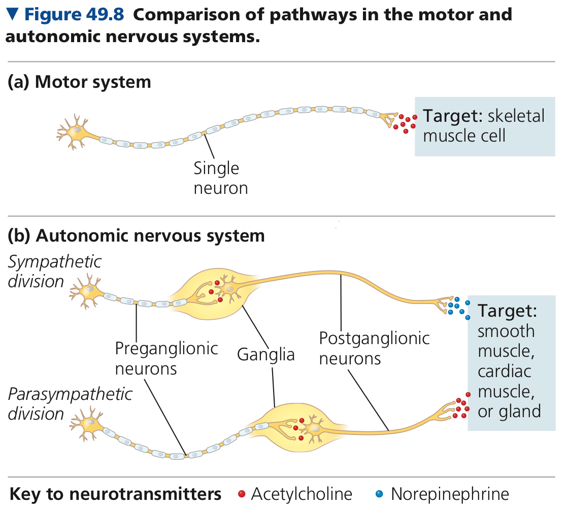
Motor System: Its neurons carru signals to skeletal muscles
Autonomic Nervous System: Involuntary regulation of smooth and cardiac muscles
Entric Nervous System: Network of neurons with direct control over digestive tract, pancreas, and galbladder
Sympathetic Division: Activation corresponds to arousal and energy generation, fight or flight
Exit CNS midway along the spinal cord and form sunapses in ganglia located just outside the spinal cord
Parasympathetic Division: Calming and returning to self maintanence functions, rest and digest
Exit CNS at the base of the brain or spinal cord, form synapses in anglia near an internal organ
Pathway for information flow typically involes a pre and post gangliogonic neuron in both divisions
Preganglionic Neurons: Cell bodies in the CNS, release acetylcholine
Postganglionic Neurons: In parasympathetic, releas acetylcholine, in sympathetic, release norepinephrine
Nervous systems of certebrates and most invertebrates have neurons and glia/glial cells
Sympathetic, since its fight or flight
49.2: The vertebrae brain is regionally specialized
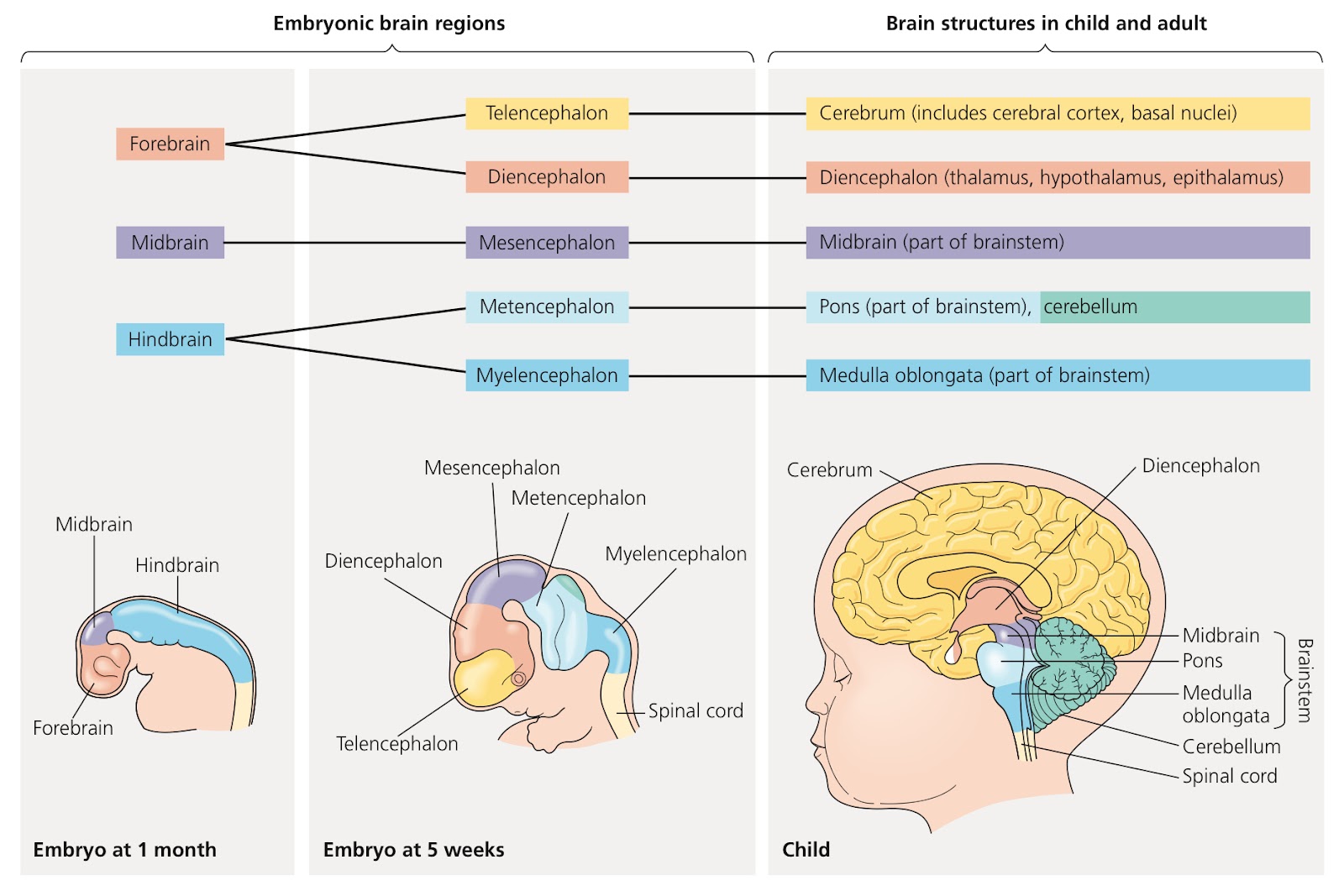
Forebrain: Activities with processing and olfactory (smells), regulation of sleep, learning, and complex processing. Contains olfactory bulb and cerebrum
Midbrain: Routing of sensory input
Hindbrain: Controls involuntary activities
As an embryo develops there are three anterior bulges, the forebrain, midbrain, and hindbrain
Brainstem: Stalk that joins with the spinal cord at the base of the brain, midbrain and portions of the hindbrain, pons and medulla oblongata, recieves and integrates several types of sensory info and sends it to specific regions of the forebrain
Pons: Transmits signals between forebrain and cerebellum
Medulla Oblongata: Connection between brainstem and spinal cord, regulates blood pressure, heart rate, etc.
Cerebellum: Behind the brainstem, controls movement and balance and helps in learning and remembering motor skills
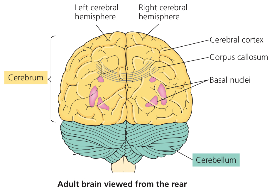
Cerebrum: Develops from telencephalon (from forebrain), controls skeletal muscle contraption and learning, emotion, memory, and perception
Two hemispheres, left and right
Cerebral Cortex: Outer layer of cerebrum, vital for perception, voluntary movement, and learning
Corpus Callosum: Thick band of axons, enables right and left cerebral cortices to communicate
Diencephalon: Gives rise to thalamus, hypothalamus, and epithalamus
Thalamus: Main input center for sensory information going to the cerebrum
Hypothalamus: Control center, the body’s thermostat and biological clock
Biological Clock: Molecular mechanism that directs periodic gene expression and cellular activity
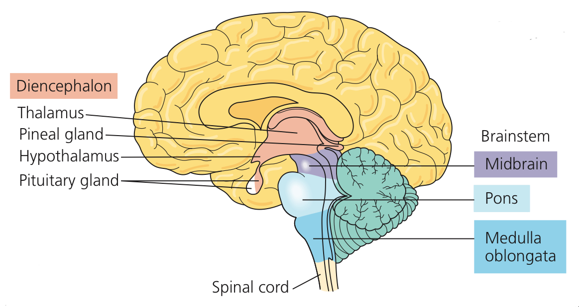
Suprachiasmatic Nucleus (SCN): Clustered neurons in the hypothalamus that coordinate circadian rhythms
Amygdala: Almond shaped brain strucutre near the base of the cerebrum for storage and recall of emotional memory
Cerebellum?
Frontal lobe
49.3: The cerebral cortex controls voluntary movement and cognitive functions
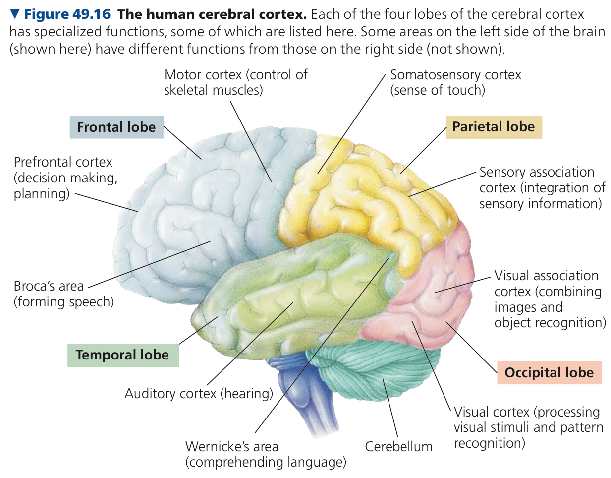
In the cerebral cortex, there are sensory areas to recieve and process sensory info, association areas to integrate the info, and motor areas to transmit instructions to other parts of the body
Four major lobes, frontal, temporal, occipital, and parietal
Frontal lobe injuries damage decision making and emotional responses but intellect and memory are fine
Same thing when connection between prefrontal cortex and limbic system is surgically severed (frontal lobotomy)
Somatosensory receptors provide info about touch, pain, pressure, temp, and position of muscles and limbs, directed via the thalamus to primary sensory areas within brain lobes and to the prefrontal cortex to plan movement
Lateralization: Difference in function between two hemispheres of brain

When they can no longer perform a function because of an injury to a certain area then we know that the area must be for the function
Broca area, think about what you want to say then form speech. Wernicke’s area, recognize what they are saying and then comprehend it
49.4: Changes in synaptic connections underlie memory and learning
Neuromal Plasticity: Capacity for nervous system to be remodeled and connects between neurons to be modified in the CNS
Most occurs at synapses
In memory, storage of information is in the cerebral cortex
Short Term Memory: Holds information for a short time and then is released if it becomes irrelevant
Information accessed via temporary links formed in the hippocampus
Long Term Memory: Long term knowledge
Links in hippocampus are replaced by connectons within the cerebral cortex
Hippocampus is responsible for aquiring long term memory but not to maintain it
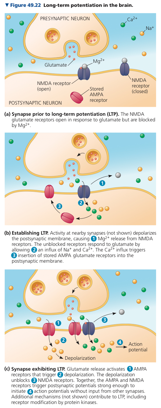
Long Term Potentation (LTP): Lasting increase in the strength of synaptic transmission
Presynaptic neuron releases excitatory neurotransmitter glutamate, and involves two types of glutamate receptors, NMDA or AMPA
Information moving from short term to long term memory and long term potentation
I don’t understand the question
49.5: Many nervous system disorders can now be explained in molecular terms
Schizophrenia: Psychotic episodes where patients have a distorted sense of reality, affects neuronal pathways that use dopamine as a neurotransmitter
Major Depressive Disorder: Periods where once enjoyable activities provide no pleasure
Bipolar Disorder: Extreme swings of mood
Alzheimers and Parkinsons are neurodegenerative
Chapter 50: Sensory and Motor Mechanisms
50.1: Sensory receptors transduce stimulus energy and transmit signals to the central nervous system
Sensory Reception: First step of a sensory pathway, the detection of a stimulus by specialized sensory cells
Sensory cell is either a neuron or cell that regulates a neuron
Sensory Receptor: Sensory cell or organ
Result of detecting any stimuli is to open or close ion channels
Receptor Potential: Change in membrane potential from sensed stimulus
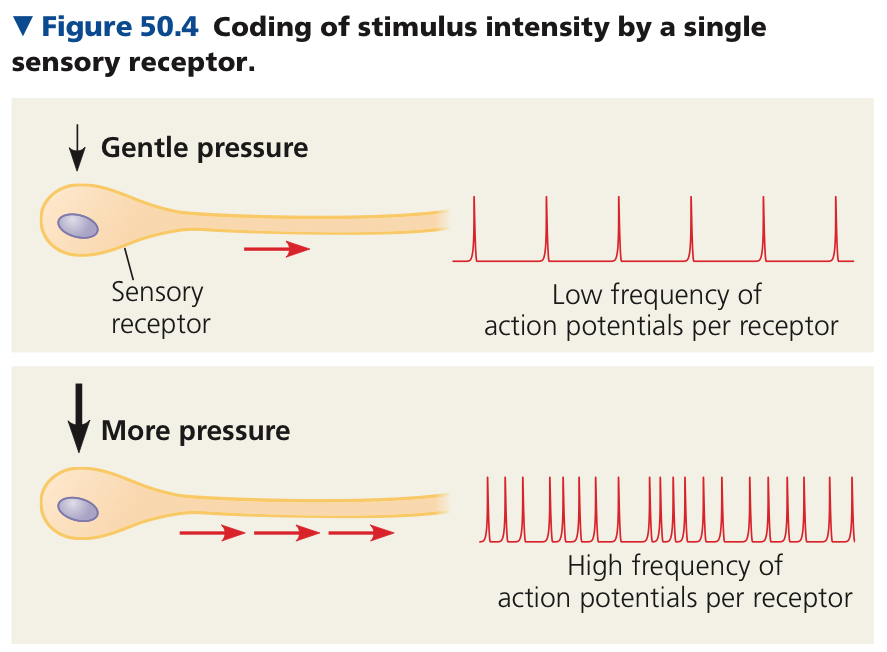
Size increases with intensity of stimulus
Usually sensory neurons spontaneously generate action potentials at a low rate, and alters how often action potentials are produced
Sensory Transduction: Conversion of stimulus to receptor potential
Integration begins when information is recieved
Perception: Generated when action potentials reach the brain via afferent (facing inwards) neurons and are processed by circuits of neurons
Two types of mofidication of transduction of stimuli by sensory receptors, amplification and adaptation
Amplification: Strengthening of a sensory signal during transduction
Sensory Adaptation: Decrease in responsiveness of receptors
Mechanoreceptors: Sense physical deformation caused by forms of mechanical energy, usually ion channels that are linked to structures that extend outside the cell (ex. cilia/hairs) and are anchored to internal cell structures
Chemoreceptors: Monitor internal environment, two broad categories
Transmit info about overall solute concentration or respond to specific molecules in body fluids
Detect stimuli in their diet and environment
Electromagnetic Receptor: Detects a form of electromagnetic energy (light, electricity, magnetism)
Thermoreceptors: Detect heat and cold
Nocireceptors/Pain Receptors: Detect stimuli that reflect harmful conditions (ex. extreme pressure or temperature) and trigger defensive reflexes
Mechanoreceptors
Pain receptors tell us to stop
Electromagnetically
50.2: In hearing and equillibrium, mechanoreceptors detect moving fluid or settling particles
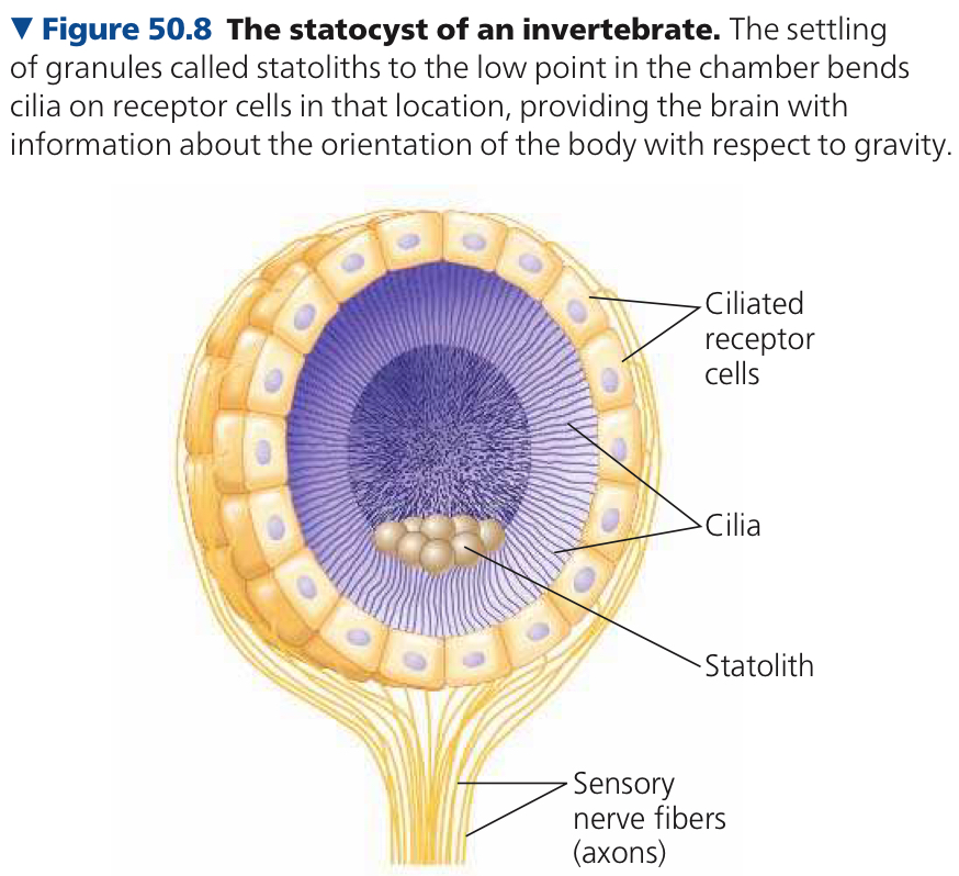
Statocysts: Organs that sense gravity and maintain equilibrium
Statoliths: Granules formed by grains of sand or other dense materials which sit in a chamber lined with cilated cells
Outer ear has external pinna and auditory canal → tympanic membrane (eardrum) which seperates the outer ear from the middle ear
In the middle ear there are three small bones, the malleus (hammer), incus (anvil) and stapes (stirrup) which transmit vibrations to the oval window (membrane beneath the stapes)
Middle ear opens into the Eustachian tube which connects to the pharynx
Inner Ear: Consists of fluid filled chambers including semicircular canals (equilibrium) and cochlea(bony chamber involved in hearing with two large canals)
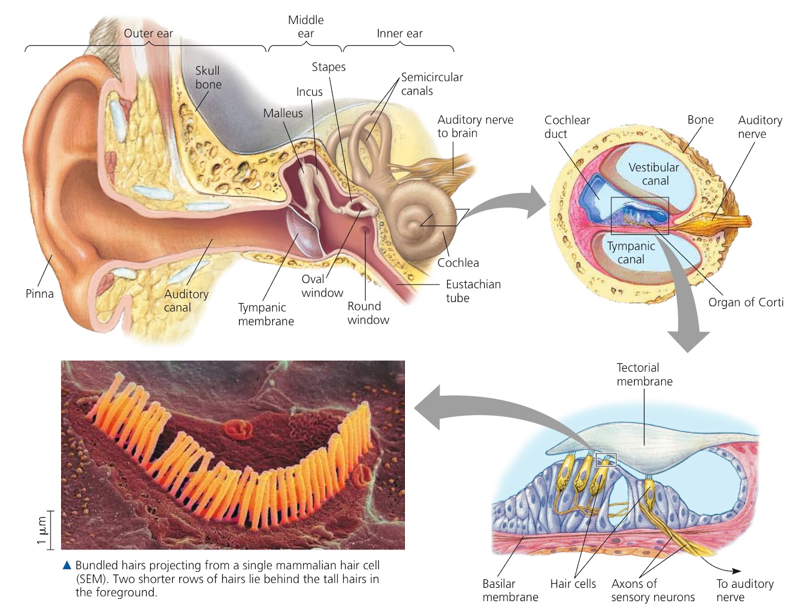
Organ of Corti: Contains mechanoreceptors of the ear, the base is the basilar membrane
Hair Cells: Sensory cells with hairlike projections that we use to detect motion
In mammals sound goes eardrum → bones of the middle ear → oval window → fluid in cochlea of the inner ear, pressure waves vibrate basilar membrane and depolarize hair cells and trigger action potententials that travel via the auditory nerve to the brain
50.3: The diverse visual receptors of animals depend on light absorbing pigments
Phororeceptors: Sensory cells with light absorbing pigment molecules, in diverse light detectors
Most animals have light detecting organs with photoreceotirs
Compound Eyes: Arthropods’ visual organ with up to several thousand ommatida
Ommatidia: Light detectors
Single Lens Eyes: Have a small opening (pupil) where light enters and the iris expands or contracts and changes the diameter of the pupil to let in more or less light. Kind of like a camera
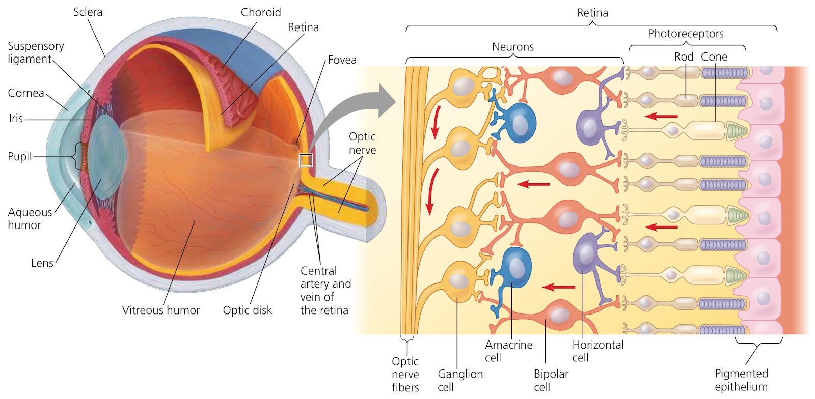
Human eyes are surrounded by the conjunctiva (a mucous membrane), the sclera (a connective tissue) and the choroid
Sclera forms the transparent cornea
Chroid forms the colored iris
Inside the choroid, the neurons and photoreceptors of the retina form the innermost layers of the eyeball
Lens: Transparent disk of protein, divides the eye into two cavities
Aqueous Humor: Clear watery substance in front of the lens
Vitreous Humor: Jellylike substance behind the lens
Bipolar cells in the retina recieve information from rods and cones, and each ganglion cell gathers input from them. Horizontal and amacrine cells integrate information across the retina
Rods: Sensitive to light
Cones: Provide color vision
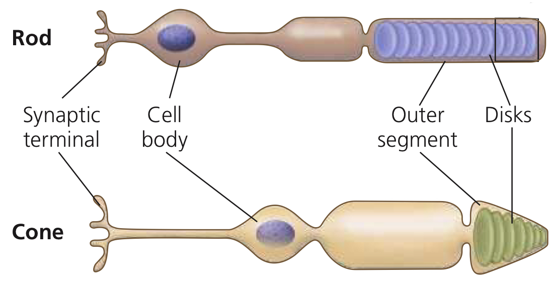
Vertebrate visual pigments have a retinal bound to an opsin
Retinal: Light absorbing derivative of vitamin A
Opsin: Membrane protein
Rhodopsin: Visual pigment of rods
Absorption of light by retinal triggers a signal transduction pathway that hyperpolarizes the receptors and makes them release less neurotransmitter
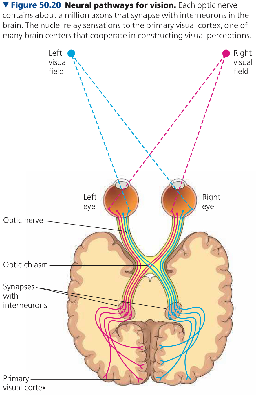
Synapses transmit info from photoreceptors to cells that integrate info and convey it to the brain along axons that form the optic nerve
50.4: The senses of taste and smell rely on similar sets of sensory receptors
Gustation: Sense of taste
Tastants: In terrestial animals the the presence of these chemicals in a solution dictate taste
Olfaction: Sense of smell
Odorants: In terrestial animals the the presence of these chemicals in the air dictate smell
Taste Buds: Receptor cells for taste in mammals, scattered in several areas of the tongue and mouth
Between five tastes, sweet, umami, and bitter each require 1+ genes encoding a GPCR
One type of sweet and one type of umami receptor
30+ bitter receptors
When an odorant diffuses into the cilia (that extends into the layer of mucus coating the nasal cavity), it binds to a specific GPCR protein, an olfactory receptor (OR) on the plasma membrane of the olfactory cilia
This causes signal transdduction which produces cyclic AMP
In olfactory cells, cyclic AMP opens channels in the plasma membrane which leads to depolarization and generates action potentials
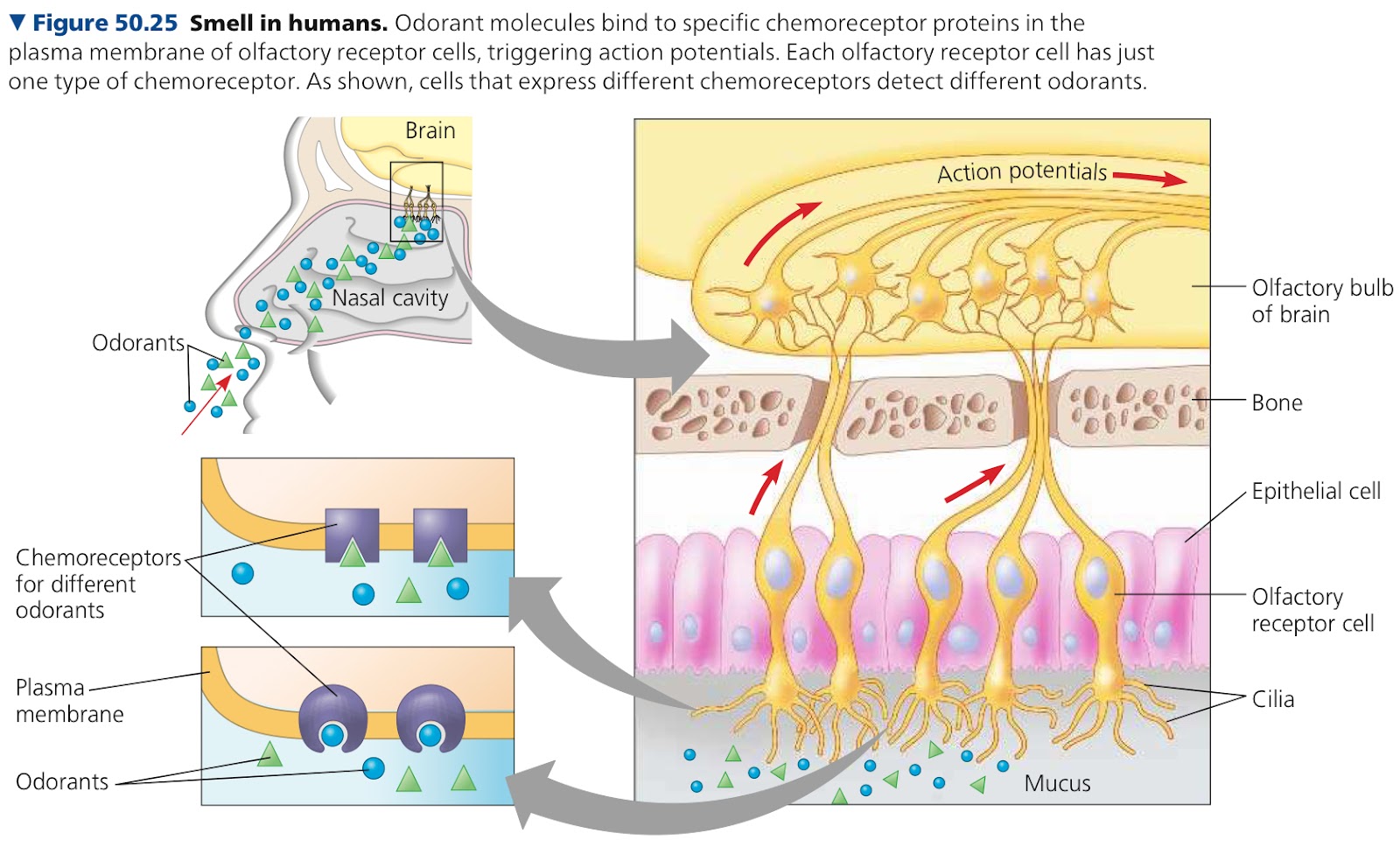
50.5: The physical interaction of protein filaments is required for muscle function
Muscle contraction relies on interaction between thick and thin filaments
Thin Filaments: Made of globular protein actin, two strands of polymerized actin are coiled around one another
Thick Filaments: Staggered arrays of myosin molecules
Skeletal Muscle: Moves bones and body, has a bundle of long fibers running along the length of the muscle, each fiber being a cell
In the cells there are multiple nuclei derived from the embryonic cells that fused to form the fiber
Myofibrils: Surround the nuclei derived from embryonic cells, consist of thick and thin filaments
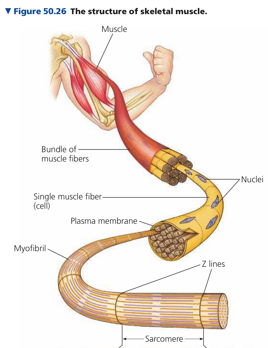
Sacromeres: Basic contractile units of skeletal muscle, make up microfibrils in muscle fibers
Sliding Filament Model: Thin and thick filaments ratchet past each other, powered by myosin molecules
Each myosin molecule has a long tail and globular head. Tail binds with tails of other myosin molecules, head can bind ATP
Hydrolysis of ATP cnverts myosin to a high energy form that bins to actin
In a muscle fiber at rest, tropomyosin and troponin complex are bound to actin strands of thin filaments
Tropomyosin: Regulatory protein
Troponin Complex: Set of additional regulatory proteins
Motor neurons cause muscle contraction in a long process triggering movement of Ca2+ into the cytosol of muscle cells
Arrival of action potential at the synaptic terminal of a motor neuron causes release of the neurotransmitter acetylcholine
Binding of acetylecholine to receptors on the muscle fiber leads to depolarization that initiates an action potential
Within the muscle fiber, action potential spreads deep into the interior, following transverse (T) tubules(infoldings of the plasma membrane)
These make close contact with the sacroplasmic reticulum (SR) (a specialized endoplasmic reticulum).
As action potential spreads along the T tubules, it triggers changes in the SR which opens Ca2+ channels
Calcium ions stored in the interior of the SR flow through open channels in the cytosol and bind to the troponin complex
This initiates the muscle fiber contraction
Relaxation starts as proteins pump Ca2+ back into the SR from the cytosol
When Ca2+ concentration drops to a low level, regulatory proteins bound to the thin filament shift back to their starting position
This once again blocks the myosin binding sites
Motor Unit: Single motor neuron and all of the muscle fibers it controls
Tetanus: Rate of stimulation is so high that muscle fiber can’t relax between stimuli and twitches fuse into a sustained contraction
Myoglobin: Oxygen sorting protein
Fast-Twitch Fibers: Develop tension 2-3 times faster than slow twitch fibers, enabling brief, powerful contraptions
Slow fibers pump Ca2+ slower so it stays in the cytosol longer and a muscle twitch lasts about 5x as long
Cardiac Muscle: Only found in the heart and is striated, some can initiate rhythmic depolarization and contraction
Smooth Muscle: Found in walls of hollow organs such as vessels and tracts of circulatory, digestive, and reproductive systems. No striations
50.6: Skeletal systems transform muscle contraction into locomotion
Hydrostatic Skeleton: Fluid held under pressure in a closed body compartment
Main type of skeleton in most cndarians, flatworms, nermatodes, and annelids
Exoskeleton: Hard covering deposited on animal’s surface
30-50% of arthropod cuticle consists of chitin
Endoskeleton: Hardened internal skeleton buried within soft tissues
Locomotion: Active travel from place to place
Chapter 51: Animal Behavior
51.1: Discrete sensory inputs can stimulate both simpe and complex behaviors
Behavior: Action carried out by muscles under control of the nervous system
Proximate causation is how a behavior occurs or is modified
Ultimate causation is why a behavior occurs in the context of natural selection
Behavioral Ecology: Study of ecological and evolutionary basis for animal behavior
Fixed Action Pattern: Sequence of unlearned acts directly linked to a simple stimulus
Sign Stimulus: Exernal trigger for a fixed action pattern
Signal: Stimulus transmitted from one organism to another
Communication: Transmission and reception of signals between animals
Pheromones: Chemical substances released by animals that communicate through odors or tastes, often relate to reproductive behavior
51.2: Learning establishes specific links between experience and behavior
Innate Behavior: Behavior that is developmentally fixed this way
Learning: Modification of behavior as a result of specific experiences
Imprinting: Establishment of long lasting behavioral response to a certain individual or object
Sensitive Period: Only time period when imprinting can take place
Spatial Learning: Establishment of memory that reflects the environment’s spatial structure
Cognitive Map: Representation in animal’s nervous system of spatial relationships between objects in its surroundings
Associative Learning: Ability to associate one environmental feature with another
Cognition: Process of knowing that involves awareness, reasoning, recollection, and judgement
Problem Solving: Cognitive activity of devising a method to proceed from one condition to another in the face of real or apparent obstacles
Social Learning: Learning through observing and interpreting behaviors
Culture: System of information transfer through social learning or teaching that influences behavior of individuals in a population
51.3: Selection for individual survival and reproductive success can explain diverse behaviors
Optimal Foraging Model: Natural selection should favor a foraging behavior that minimizes the cost of foraging and maximizes the benefits
Some mates are monogamous some are polygamous
Mate Choice Copying: Behavior where individuals in a population copy the mate choice of others
Game Theory: Alternative strategies in situations where ooutcome depends on strategies of individuals involved
51.4: Genetic analyses and the concept of inclusive fitness provide a basis for studying the evolution of behavior
Antidiuretic Hormone (ADH)/Vasopressin: Peptide released during mating and binds to a specific receptor in the central nervous system
Altruism: Behavior that reduces an animal’s fitness but increases fitness of other individuals in the population
Inclusive Fitness: Total effect an individual has on proliterating its genes by producing its own offspring and providing aid that enables other close relatives to produce offspring
Hamilton’s Rule: Natural selection favors altruism when the benefit exceeds the cost
rB > C
C, the cost, is how many fewer offspring the altruist produces
B is the average number of extra offspring for the recipient
Coefficient of Relatedness: Fraction of genes that, on average, are shared, r
Kin Selection: Natural selection that favors altruism by enhancing reproductive success of relatives
Reciprocal Altruism: Altruism that occurs between unrelated humans