Mechanoreception
Types of mechanical forces
Outer mechanical forces
Touch
Pressure
Vibration
Air currents
Water currents
Sound
Sensed by the ear and somatosensory system
Inner mechanical stimuli
Pressure on blood vessels
Contraction of muscles
Limb flexions
Proprioceptors deal with these forces
Other
Gravity
Acceleration
Sensed by equilibrium organs, found in inner ear in humans
Mechanoreceptors transduce mechanical forces and transmit the information to the brain.
Mechanical forces are directed to specific ion channels
High speed responses lack second messengers
Latency of mechanoreceptors = time between onset of a stimulus to the opening of the first ion channels. 10-6 seconds (1000x faster than photoreceptors/vertebrate chemoreceptors)
Types of mechanoreceptor cells
Receptor cells with cilia
Epithelial cells with at least one cilia
Found in
Cnidarian nematocytes
Inner ear of humans
Lateral line organ of fishes/amphibians
Wings, joints, antennae of arthropods and spiders
Receptor cells without cilia
Nerve cells
Free nerve endings
Pacinian corpuscles in human skin
Ganglion cells
Crustacean stretch receptors
Annelid and mollusc touch receptors
Mechanoreceptors in human skin + tendons
Spindle organs in human muscle
Human skin
Meissner’s corpuscles
Detect pressure on the skin
Detect slow vibration
Merkel’s disks
Detect pressure and touch
Pacinian corpuscles
Detect pressure on the skin
Detect rapid vibration (to feel texture of objects)
Ruffini’s endings
Detect stretch and sustained pressure
Free nerve endings
Detect pain, warmth, cold, tickle
Hair follicle receptors
Detect tickle and light touch
Only found in hairy skin
Somatosensory cortex
Large part of the brain
Deal with the impressions on our body surface
Are represented in somatotopic order
Neighbouring locations on the body are represented by neighbouring locations in the brain
Note that size may vary. Face, hands, and genitals are very sensitive to mechanical stimuli and thus correspond to larger parts of the somatosensory cortex
Lateral line organ
Detects wave vibration and water currents
Guiding fishes during locomotion
Allows them to detect and locate prey, predators, and potential mates
Neuromasts
Receptor cells
Usually located in canals under the body surface
Water passes through when currents flow around the fish’s body
Canals are connected to the external water by small openings
Each neuromast is a collection of hair cells with sensory hairs embedded in a jelly-like substance (cupula).
Projects upwards into the canal
Gets deformed when water flows through canal
Deformation cause hairs to bend, responding with action potentials
Proprioception
Monitor tension of gut, blood pressure, position of joints, length and tension of muscles and tendons
In vertebrate muscles, there are muscle spindle organs, which are free nerve endings wrapped around specialised thin muscle fibres.
The afferent neuron senses stretch and sends action potentials to the spinal chord, where it synapses with the motor neuron controlling the muscle.
The efferent neuron sends its signal back to the muscle, maintaining the tone.
Balance
Sensed by statolith organs
Sense body angle relative to gravity
Sense angular acceleration
Both of these are in humans called the vestibular organ
Based on hair cells, like hearing
Mist animals use statocyst organs to sense direction of gravity
Statoliths are small stonelike concretions in a fluid-filled chamber covered by ciliary mechanoreceptors
When tilting, the statoliths move and cause deformation to the hair cells, that respond with action potentials
This organ can be made in two ways
The statoliths move freely in the liquid-filled chamber
Entire chamber is lined with hair cells and filled with statoliths
The statolith is connected to the mechanoreceptor hairs
The direction and strength of bending of the cilia codes body angel
In vertebrates, the statoliths are called otoliths
The otolith organ consists of the utricle and saccule
In insects, gravity can be sensed also by
Weight of body parts (flying insects) instead of statoliths.
Air bubbles (water-living insects)
Vertebrate otolith organ
Part of the vestibular system in the inner ear
Two parts (saccule and utricle), both filled with endolymph
The hair cells are attached to a gelatinous mass (macula), where the otoliths are embedded
In mammals:
The utricular macula is oriented horizontally
The saccular macula is oriented vertically
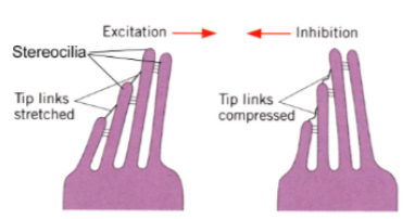
The hair cells have hair bundles embedded in a gelatinous matrix and connected to each other at the tips by tip-links. Bending of the hair bundle in one direction causes the opening of ion channels and a depolarisation of the cell (excitation). Bending in the opposite direction causes the closing of ion channels and a hyperpolarisation of the cell (inhibition).
 Utricular macula
Horizontal orientation
Monitor direction of gravity due to changes in head angle
Saccular macula
Vertical orientation
Monitor linear acceleration
Either side-to-side movements or up-and-down movements
Animals that perform fast body movements often have separate organs to sense angular acceleration. In the highly evolved equilibrium organs of vertebrates, crustaceans and cephalopods, angular acceleration is sensed in a similar way to gravity. In humans, the three semicircular canals, filled with endolymph, each have an enlarged region called the ampulla. Inside the ampulla, a field of hair cells (similar to the hair cells in the otolith organs) project their cilia into a cupula. If the channel rotates, the endolymph – due to inertia – tends to remain stationary, thus deforming the cupula and its cilia. The hair cells to which the cilia are attached then indicate the direction of head rotation. If you turn your head in one of the three planes (X, Y or Z), the walls of the canal will move with the head and thus relative to the endolymph. This is analogous to turning a glass of water: the glass moves but the water remains stationary.