Musculoskeletal System
Musculoskeletal System - A system of muscles, joints, tendons, and ligaments providing movement, form, strength, warmth, and protection.
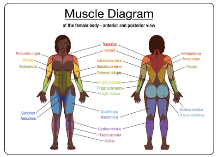
Muscles - provide form and produce heat for the body. Contract to help bones move.
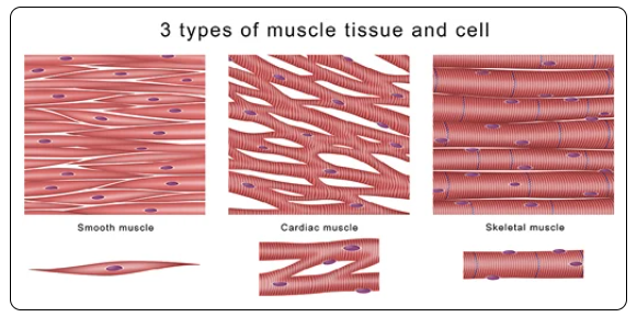
Skeletal muscle - Striated muscle that is attached to the skeleton by tendons; contraction of skeletal muscle is under voluntary control
Bicep - Large muscle on front of upper arm between the shoulder and elbow. Flexes the elbow and rotates the forearm.
Deltoid - Thick triangular muscle covering shoulder joint. Flex the arm away from the body (abduction)
Gastrocnemius - two-headed muscle in the back of the lower leg. It flexes the knee and helps flex the foot (plantar flexion)
Hamstring - One of three thigh muscles on the back of the leg between the hip and knee. Semimembranosus, semitendinosus, and biceps femoris. Flex the knee and extend the hip (extension)
Pectoral - One of two muscles that connect the front walls of the chest to the bones of the upper arm and shoulder. Pectoralis major and pectoralis minor. Help with moving the arm toward the body (adduction) and rotating the arm inward
Quadricep - 4 muscles that cover the front and the sides of the thigh. Keeps the knee stable and helps it straighten
Triceps - large 3 headed muscle on the back of the arm. Help extend the elbow joint and keeps the humerus bone secured in the glenohumeral joint.
Cardiac Muscle - Heart muscle that consists of Interlocking Involuntary straited muscle, which allows the electrical impulses to pass quickly across the muscle fibers.
Smooth Muscle - in the walls of all the hallow organs of the body (except heart). It's contraction reduces the size of these structures; movement generally is involuntary
Tendons, Ligaments, and Fascia
Tendons - Made of fibrous connective tissue, connect muscle to bone.
Help bones move by transmitting the mechanical force of muscle contraction to the bones
Strong and able to stretch to withstand normal movement
Can be injured when overused from age
Tendon strain is when it is overstretched or torn
Tendonitis is inflammation of the tendon and can happen from trauma, overuse, or degeneration from age
Extensor tendons - allow a joint to open or straighten out
In arms, legs, hands, and feet
Flexor tendons - allow a join to close or contract
In arms, legs, hands, feet, and hips
Ligaments - Made of fibrous connective tissue, connect bone to bone.
Band bones together to help stabilize joints
Have elastic fibers that allow movement of the joint but keep the joint from moving too far (right/left/behind/forward)
An injured ligament is a sprain - classified by grade (slightly overstretched -> complete tear)
Around knees, ankles, elbows, shoulders, and other joints
Knee Ligaments - Connecting Femur (thigh bone) to tibia (Shin bone)
Anterior cruciate ligament (ACL) - most common injury
Medial collateral ligament (MCL)
Posterior cruciate ligament (PCL)
Lateral collateral Ligament (LCL)
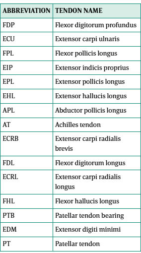
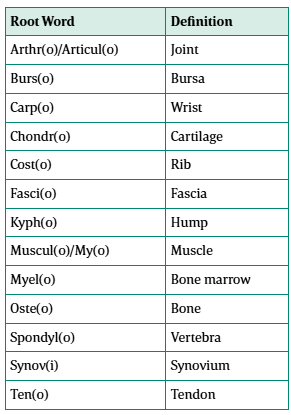
Surgeons will use acronyms in the operative report to identify the joint they are working on
Fascia - this casing of connective tissue that surrounds and holds in place the body's organs, bones, and muscles
Can hold muscle groups together, creating planes between groups of muscles
Human Skeleton - Made of bone, support system for body
2 parts:
Axial - Skull, hyoid, rib cage, sternum, vertebrae (spine bones), and sacrum
80 bones in total
Appendicular - shoulder bones, pelvic bones, arms, legs, hands, and feet
126 bones in total
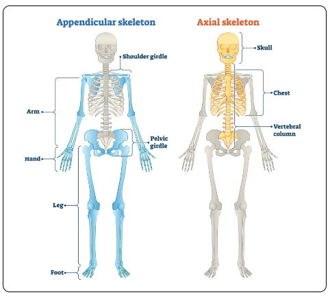
Axial Skeleton:
Skull - Bony housing for the brain that's a closed system, except for the Foramen of Magnum (where the spinal cord exits)
Contains:
1 frontal bone
1 occipital bone
1 ethmoid bone
1 sphenoid bone
2 parietal bones
2 temporal bones
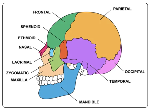
Facial Bones:
Maxilla
2 zygomatic
Mandible
Nasal
Palatine (2)
Inferior nasal concha (2)
Lacrimal (2)
1 vomer (1)
Cranial bones of the auditory system:
2 incudes
2 mallei
2 stapes
Hyoid Bone (lone bone under chin)
Bones of the thorax
24 ribs
1 sternum
Spinal column
7 cervical
12 thoracic
5 lumbar
Sacrum
Appendicular Skeleton - 126 bones
Shoulder girdle
2 clavicles
2 scapulae
Pelvic girdle
2 hip bones
Upper Extremity -
2 humeri
2 radii
2 ulnae
16 carpals
10 metacarpals
28 phalanges
Lower Extremity -
2 femurs
2 tibiae
2 fibulae
2 patellae
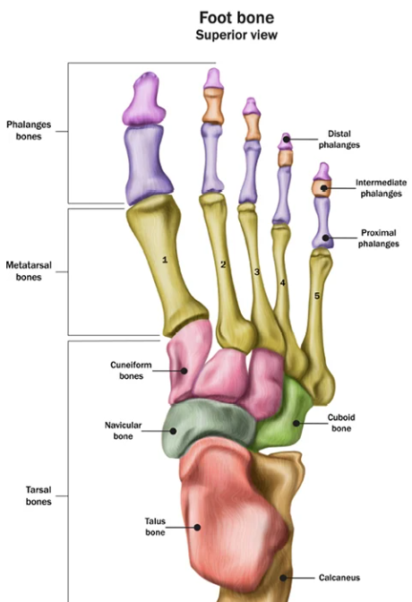
14 tarsals
10 metatarsals
28 phalanges
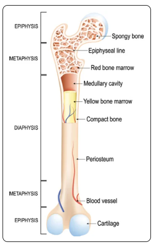
Bones - give bodies shape, protect organs, support weight
Work with muscles & tendons to help the body move, and the marrow of the bones produces blood cells for the body
Store calcium, phosphorus, and magnesium salts
Made of rigid connective tissue
Bone Marrow - produces stem cells which make the body's red blood cells, platelets, and some types of white blood cells
Classifications:
Long - bones longer than they are wide and found in the limbs (ex. Femur & humerus).
Tubular - long bones
Short - Roughly cube-shaped bones such as carpal bones of the wrist and tarsal bones of the ankle
Sesamoid - short bone shaped like a sesame seed, formed within tendons, cartilaginous in early life and osseous (bony) in the adult. (ex. Patella is the largest sesamoid bone)
Cuboidal - Short bones
Flat - Consist of a layer of spongy bone between two thin layers of compact bone; cross-section is flat, not rounded. Flat bones have marrow but lack a bone marrow cavity. (Ex. Skull & ribs)
Irregular - These are bones in the body not fitting into the above categories; several are found in the face, such as zygoma and mandible. Vertebrae are irregular bones.
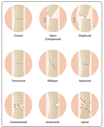
Bone Fracture Types:
Closed Fracture - Does not involve a break in the skin
Colles' fracture - Occurs in wrist and affects the distal radius bone
Comminuted fracture - more than two separate bone components (fragments)
Compound fracture - projects through the skin with a possibility of infection
Compression fracture - Vertebrae collapse due to trauma, tumor, or osteoporosis
Epiphyseal fracture - occurs when matrix is calcifying, and chondrocytes are dying; usually seen in children
Greenstick fracture - Only one side of shaft is broken and other is bent, common in children
Transverse Fracture - Breaks shaft of a bone across the longitudinal axis
Spiral Fracture - Spread along length of bone and produced by twisting stress Lefort fractures
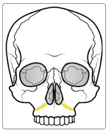
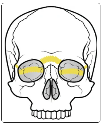
Le Fort 1 fractures (horizontal) - usually result from blunt force trauma occurring low on the maxillary alveolar rim in the downward direction. Extends from the nasal septum to the lateral pyriform rims, travels horizontally above the teeth apices, then crosses below the zygomaticomaxillary junction and traverses the pterygomaxillary junction, interrupting the pterygoid plates.
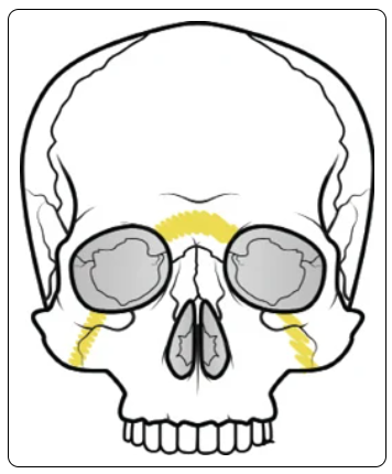
Le Fort 2 (Pyramid) - usually result from a blow to the lower or mid maxilla. Extends from the nasal bridge at or below the nasofrontal suture through the frontal processes of the maxilla. This will extend through the lacrimal bones and inferior orbital floor and rim through or near the inferior orbital foramen. Progresses inferiorly through the anterior wall of the maxillary sinus. Travels under the zygoma, across the pterygomaxillary fissure, and through the pterygoid plates.
Le Fort 3 (transverse) - total craniofacial separation. Can follow impact to the nasal bridge or upper maxilla. Essentially the lower face is no longer attached to the skull when this type of fracture occurs. These fractures start at the nasofrontal and frontomaxillary sutures and extend posteriorly (Backward) along the medial wall of the orbit through the nasolacrimal groove and ethmoid bones. The fracture continues along the floor of the orbit along the inferior orbital fissure and continues superolaterally through the lateral orbital wall, through the zygomaticofrontal junction and the zygomatic arch. Intranasally, a brand of the fracture extends through the base of the perpendicular plate of the ethmoid, through the vomer, and through the interface of the pterygoid.
Cartilage & Joints
Cartilage - Flexible connective tissue that is nonvascular (has no blood vessels)
Can be found in joints between bones
Ear, nose, knee, ankle, and rib cage
Cushion between bony surfaces or where there is a need for flexibility
Heals slowly due to no blood vessels
Conditions:
Chondrosis - When cartilage breaks down or deteriorates.
Example: Osteoarthritis, where cartilage wears away and the bones of a joint rub against each other causing inflammation
Chondritis - Inflammation of cartilage.
Example: costochondritis where the cartilage of the ribs becomes inflamed
Chondromalacia - Cartilage becomes soft. May be seen in all types of joints, but is often in the knee joint at the patella (kneecap)
Joints - 3 types:
Fibrous
Cartilaginous
Synovial (most joints)
Articular cartilage that covers the bone ends
Joint cavity lined with a synovial membrane, which secretes a thick, viscid, slippery mucous that cushions the joint and allow smooth motion
Joint capsule of fibrous connective tissue that surrounds and provides stability of the joint
Accessory ligaments that provide reinforcement and stability
Types:
Ball-and-socket - Shoulder or hip, allows a full range of movement
Hinge - knee or elbow, allows only bending and straightening
Pivot joints - Neck, allows it to swivel and bend
Salter-Harris Fractures - aka Physeal fractures. Fractures that occur through a growth plate (physis). Specific to children who are relying on the growth plate to continue to support their growth to full height. (Most common: epiphyseal plates, or physis)
4 types:
Type I - fracture occurs through the growth plate. Fracture line extends within the growth plate (physis)
Type II - Most common Salter-Harris fracture. Occurs through both the metaphysis and the growth plate
Type III - fracture in the joint (intra-articular fracture) extending through the growth plate and the epiphysis. This type of fracture is rare, but most commonly found at the distal end of the tibia (by the end of the shin bone, by the ankle)
Type IV - Fractures extend through the growth plate, epiphysis, and metaphysis
Type V - Usually the result of a crushing or compression injury of the growth plate. The amount of force has the potential to cause disruption on the germinal matrix, hypertrophic region, and vascular supply to a growth plate. Extremely rare. Due to the severe nature of the injury, these typically have a poor prognosis and can result in bone growth arrest.
The Spine - Extremely challenging interpreting the operative note since spinal surgery can occur at the boney vertebra level or the cartilaginous disc level, or both.
The surgeon will either count the # or vertebrae works on, or # of intervertebral spaces (disc spaces) worked on
Since there is a vertebra above and below each intervertebral space/disc space, the units of service for work at the intervertebral level is equal to the number of vertebrae minus one
Example: Intervertebral/disc level - surgeon cites diskectomies at L1-L2 and L2-L3. 3 vertebral bodies listed, but only 2 disc spaces operated on (3 vertebrae - 1 = 2 discs). 2 units of work.
Example: Vertebral level - surgeon cites laminectomies on L1-L4. Surgical removal of lamina for those 4 vertebrae. 4 units of work.
The Disc - Fibrocartilaginous material in the spine; 24 discs.
Annulus Fibrosus - outer rim of the spinal disc, made of strong material
Mucoprotein Gel - jelly-like substance, protected by the nucleus pulposus. The gel moves inside the annulus fibrosus and redistributes itself to absorb impact, as the spine is subjected to pressure.
Nucleus Pulposus - protective covering that surrounds gel (mucoprotein gel) in the disc
Sits between 2 vertebra (intervertebral disc) and work on the disc can dictate the surgical approach - anterior approach provides the best access to the intervertebral disc
Requires dissection through the thorax or abdomen by a cardiothoracic or general surgeon
Anterior access to the cervical spine can often be achieved through an anterolateral approach through the neck by an orthopedic or neurosurgeon - patients lie face up (supine)
Posterior or posterolateral approach to the spine - discs can be removed. Less common. Surgeon dissects through the back of the patient. Patient is lying face down (prone)
Vertebra - interlocking bones that make up the vertebral column (spine)
3 functional components:
Vertebral body is specifically designed for load bearing, since the weight of the body depends on it
Vertebral arch existed to protect the spinal cord
Transverse processes are designed to allow for ligament attachment. This provides stability
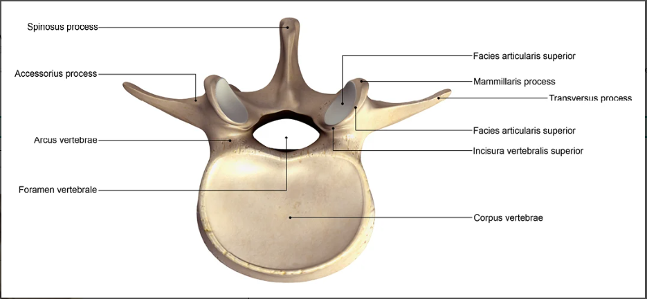
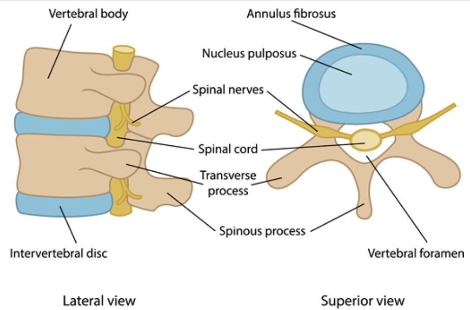
Facet Joint - symmetrical synovial-lined joints that connect the articular facets of the vertebrae. Connect the vertebra above and below, and allow some motion in the spine.
Surrounded by a fibrous capsule
Located between the pedicle and lamina of the same vertebra and form articular pillars, which provide structural stability to the entire vertebral column
Composed of 2 articular processes of adjacent vertebrae
Joint consists of the superior articular process of one vertebra located immediately anterior (above) to the inferior (below) articular process of the vertebra below
Bone Conditions:
Ankylosis - Condition of stiffening of a joint
Arthralgia - Pain in the joint
Arthritis - Inflammation of a joint
Arthropathy - joint disease
Bursitis - Inflammation of the bursa
Chondralgia - paint around and in the cartilage
Kyphosis - Abnormal curvature of thoracic spine (humpback)
Lordosis - Abnormal curvature of spine, usually lumbar (swayback or hollow back)
Osteosarcoma - Cancerous tumor of the bone
Osteochondritis - Inflammation of bone and cartilage
Osteopenia - Lower than average bone density, can be a precursor to osteoporosis
Scoliosis - Lateral curvature of the spine
Tendonitis - Inflammation of the tendon
Achilles rupture - a tear of the Achilles tendon.
Achilles Tendon - a strong cord that connects the back of the calf to the heel bone (calcaneus).
Can tear due to overuse or injury. Treatment: surgical & Non-surgical.
Bursa - A fluid-filled sac that works like a cushion to reduce friction. Most bursa are near tendons of major joints like shoulders, hips, and knees.
When inflamed, causes pain, swelling, and stiffness. Treatment: rest, ice, pain relievers
Carpal Tunnell Syndrome (CTS) - compression on the median nerve in the carpal tunnel.
Numbness, tingling, and weakness in the hand and arm. Treatment: Surgery or non-surgical wrist splits worn at night, cortisone injections, and ice
Compression Fracture - fracture of the vertebra typically caused by the loss of bone mass. Caused by disease or injury.
Back pain, loss of height, hump in the back. Surgery (vertebroplasty or kyphoplasty). Non-surgical rest, pain meds, and ice/heat at the area of fracture.
Fibromyalgia - Chronic disorder that causes widespread pain, sleep problems, fatigue, and emotional/mental distress.
Treatment: medications, stress reduction, psychotherapy
Osteoarthritis (OA) - Degenerative joint disease (DJD). Most common type of arthritis. Inflammation and injury to the joint cause the protective cartilage that cushions the ends of bones to gradually deteriorate over time, causing the bones within the joint to rub together. Can cause joints to get painful and swollen, limit mobility. Aka wear and tear arthritis
Can affect any joint, most common: hips, knees, and spine, neck, small joints of the fingers, base of the thumb, and the hallux (big toe)
Characterized by cartilage breakdown, bony changes, deterioration of tendons and ligaments that hold joint together, and various degrees of inflammation of the synovium (joint lining)
Osteoporosis (porous bone) - Chronic progressive disease that causes bones to become weak, thin, brittle - a fracture may occur with only mild stress such as coughing or bending over. Most common type of bone disease
Osteopenia - Low bone density
Plantar Fasciitis - the fascia covering the muscles and tendons of the bottom of the foot becomes inflamed.
Stabbing pain in the heel and is one of the most common reasons for heel pain. Worse in morning. Treatment: physical therapy, shoe inserts, injections, and surgery.
Rheumatoid Arthritis (RA) - Chronic systemic inflammatory disease that causes joint pain and damage throughout the body. Exact cause has not been identified, result of an autoimmune response where immune system mistakenly attacks the joints and other healthy tissues in the body.
Trigger Finger - Finger or thumb catches or locks when bent. Usually worse in morning.
Treatment: splinting, injections, medications, or surgery
Tennis elbow (lateral epicondylitis) - Inflammation of forearm muscles from overuse. Pain is usually on the outside of the elbow and sometimes into the forearm and wrist.
Treatment: rest, physical therapy, and pain relievers, ,or surgery.
Musculoskeletal Surgical Approaches - Less invasive procedures
Percutaneous - procedure through the skin. Ex: puncturing the skin to gain access to a body cavity or organ with a small catheter or probe
Per - through
Cutaneous - skin
Endoscopic/arthroscopic - percutaneous approach using an endoscope (flexible tube with a light source and camera) inserted into a joint for either therapeutic or diagnostic procedures.
Open surgical - surgeon uses a scalpel to open the area under treatment and dissects down to the area of concern.
Depth of dissection is important to note.
Go to muscles, tendons or ligaments?
Open the periosteum (a dense layer of vascular connective tissue surrounding bones that does not extend into the surfaces of the joint)?
Intra-articular (inside the joint capsule)?
Extra-articular (outside the joint capsule)?
Surgery Suffixes:
-centesis - to puncture
-clasis - Surgical break or fracture
-desis - surgical fixation of bone or joint
-ectomy - surgical removal
-graphy - imaging
-lysis - to free up
-orraphy - surgical suture
-opexy - surgical fixation of an organ
-otomy - to cut a part of the body, but not necessarily remove the organ
-scopy - observation often related to visual observation with an endoscope
-plasty - remodel or repair
Diagnostic Tests:
Arthrogram - Imaging of inside of joint using contract dye; may use x-ray, CT, or MRI depending on problem
Computed tomography (CT/CAT) scan - Imaging to diagnose problems with bones or muscles
Dual-energy X-ray absorptiometry (DEXA) Scan - Measures density and mass of structures in the body (ex: bone mass)
Electromyography (EMG) - Measure electrical activity of muscle
FABER Test (Patrick's test) - Positive sign identifies sacroiliac dysfunction (FABER = Flexion, abduction, external rotation)
Fluoroscopy - imaging that uses continuous x-ray images to look at a body part or system
Finkelstein test - Positive sign identifies de Quervain's tenosynovitis as the cause of wrist pain
Homan's sign test (Dorsiflexion Test) - positive test indicates possible deep vein thrombosis (DVT)
Magnetic resonance imaging (MRI)- radio waves and magnetic fields to capture soft tissue or joint damage
Tompson test - Tests for Achilles tendon rupture. Squeezing the calf produces a foot flex when the Achilles tendon is intact. If no movement, the Achilles tendon is likely injured
Tinel's sign - Positive sign indicates carpal tunnel syndrome. Performed by tapping over the carpal tunnel at the wrist.
Ultrasound - Soundwaves to image soft tissues
X-ray - Imaging using electromagnetic waves to diagnose problems (primarily bones)
Musculoskeletal Procedures:
Arthrocentesis - Fluid is aspirated from an affected joint using a needle and examined under the microscope.
Can detect synovial inflammation and exclude other causes of arthritis
Removal of joint fluid and injection of corticosteroids into the joints during arthrocentesis can help relieve pain, swelling, and inflammation
Arthroscopy - A surgical technique where a small scope (long narrow tube with a light and camera on the end of it) is used to examine the inside of a joint. Camera transmits pictures to a video monitor
can detect signs of RA and other joint diseases
Kyphoplasty - Minimally invasive surgery to treat a spinal compression fracture by using a cannula to insert a balloon into the fracture to create a cavity, then uses bone cement to reinforce the vertebra where the compression fracture is
Fasciotomy - Cutting into the fascia to release, not remove, fascia.
Example: plantar fasciotomy - the fscia over the muscles and tendons on the bottom of the foot is incised. When the fascia heals, it effectively has lengthened the fascial area and relieves the fasciitis
Trigger Finger release - Tenolysis at A1 pulley to allow more movement of flexor e=tendon through tendon sheath
Trigger point injection - Injections into a muscle trigger point with a small amount of anesthetic and/or steroid may relieve pain. Trigger points are painful "knots" in muscles.
Vertebroplasty - Minimally invasive surgery to treat a spinal compression fracture. A cavity is not created before using bone cement to reinforce the vertebra where the compression fracture is.
Joint Replacement -
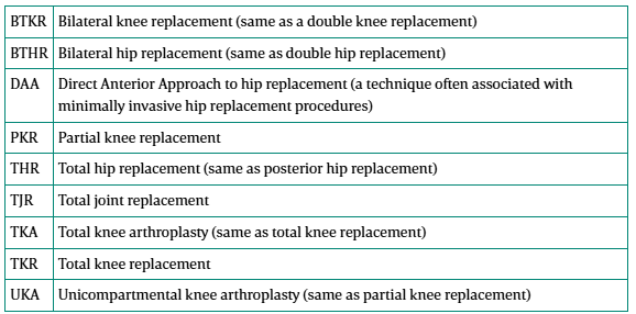
Total Knee Replacement (TKR)/Total knee arthroplasty (TKA)
All of the damaged joint, including bone, cartilage, and osteophytes are removed and replaced with artificial components.
3 replacement components: tibial prosthesis, femoral prosthesis, and an artificial patella
Partial Knee Replacement (PKR)/Unicompartmental Knee Arthroplasty (UKA)
When the healthy bone is left in place and only the diseased parts are resected and replaced
Total Hip Replacement (THR) - commonly approached from the posterior aspect of the joint. The incision is made at the upper thigh/buttock area, while patient is lying on their side.
Bilateral hip & knee replacement (BTHR, BTKR) - two paired structures such as two hips or two knees. Having both replaced during a single same-day operation. Contrast would be unilateral joint replacement, each being replaced during separate surgical sessions.
Joint-related Acronyms:
Body Directions:
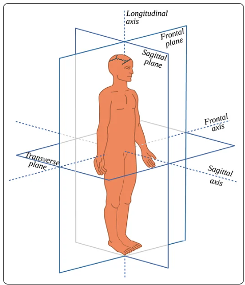
Anterior/Ventral - toward the front of the body or body part (front view)
Opposite: Posterior/Dorsal
Posterior/Dorsal - toward the back of the body or body part (back view)
Opposite: Anterior/Ventral
Medial - toward the midline of the body (side view)
Opposite: Lateral
Lateral - toward the outside of the body away from the midline (side view)
Opposite: Medial
Dorsoradial - both dorsal and radial in direction
Inferior - Something below (feet are inferior to the knees)
Opposite: Superior
Midline - an imaginary vertical line that divides the body equally in half
Prone - Lying face down
Opposite: Supine
Radial - Structures closer to the radius
Opposite: Ulnar
Superior - Something above (head is superior to neck)
Opposite: Inferior
Supine - Lying face up
Opposite: Prone
Transverse - Lying in a crosswise direction (horizontal)
Distal - The direction away from the origin of the structure
Opposite: Proximal
Volar - relating to the palm of the hand or the sole of the foot
Valgus - This refers to angulation (or bowing) within the shaft of the bone or at a joint in the coronal plane. Whenever the distal end of the long bone is pointing outward or is more lateral, it's called valgus
Varus - This refers to angulation (or bowing) within the shaft of a bone or at a joint in the coronal plane. Whenever the distal part of the long bone is pointing inward or is more medial, it Is called varus.
Ventral - The front or lower side
Proximal - Closer to the origin of the structure
Opposite: Distal
Musculoskeletal Terms :
A1 pulley - Band of tissue holding flexor tendon closely to finger bones, near palm
Arthrocentesis - Surgical puncture of a joint for aspiration of fluid
Arthrodesis - surgical fixation of a joint
Aspiration - Withdrawal of fluid or tissue from the body, typically using a needle
Calcaneus - Large bone of the heel
Carpal - Pertaining to the write bones
Concentric reduction - Putting a dislocated joint back to its normal position allowing the joint to move freely
Chondral - Pertaining to cartilage
Coccygeal - pertaining to the coccyx
Connective - Tissue connecting or binding together
Crystalloid (solution) - used to increase intravascular volume caused by loss of fluid during surgery
Dactylic - Pertaining to finger or toe
Femoral - pertaining to the femur (thigh bone)
Hallux - refers to the big toe
Hallux Rigidus - refers to a stiff big toe, usually due to osteoarthritis and bone spur formation of the MTP joint
Hallux Valgus - progressive deformity of the metatarsophalangeal (MTP) joint and is the most common foot deformity. Over time this becomes swollen and painful making it difficult to walk. The joint will gradually sublux, pulling the phalanges in (adduction) and pushing out the MTP joint (abduction). The boney prominence on the outside of the foot at the metatarsal head is referred to as a bunion.
Hammer Toe - deformity of the toe, commonly caused by arthritis or wearing ill-fitting shoes. The toe or toes curl downward instead of laying flat. Commonly found in second and third toes.
Iliac - Pertaining to the ilium
Metacarpal - Long bones of the hand that form the skeletal structure of the palm
Metatarsal - area of the foot between the angle and toes, five bones extending from tarsus to phalanges
Osteoblast - cells that form bone tissue
Patellar - Pertaining to the patella (kneecap)
Phalanges - Bones of the fingers and toes
Sternotomy - Surgical incision of sternum
Tarsal - pertaining to the tarsal bones in the foot
Tuberosity - large prominence on bone for attachment of muscles or ligaments
Tendon Abbreviations:
