HILLEGAS CHAPTER 1
Chapter 1
The respiratory system
The muscles of ventilation (inspiration + expiration)
Pulmonary ventilation
The respiratory system = Bony thorax + muscles of ventilation + upper and lower airways + pulmonary circulation (interaction)
Main functions:
gas exchange
fluid exchange
keep blood reserves
filtration
metabolism
The muscles of ventilation
Ventilation/breathing = inspiration + expiration.
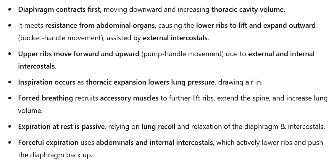
Inspiration: Muscles of the thorax and abdomen create changes in volume → There is a reduction of intrathoracic pressure (ITP)
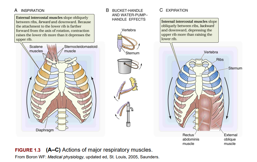
They increase the volume of the thoracic cavity by producing bucket-handle and pump-handle movements of the ribs and sternum = ITP is below atmospheric pressure, forcing air into the lungs to help normalize pressure differences.
Active processes of inspiration at rest = diaphragm + external intercostals.
Forceful inspiration during exercise or cardiopulmonary distress → accessory muscles assist (sternocleidomastoid, scalenes, serratus anterior, pectoralis major and minor, trapezius, and erector spinae muscles)
A more detailed look at the muscles
Muscles for inhalation
Diaphram
The primary muscle of inspiration, forms the floor of the thorax, separating thoracic and abdominal cavities.
Divided into right and left hemidiaphragm.
Right hemidiaphragm is stronger due to liver support; the left is more prone to rupture/hernia.
It is composed of sternal, costal, and lumbar portions converging into the central tendon.
Three major openings:
Vena caval opening (inferior vena cava)
Esophageal opening (esophagus & gastric vessels)
Aortic opening (aorta, thoracic duct, azygos veins)
The phrenic nerve (C3–C5) controls diaphragm contraction.
The resting position is arched high in the thorax; movement varies with body position, obesity, and organ size.
Inspiration:
The diaphragm contracts, pulling the central tendon down & forward.
Flattens, increasing thoracic volume & decreasing pressure.
Abdominal cavity volume decreases, increasing intra-abdominal pressure.
“if you don’t remember how pressures work, think about what happened to the titan at the bottom of the ocean, the pressure surrounding it decreased, and pressure in the titan increased kaboom“
Lower ribs elevate, and the sternum moves forward due to resistance from abdominal muscles.
The right hemidiaphragm meets more resistance (liver), making it stronger than the left (stomach).
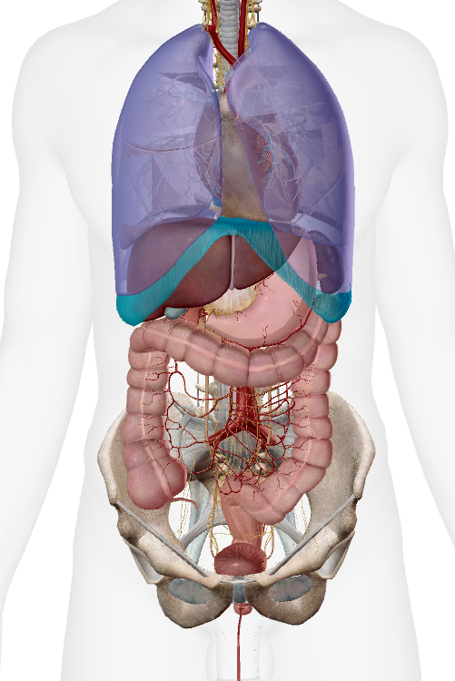
In PT with COPD
expire ability is compromised → diaphragm flattens due to hyperinflated lungs
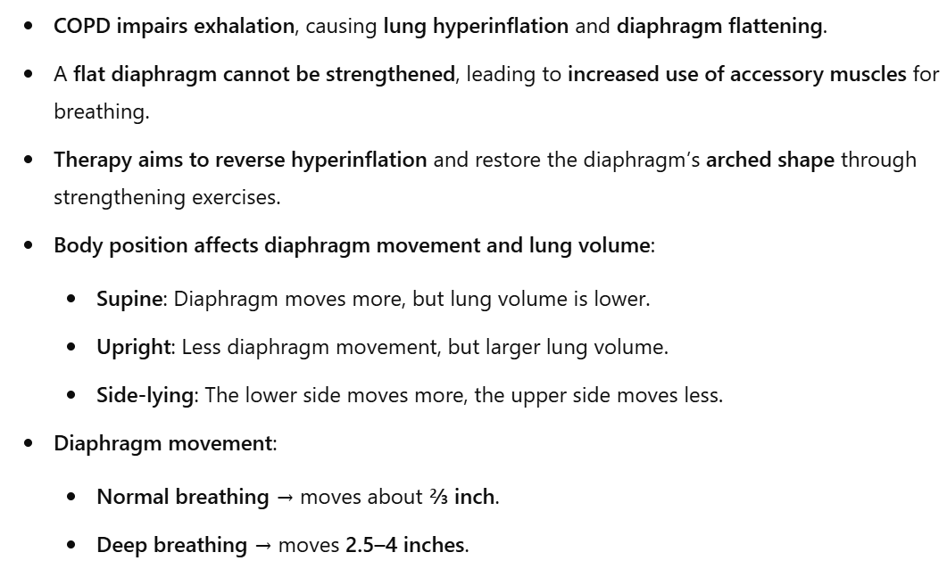
Supine position → is generally preferred for diaphragmatic strengthening because:
✅ Gravity is reduced, allowing the diaphragm to move more freely.
✅ Increased diaphragm excursion, meaning it can contract and relax more effectively.
✅ Less reliance on accessory muscles, and better breathing mechanics.
External Intercostal muscles
11 on each side of the sternum
role: contraction elevates the rib, therefore expanding the chest
Accessory muscles
I know them, just their function in inspiration
1) sternocliedomastoid
For this muscle to facilitate inspiration, the head and neck must be held stable by the neck flexors and extensors.
This muscle is a primary accessory muscle and elevates the sternum, increasing the anteroposterior diameter of the chest.
2) scalene
elevate first and second rib
3) Upper trapezius
This muscle assists with ventilation by helping to elevate the thoracic cage.
4) Pec major and minor
Pec major → When the arms and shoulders are fixed, by leaning on the elbows or grasping onto a table, the pectoralis major can use its insertion as its origin and pull on the anterior chest wall, lifting the ribs and sternum, facilitating an increase in the anteroposterior diameter of the thorax.
Pec minor → assists in forced inspiration by raising the ribs and increasing intrathoracic volume.
5) Serratus anterior and rhomboid
The serratus can only be used as an accessory muscle in ventilation when the rhomboids stabilize the scapula in adduction.
The action of the rhomboids fixes the insertion, allowing the serratus to expand the rib cage by pulling the origin toward the insertion.
6) Latissumus Dorsi
The posterior fibers of this muscle assist in inspiration as they pull the trunk into extension.
7) Serratus posterior superior
It assists in inspiration by raising the ribs to which it is attached and expanding the chest.
8) Thoracic erector spinae
It raises the rib cage to allow greater expansion of the thorax.
Muscles of expiration
1) abdominal muscles
These muscles work to raise intraabdominal pressure when a sudden expulsion of air is required in maneuvers, such as hung and coughing. Pressure generated within the abdominal cavity is transmitted to the thoracic cage to assist in emptying the lungs.
2) internal Intercostal muscles
11 muscles on each side of the sternum
The posterior aspect on the internal intercostal muscles is termed the interosseus portion and depresses the ribs to aid in a forceful expiration.
The inter cartilaginous portion of the internal intercostals elevates the ribs and assists in inspiration.
Pulmonary ventilation
Pulmonary ventilation = aka breathing
Definition: “PV is the process in which air is moved in and out of the lungs”
Inspiration = is an active process at rest and during exercise
it involves contraction of the diaphragm and external intercostal muscles.
Expiration = at rest, it is a passive process and achieved through the elastic recoil of the lung and relaxation of the external intercostal and diaphragm muscle
Pleurae
Summary in bullet points from book:
Treatment:
Chest tube insertion → Drains fluid/air & restores negative pressure.
Thoracocentesis → Needle aspiration of excess fluid.
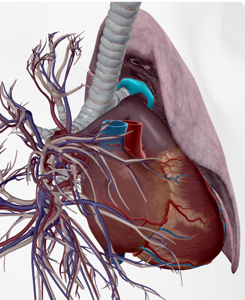
Lungs
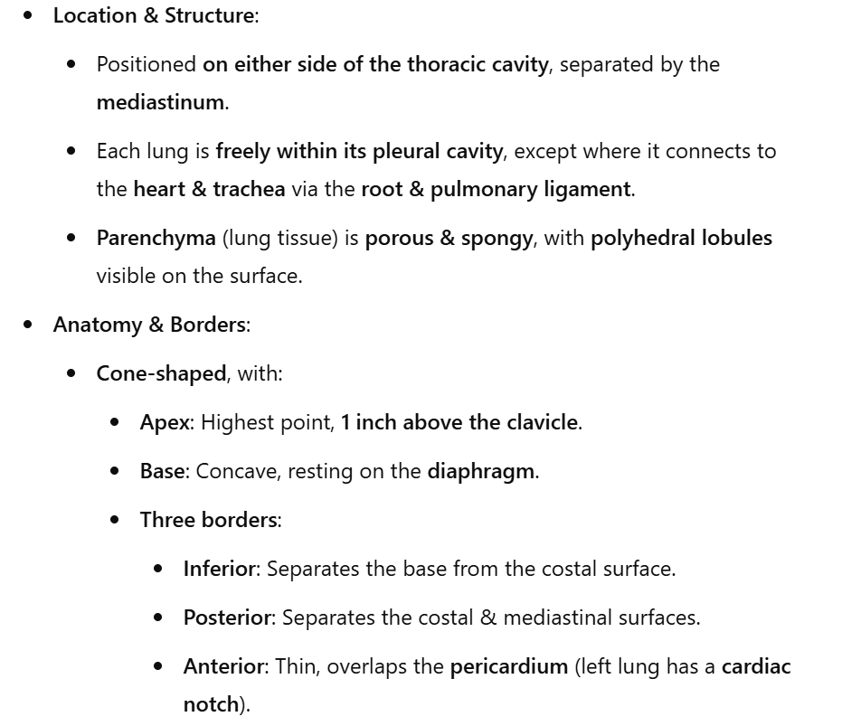
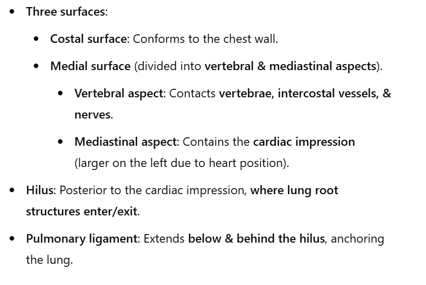
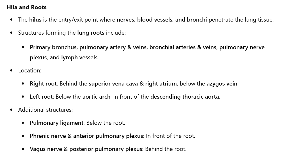
Hila and roots (lungs)
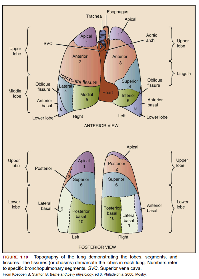
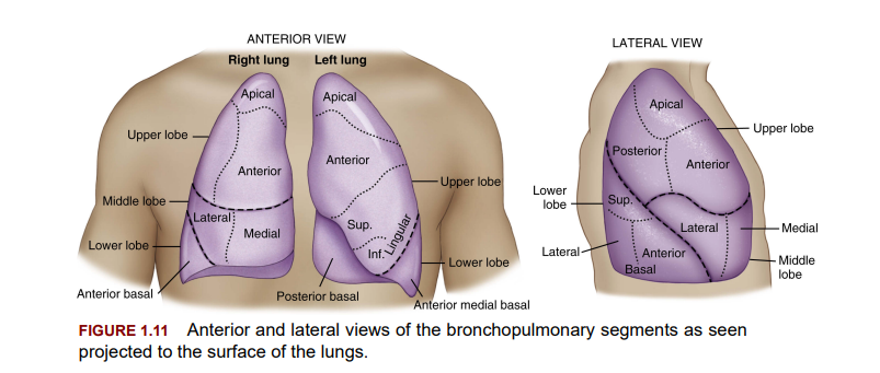
Lobes, fissures and segments (lungs)
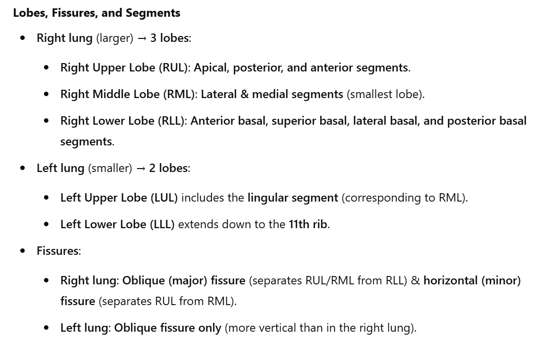
Only view of anterior side (for posterior look at book image below)
In the right side, there are 3 lobes
in colors you find the segments
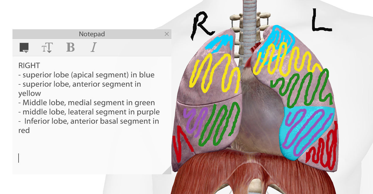
In the left lung, there are 2 lobes
In colors you find the segments
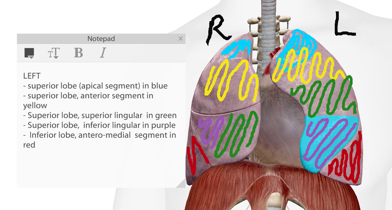
Fissures:
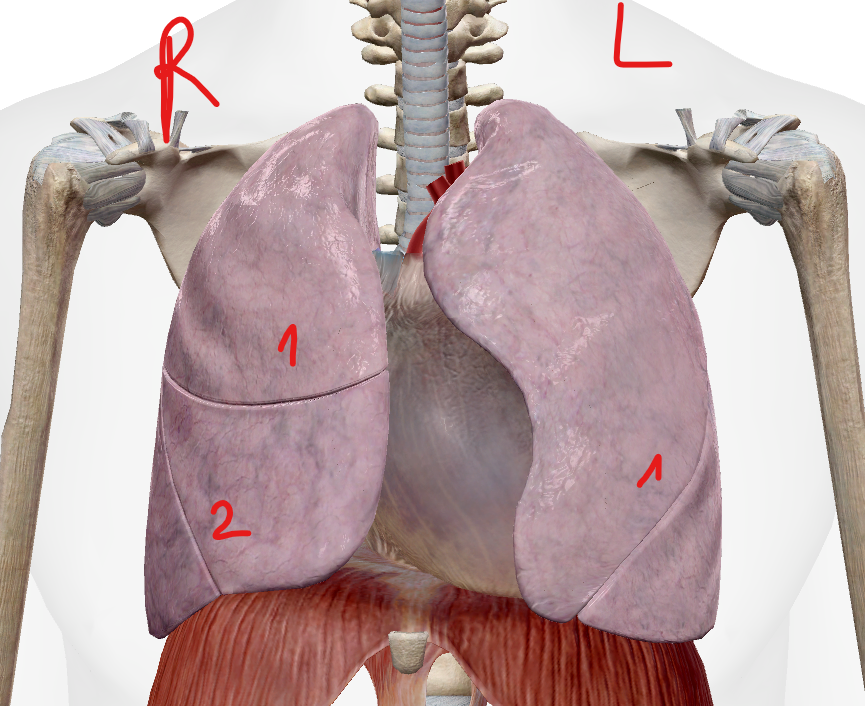
View anterior and posterior with nomenclature (important)
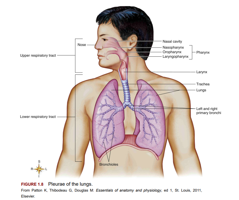
Upper respiratory tract
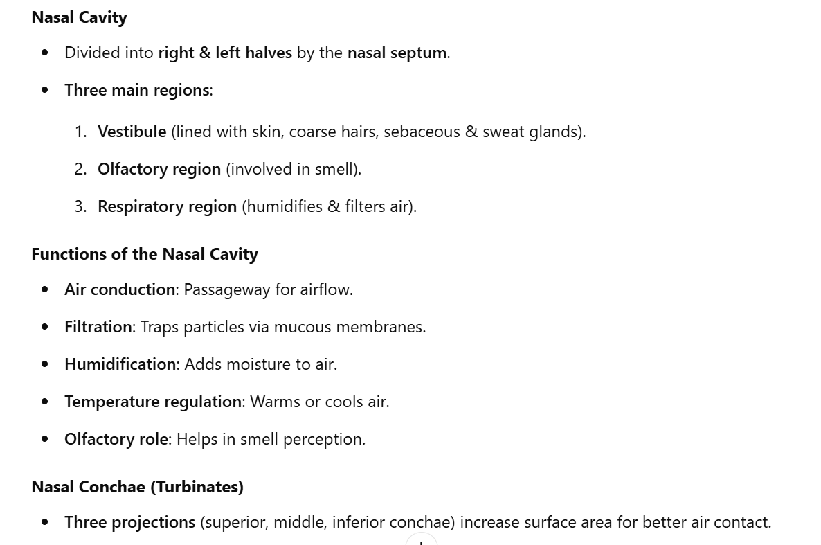
Nose
Pharynx
Length: 5 to 6 inches long, located posterior to the nasal cavity.
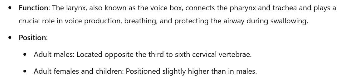
Position: Extends from the base of the skull to the esophagus, corresponding to the line from the sixth cervical vertebra to the lower border of the cricoid cartilage.
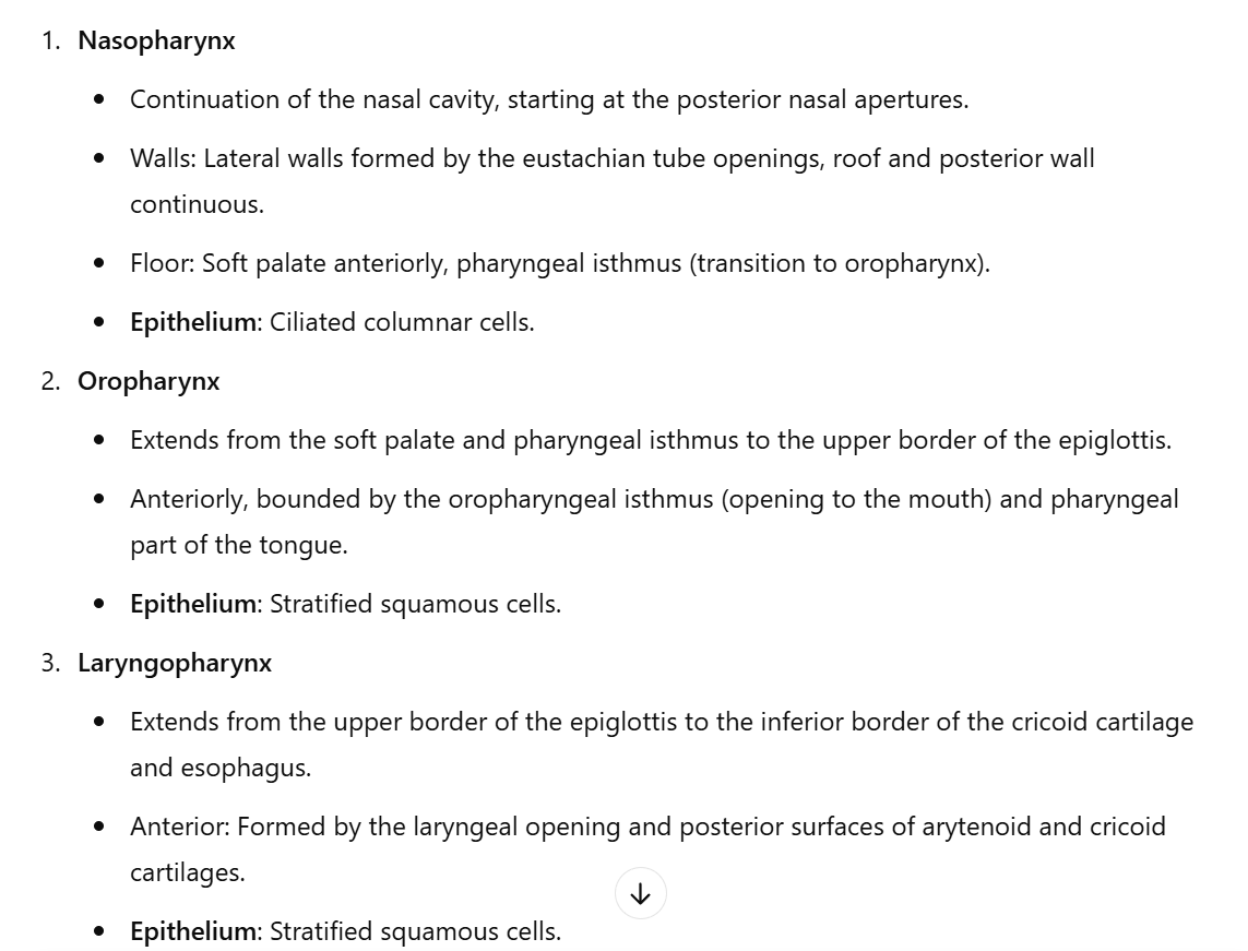
Divided into three parts:
Nasopharynx
Oropharynx
Laryngopharynx
Larynx
Larynx aka voice box
How is the voice created? → Sounds are created when expired air passes over contracting vocal cords, causing them to vibrate
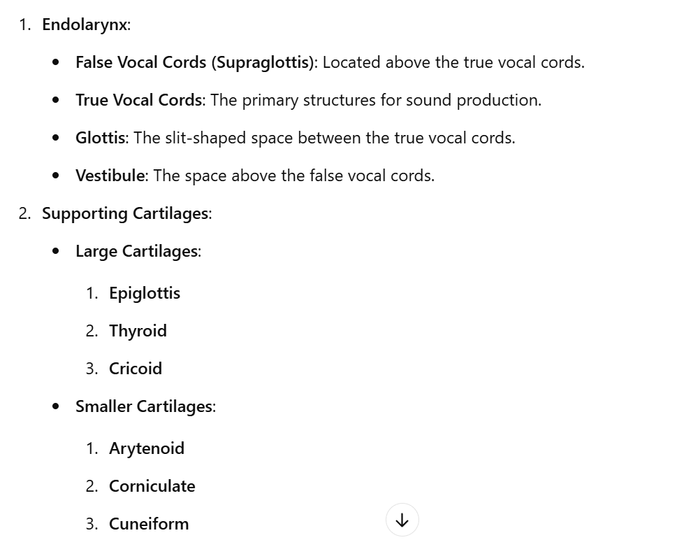
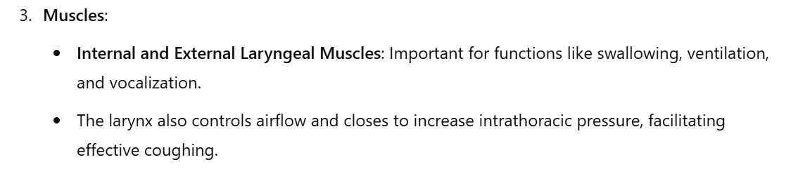
Anatomy of the Larynx
Low respiratory tract
The lower respiratory tract starts from the true vocal cords in the larynx down to the alveoli in the lungs.
It is divided into two primary sections:
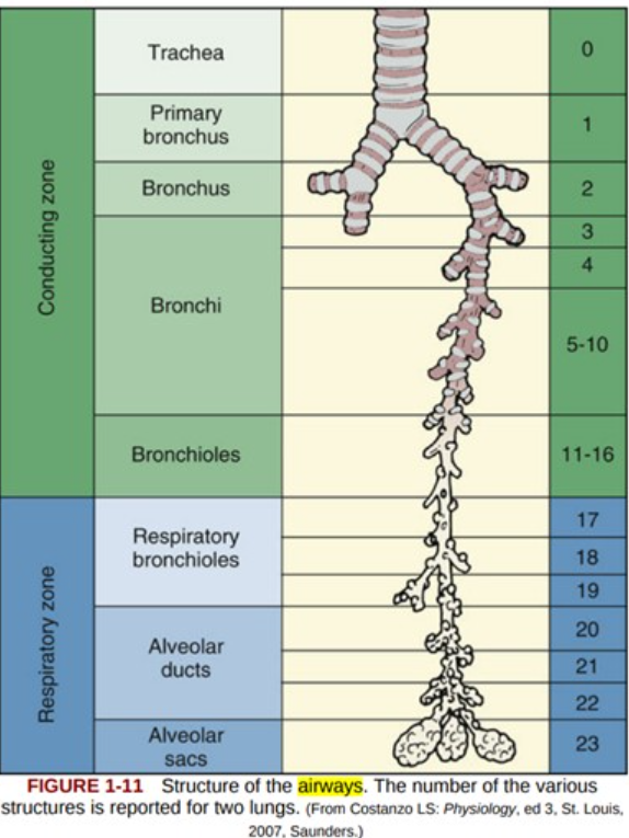
1) the tracheobronchial tree (conducting airways)
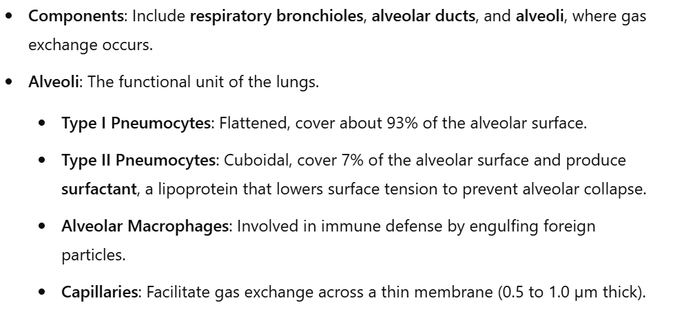
2) the acinar or terminal respiratory units.
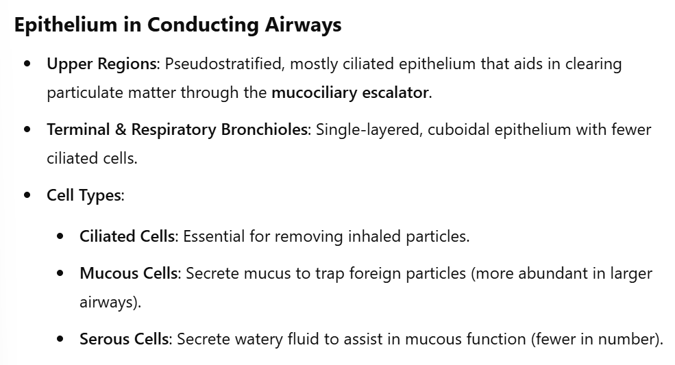
The tracheobronchial tree + conducting airways
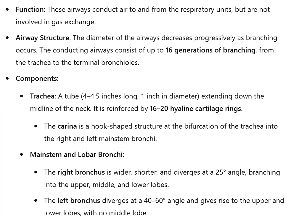
Segmental and Subsegmental Bronchi: These continue to branch from the lobar bronchi, leading to more specialized regions of the lungs.
Terminal respiratory acinar units