Cell Structure & Microscopy Review
Prokaryotes vs. Eukaryotes:
Similarities:
all have cytosol → a semifluid jellylike substance inside the cell.
Contain chromosomes → carry genes in the form of DNA.
Have ribosomes → make proteins based on the genes
Both have a cytoplasm → in eukaryotes, this means the region inside the cell that is between the nucleus and membrane.
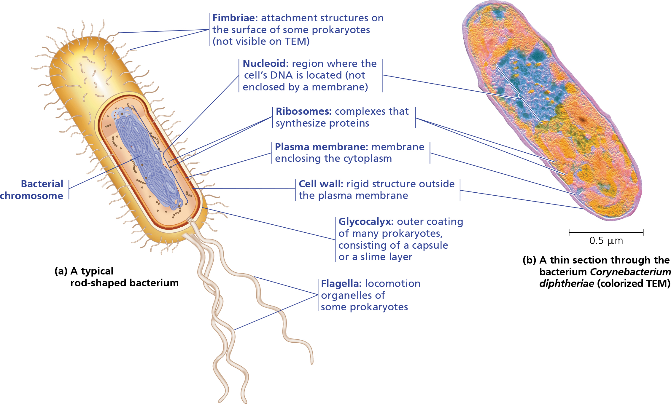
Prokaryotes:
DNA is concentrated in a region called the nucleoid.
The nucleoid is non membrane enclosed.
DNA is in the form of circular chromosomes.
Was the early form of cells before eukaryotes.
Examples of prokaryotes are bacteria and archaea.
Is known to be “simpler” due to it lacking an actual nucleus.
Are much smaller, only 1-5 micrometers in diameter.
Eukaryotes:
Most DNA is concentrated in the nucleus, which is bounded by a double membrane.
DNA is in the form of rod shaped chromosomes.
More complex since they have many membrane bound organelles that prokaryotes don’t have.
Examples of eukaryotes are plant and animal cells.
Are generally much larger than prokaryotes, 10-100 micrometers in diameter.
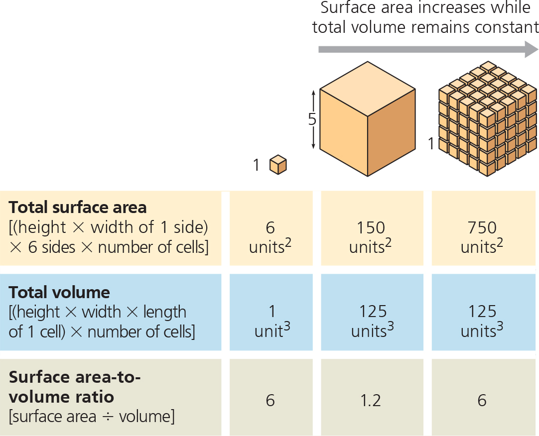
Surface Area & Volume of a Cell Relationship:
As a cell increases in size, it’s surface area grows proportionally less than its volume.
This means that the need for nutrients increases proportionally more as the surface area increases → lower surface area to volume ratio.
Multicellular organisms grow by creating more cells.
This means that while the surface area increases, the volume stays constant. This creates a higher surface area to volume ratio.
An increase in surface area allows for more reactions.
 Cell parts & Functions:
NOTE: organelles only found in the animal cell are denoted in RED. Organelles only found in the plant cells are denoted in GREEN. Organelles found in both plant & animal cells are denoted in BLUE.
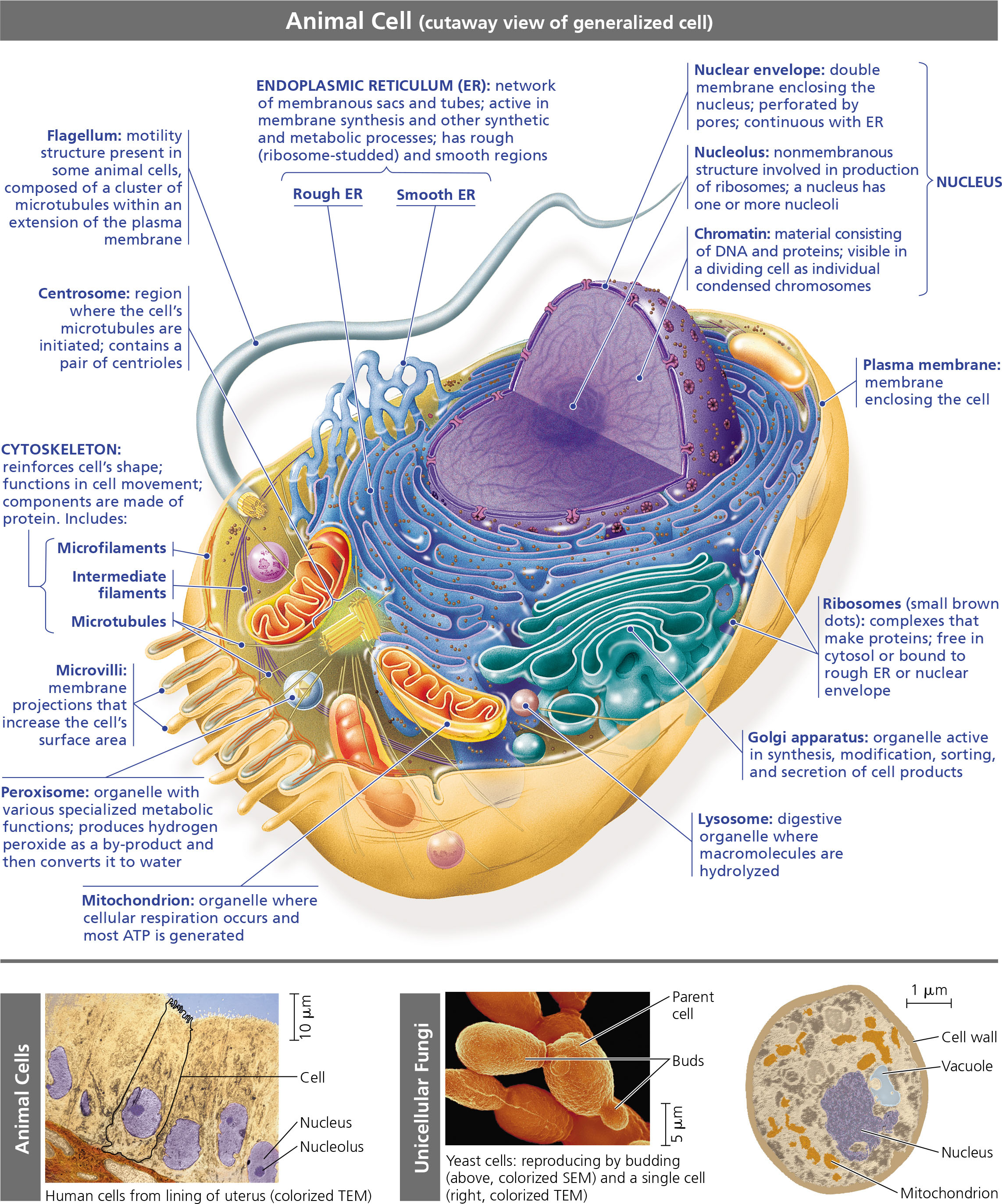
Animal Cell:
 Plant Cell:
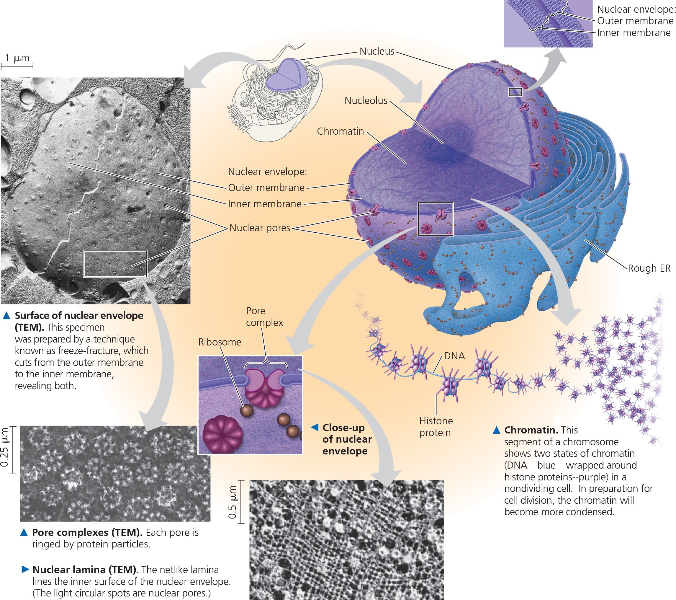
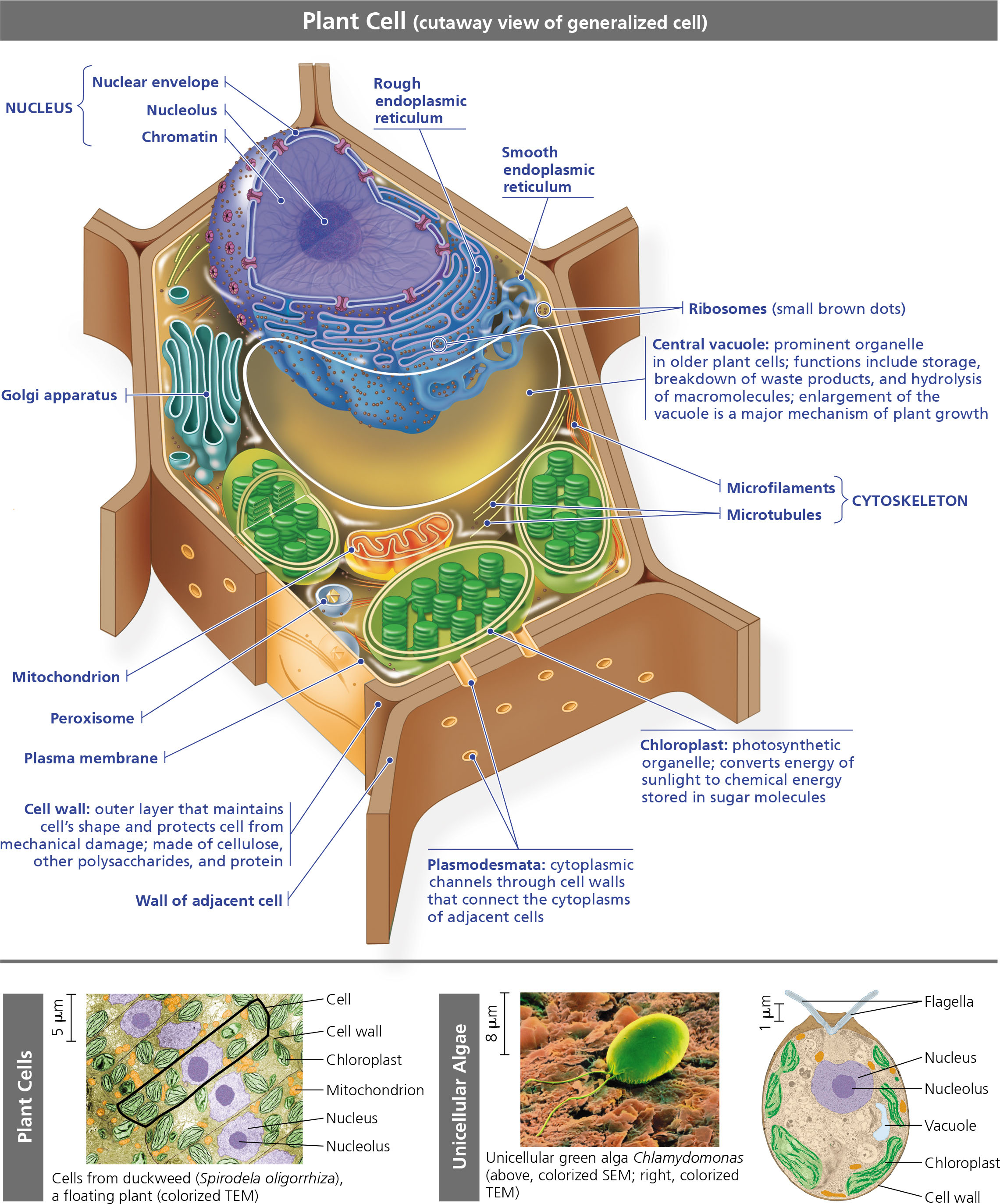 Nucleus:
Is the information center of the cell, contains MOST genes (not ALL!).
Is enclosed by the nuclear envelope
the nuclear envelope is the double membrane that surrounds and protects the nucleus.
It is attached to the endoplasmic reticulum.
Lined with pores called pore complexes that regulate the entry and exit of proteins and RNA.
Nuclear lamina and the nuclear matrix help hold the shape of the nuclear envelope.
Nuclear lamina → array of protein filaments
Nuclear matrix → framework of protein fibers.
DNA is organized in chromosomes; carries genetic information.
Chromosomes contain 1 long DNA molecules with small proteins called histones.
These DNA and proteins together are called chromatin.
Nucleolus → the primary structure inside the nucleus.
Synthesizes ribosomal RNA (rRNA)
Proteins + rRNA = ribosomes, which exit the nucleus via the pores.
Directs protein synthesis by creating mRNA and sending it out to the ribosomes.
The mRNA has instructions to make the proteins, so the mRNA gives the instructions to the ribosomes.
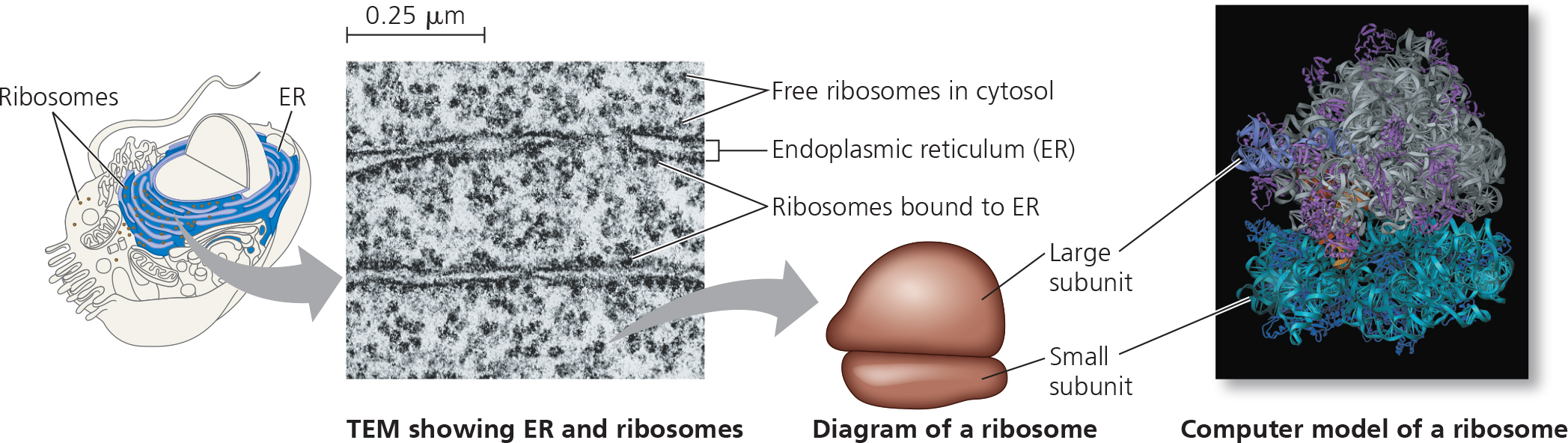
 Ribosomes:
Carries out protein synthesis.
Technically not organelles.
Cells w/ high protein synthesis has more ribosomes, human pancreatic cells have 10 million ribosomes in each cell.
Build proteins in 2 cytoplasmic regions.
Free ribosomes are suspended in the cytosol.
Most free ribosomes synthesis proteins for function in the cytosol, like enzymes that catalyze for sugar breakdown.
Bound ribosomes are attached to either the rough endoplasmic reticulum or the nuclear envelope.
Synthesize proteins for insertion into the membrane, packaging for lysosomes, & excretion.
 The Endomembrane System:
Includes the nuclear envelope, endoplasmic reticulum, Golgi apparatus, lysosomes, vesicles, and the plasma membrane.
carries out a variety of tasks:
Synthesizes proteins
Transports proteins into membranes & organelles or out of the cell.
Metabolism & movement of lipids.
Detoxification.
Related either through physical attachment or movement of vesicles (sacs of membranes).
Endoplasmic Reticulum:
Extensive network of membranes
Consists of a network of tubules & sacs called cisternae.
The ER membrane protects the interior compartment called the ER lumen.
is continuous w/ the nuclear envelope.
2 distinct types of the ER.
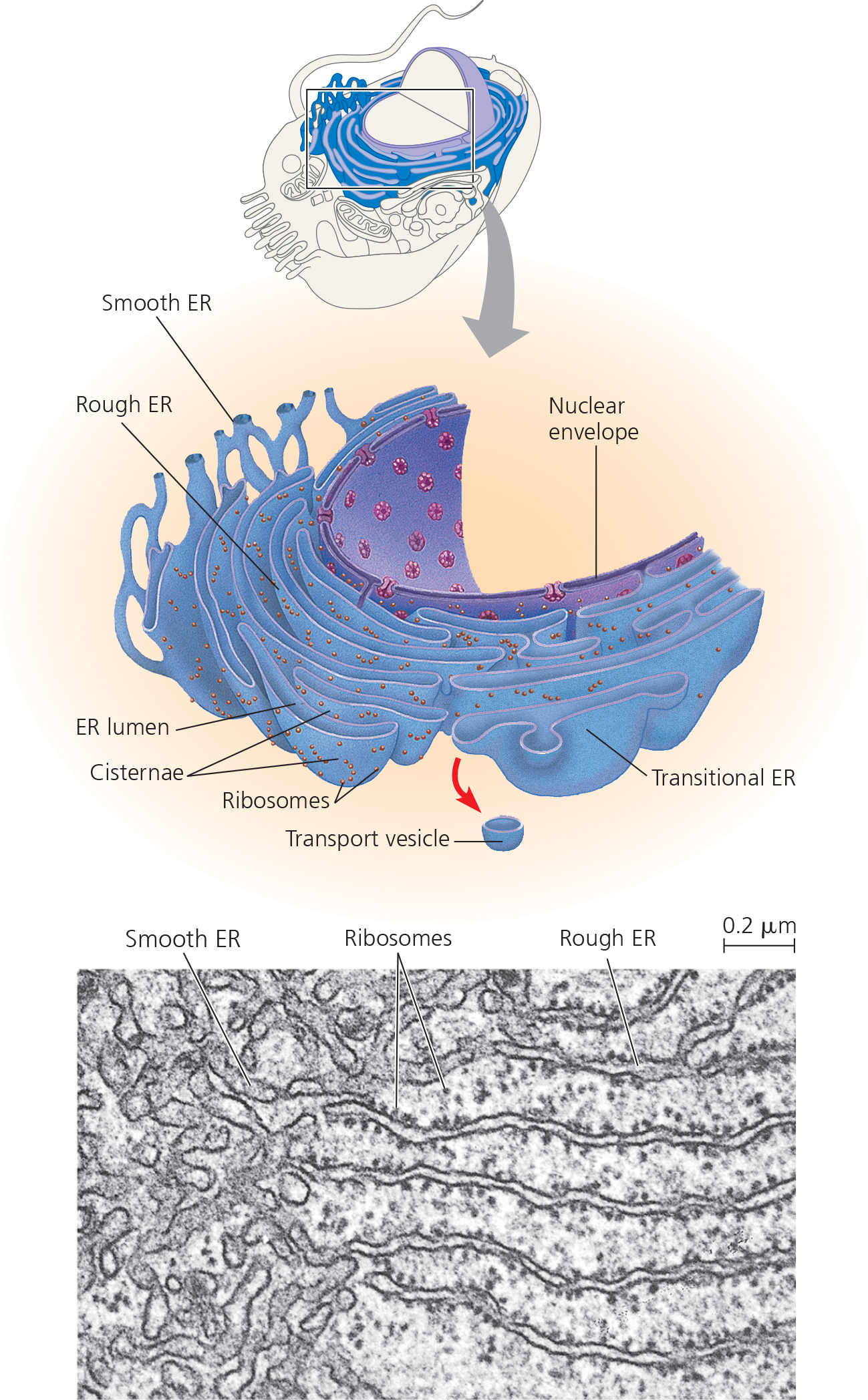
Smooth ER:
lacks ribosomes on the surface.
Enzymes in the ER synthesize lipids like oils, steroids, & phospholipids.
Sex hormones in animal cells are also synthesized through steroid synthesis.
Detoxifies toxins like alcohol, particularly in liver cells.
Adds hydroxyl groups to the drug molecules in order to flush them out.
As more barbiturates & toxins are introduced, the rate of detoxification increases, increasing tolerance for the toxins. ‘
Stores calcium ions.
Rough ER
Has ribosomes bounded on the surface.
Synthesizes secretory proteins by having a polypeptide chain threaded into the ER lumen.
Adds carbohydrates to the secretory proteins, creating glycoproteins.
Keeps the secretory proteins away from proteins in the cytosol.
The secretory proteins leave the rough ER in a region called the transitional ER wrapped in transport vesicles.
Also creates membrane phospholipids.
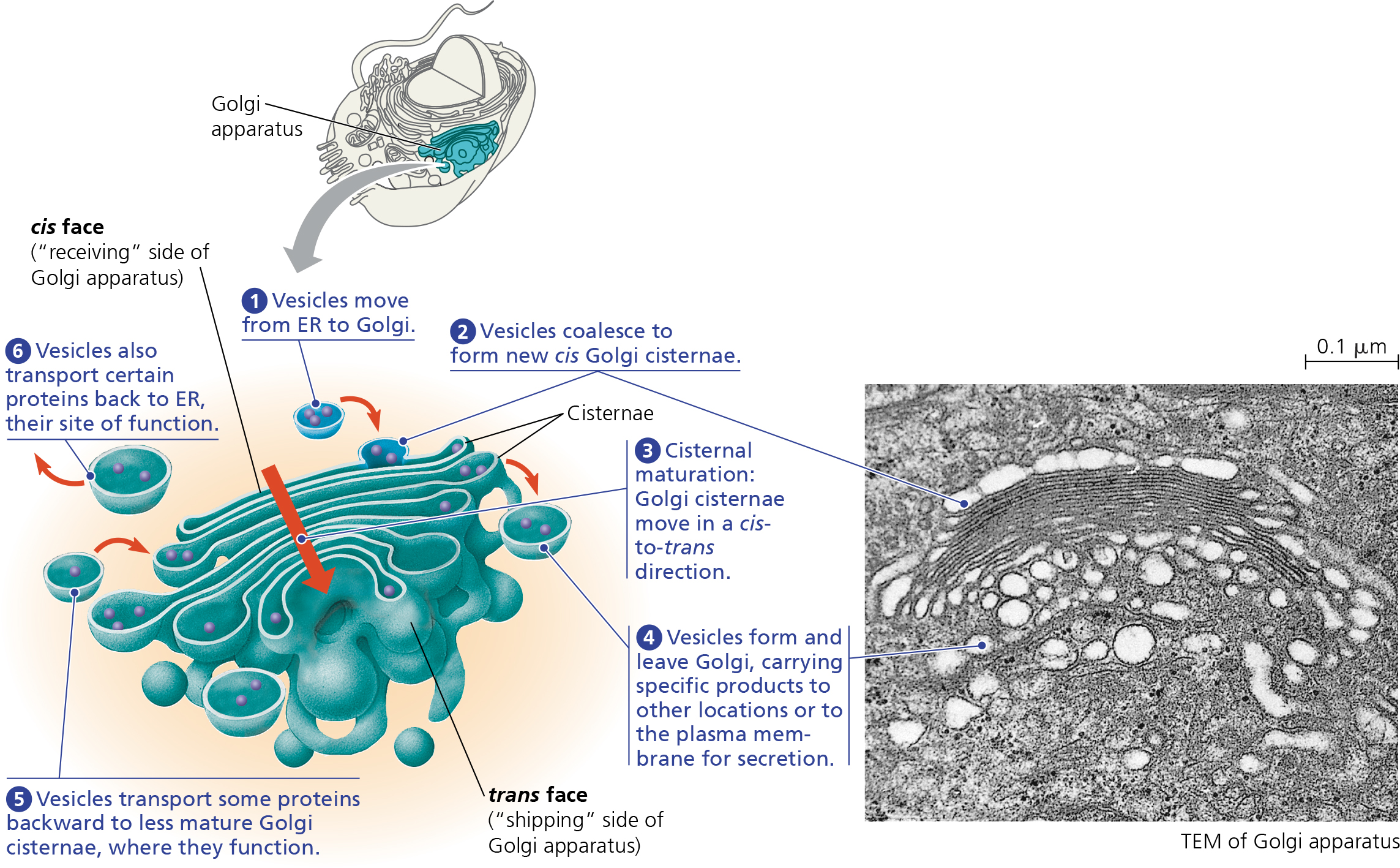
Golgi Apparatus:
Transport vesicles that contain the proteins go to the Golgi apparatus.
Is a warehouse for receiving, sorting, shipping the ER products.
Consists of a group of flattened membranous sacs called cisternae.
Transport vesicles from the ER enter the apparatus through the “cis” face.
cis face points towards the ER.
Vesicle membrane fuses w/ the cis face membrane & dumps the contents.
The contents then passes through all of the other faces inside, undergoing modification
Ex. glycoproteins from the ER have their carbohydrate components modified.
At the final trans face, the products are sorted & targeted.
Creates molecular identification tags, which act like zip codes.
The products leave by being put into another transport vesicle.
Manufactures some macromolecules, like pectins in plant cells.
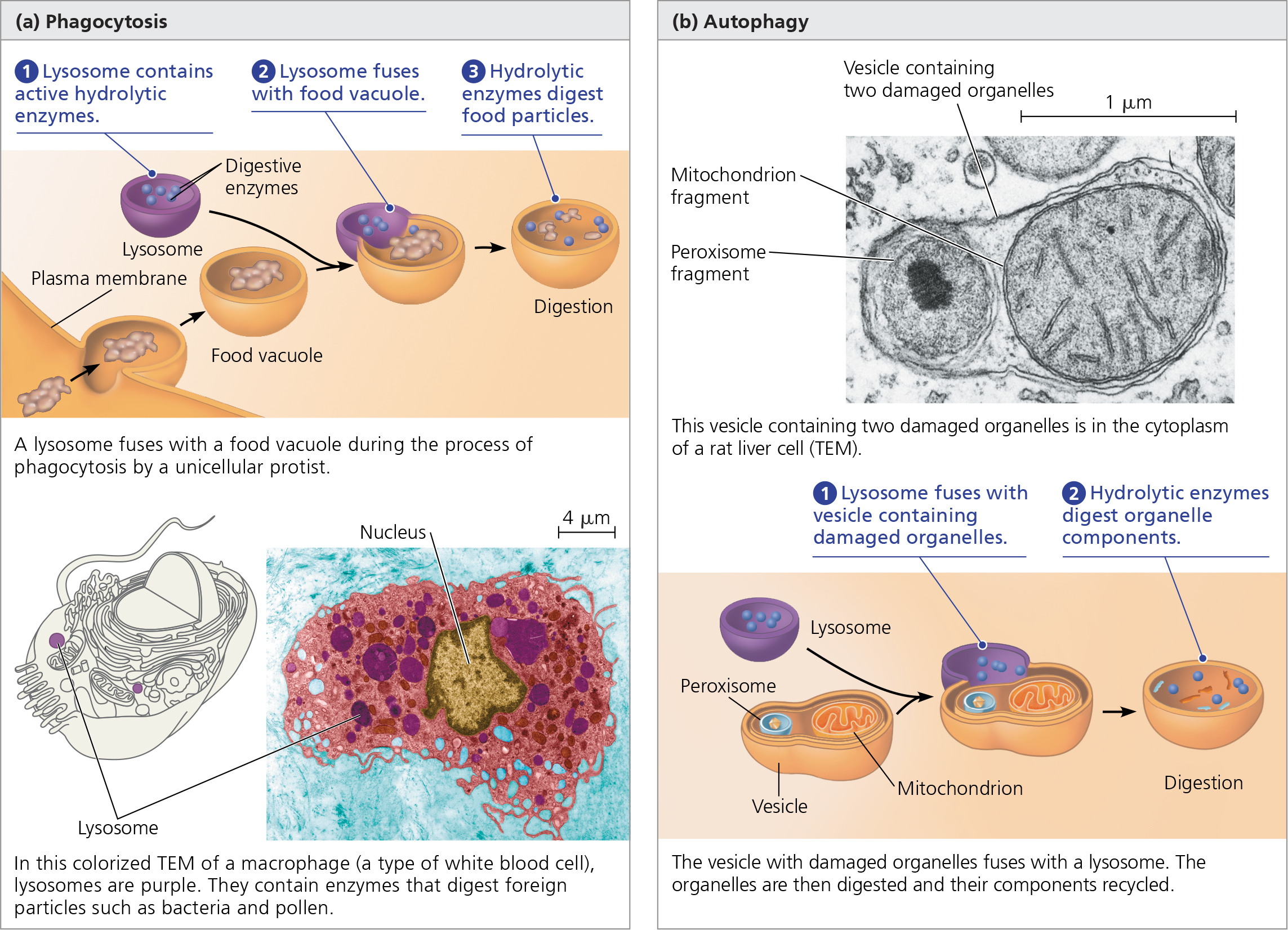
Lysosomes:
Membranous sac of enzymes that digest/hydrolyze macromolecules.
The enzymes & lysosome membrane is created by the rough ER & packaged into vesicles by the Golgi apparatus.
Fuses w/ the food vacuole containing the macromolecules that were obtained using phagocytosis.
Amoebas & other unicellular protists do this.
Recycles the cell’s own organic material in a process called autophagy.
A damaged organelle becomes surrounded in a vesicle & the lysosome fuses w/ this vesicle. The lysosomal enzymes then dismantle the membrane & the organic materials inside are released to the cytosol for reuse.
The cell continually renews itself due to this.
Lysosomal storage diseases, like Tay Sachs, lack the functioning enzymes present in lysosomes.
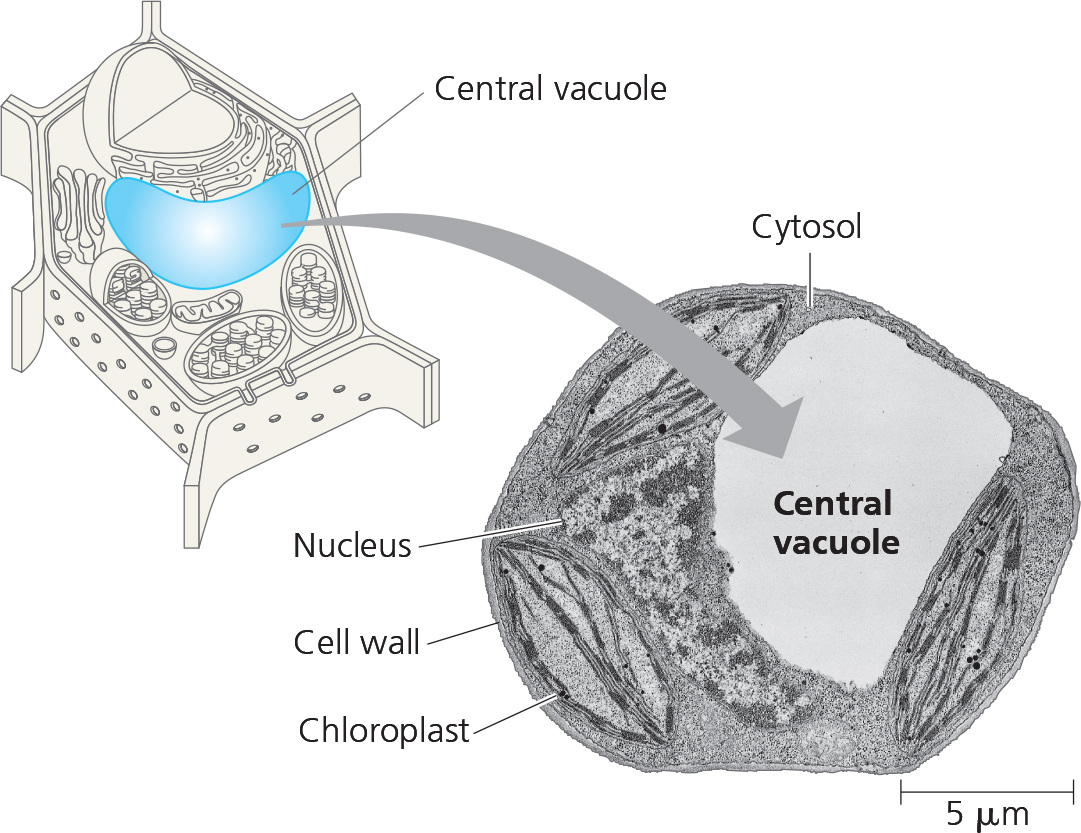
Vacuoles:
large vesicles derived from the endoplasmic reticulum & the Golgi Apparatus.
Has a membrane called the tonoplast.
Food vacuoles → stores food macromolecules obtained by the cell via phagocytosis.
Contractile vacuoles → pumps excess water out of a cell in order to regular a stable concentration of ions & molecules.
Plant cells have a large central vacuole.
The solution inside is called cell sap, is the plant’s repository of inorganic ions like potassium & chloride.
Plays a large role in plant cell growth, expands as the vacuole absorbs water.
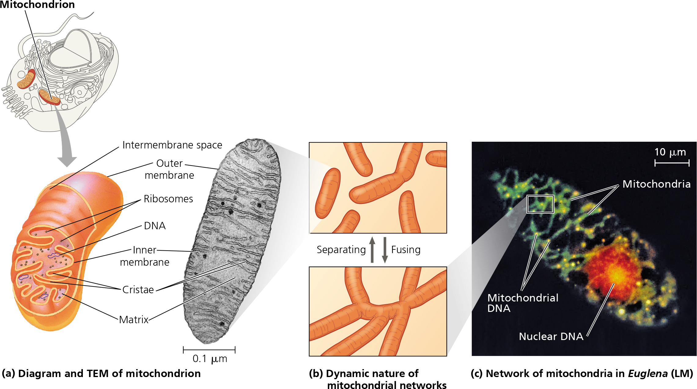
 Mitochondria:
Has a double membrane system with each of them having a phospholipid bilayer.
The inner membrane is folded inside, with the folds known as cristae.
The cristae gives the mitochondria a bigger surface area to increase the productivity of the cellular respiration.
Divides the mitochondria into 2 compartments - the intermembrane space & the mitochondrial matrix.
Contains many different enzymes.
The enzymes in the matrix catalyze the steps of cellular respiration, including the enzyme that makes ATP.
IS THE POWERHOUSE OF THE CELL!!!
 Chloroplasts:
Contain chlorophyll that function in photosynthesis.
Found in leaves & other green organs in plants.
Has a double membrane.
Has a membranous system in the form of flattened sacs called thylakoids.
Can be stacked into grana.
The fluid surrounding, called stroma, consists of chloroplast DNA and ribosomes.
Is part of a family of plant cell organelles called plastids.

Evolutionary Origins of Mitochondria & Chloroplasts:
Both display similarities w/ bacteria & prokaryotes, leading to the endosymbiotic theory.
Have a double membrane.
Have circular chromosomes.
Can grow & reproduces on their own.
A prokaryotic cell was engulfed by an eukaryotic cell, and ended up living inside it, making it an endosymbiont.
This prokaryote could have evolved to be a mitochondria or a chloroplast.
Peroxisomes:
Specialized metabolic component bounded by a single membrane.
Contains enzymes that adds hydrogen ions to oxygen, creating hydrogen peroxide as a byproduct which is toxic.
Enzymes break down the hydrogen peroxide byproducts by converting them to water.
Cytoskeleton:
Network of fibers that extend throughout the cytoplasm.
Gives mechanical support to the cell and maintain shape.
Some types of cell motility (changes in cell location & movement) involve the cytoskeleton.
Motor proteins interact w/ the cytoskeleton by “walking” to their destination.
Manipulates the plasma membrane.
Includes microtubules, microfilaments, & intermediate filaments.
Microtubules:
hollow rods made from globular proteins called tubulins.
Each tubulin protein is called a dimer.
Each end of a tubulin are slightly different from each other.
One end can accumulate & release tubulin dimers much faster, called the plus end.
Shape & support the cell, serve as tracks where organelles w/ motor proteins can move along the cell.
Guide vesicles from the ER to the Golgi apparatus and from the Golgi to the plasma membrane.
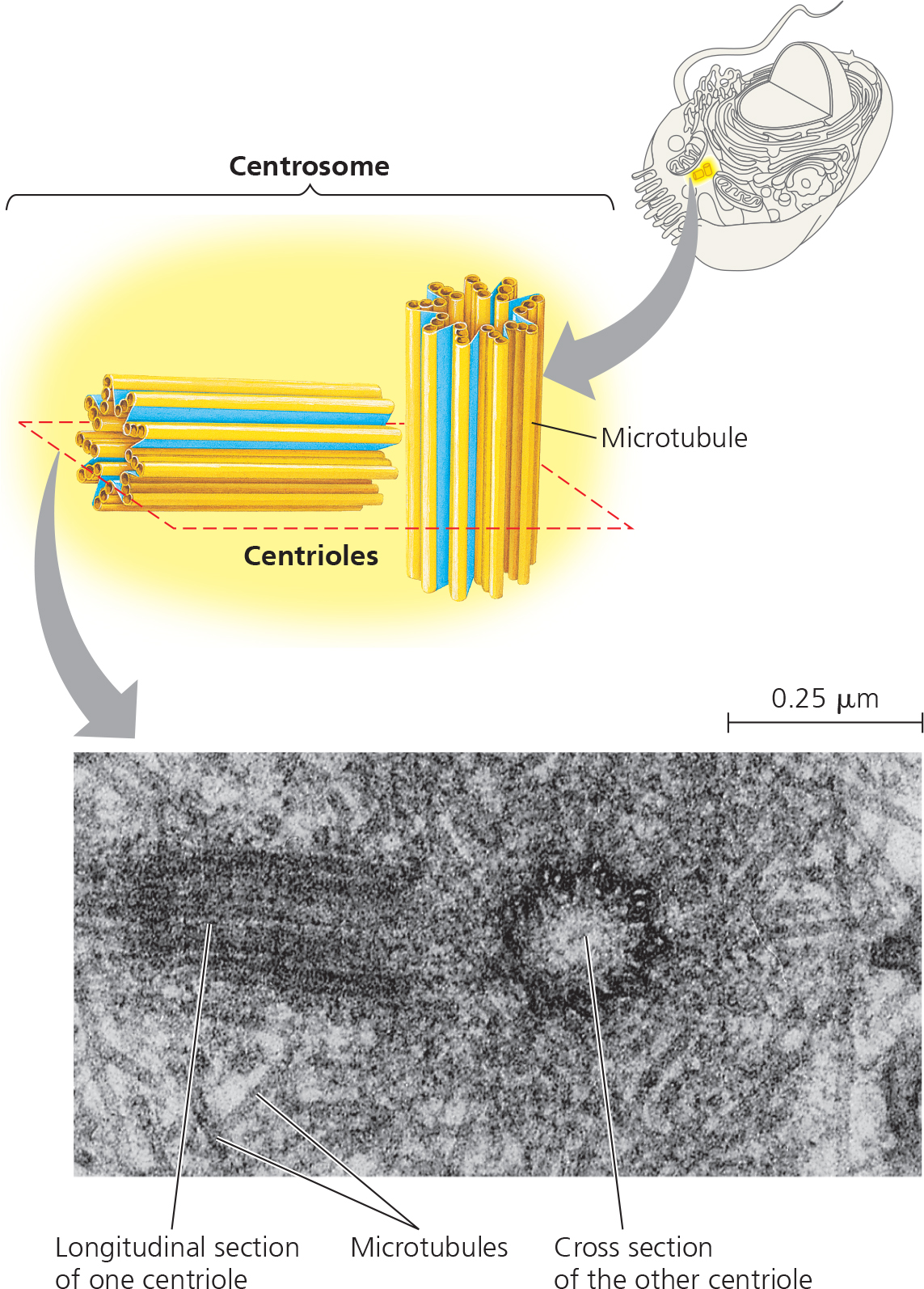
Centrosomes & Centrioles:
Microtubules grow out of a centrosome, region near the nucleus of animal cell.
Pair of centrioles, which consist of a ring of microtubules, are in the centrosome.
Cilia & Flagella
Cellular extensions that contain microtubules.
They can move fluidly.
Found in large batches on the cell surface.
 Microfilaments:
Thin solid rods made from actin.
Can form structural networks.
Mainly known for their role in cell motility.
Filaments interact to cause contraction in muscle cells.
Actin-protein interactions contribute to cytoplasmic streaming in plant cells.
Intermediate filaments:
Larger than microfilaments, but smaller than microtubules.
Are more permanent fixtures of the cells.
Play an important role in reinforcing the cell shape & fix the position of certain organelles.
Ex. nucleus sits in a cage made of intermediate filaments.
Cell Wall:
Extracellular structure of plant cells.
Protects the plant cell, maintains shape, & regulates flow of water.
Holds the plant cell against gravity.
Are much thicker than the plasma membrane, is made of cellulose.
Plant cells first have a primary cell wall, which is thin, but then adds a secondary thicker cell wall layer. These layers are held up by pectins.
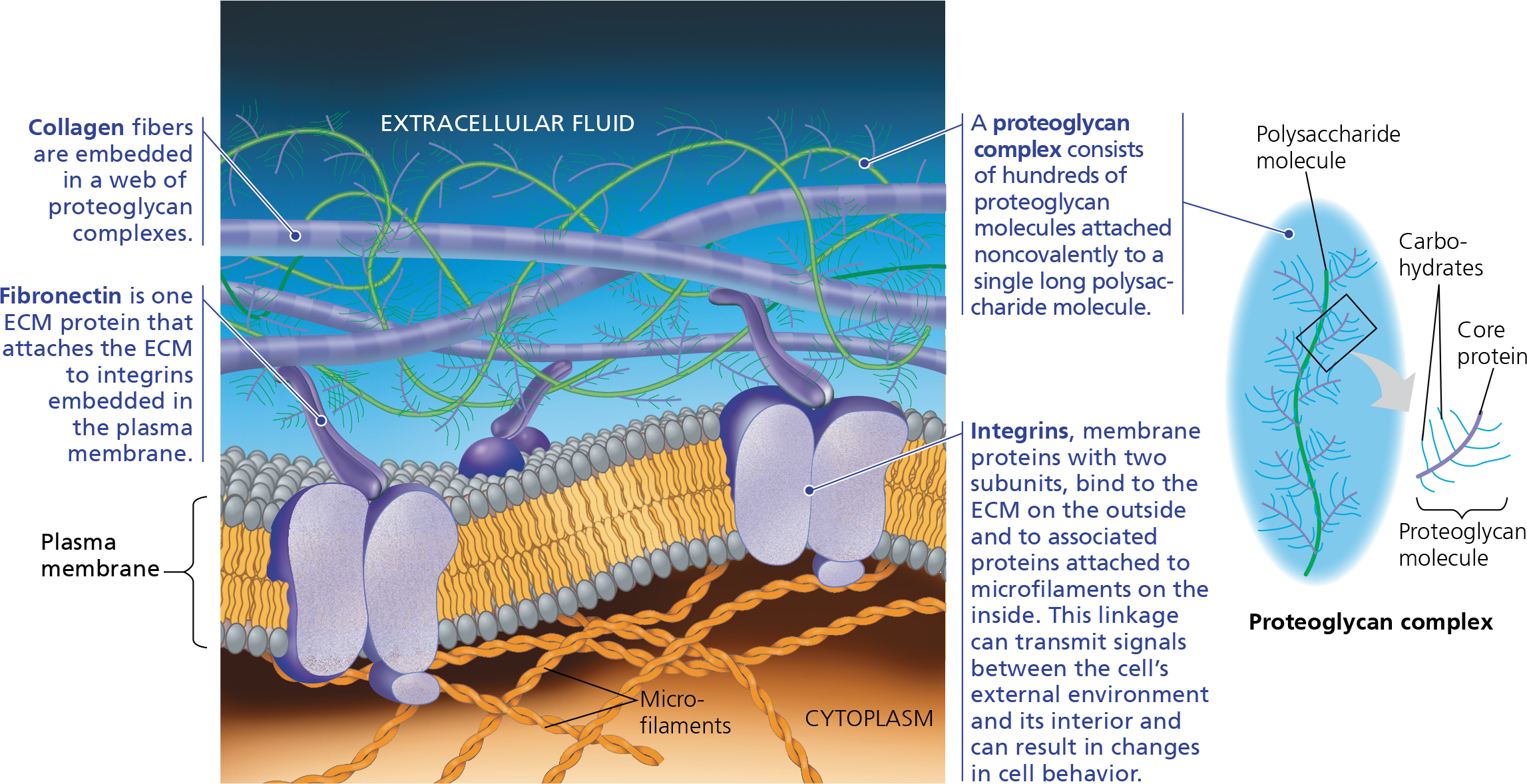
Extracellular Matrix (ECM)
Main components are glycoproteins & other carbohydrate containing molecules.
Collagen is the most abundant glycoproteins.
Bind to cell surface proteins called integrins that are built into the plasma membrane.
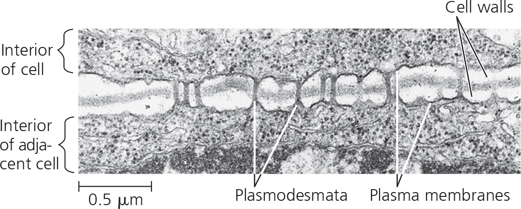
Plasmodesmata:
Many plant cell walls are filled with plasmodesmata channels that connect cells.
The channels are filled with cytosol.
These channels help cells connect to each other, which unifies the plant.
They allow for water and other solutes to travel from cell to cell.
 Parts of an Eye:
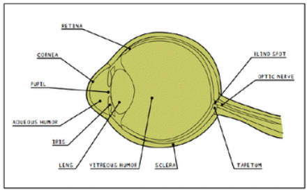
Retina:
Layer of light sensitive cells at the back of the eye.
Detects images focused by the cornea & the lens & converts them to nerve impulses w/ a system of rods & cones.
Connected to brain via optic nerve.
Cornea:
Tough clear covering over the iris & pupil that protects the eye.
Bends light as it enters the eye, first step in creating an image.
Pupil:
Hole that lets light into the inner eye.
Oval in cow’s eye.
Aqueous Humor:
Clear fluid that keeps the cornea round.
Iris:
Muscle that controls how much light enters the eye.
Suspended between the cornea and the lens.
Lens:
Clear & flexible structure that makes an image on the eye’s retina.
Can change shape, focus on faraway or near objects.
Vitreous Humor:
Jellylike substance that gives the eyeball its shape.
Sclera:
Thick & tough white outer covering of the eyeball.
Blindspot:
Place where the optic nerve leaves the retina. No light sensitive cells are found here.
Optic Nerve:
Bundle of nerve fibers that carry info from the retina to the brain.
Tapetum:
Colorful & shiny material that is located in the back of the eye. Allows for night vision (humans don’t have this).
Macula:
Region in the retina that gives fine pinpoints & peripheral vision.
Cell Theory:
Robert Hooke:
Was the first person to observe cells using a microscope that he designed himself.
Looked at a small piece of cork under light
Observed rows of boxes, similar to monasteries cells, so dubbed them “cells”.
Discovered that the cells had a lot of air in them, which is why cork floats.
Concluded that structure of cells determine function.
Lorenz Oken
Claimed that all organic & living things originate from cells.
Was the foundations of cell theory.
Jean Baptiste Lamarck
First to hypothesize a mechanism for how evolution occurs.
Life cannot exist if its constituent parts are not cellular tissue or is not formed by cellular tissues.
Mathias Schleiden & Theodore Schwann:
Unified cell theory
Claimed that all living things are made of cells, cell is the basic unit of life, & new cells originate from preexisting cells.
Rudolf Virchow:
Claimed that the current theory of spontaneous generation was wrong & claimed that cells came from preexisting cells in a theory called biogenesis.
Spontaneous generation → the belief that life comes from non life.
Example: If meat is left out it would generate maggots.
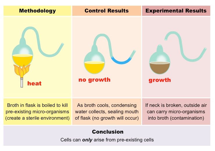
Louis Pasteur:
Disproved spontaneous generation
Had two flasks; in a regular flask, he boiled the liquid inside, while he did the same with a swan necked flask.
The regular liquid grew live bacteria, while the swan necked one didn’t.
 Parts of a Microscope & their Functions:
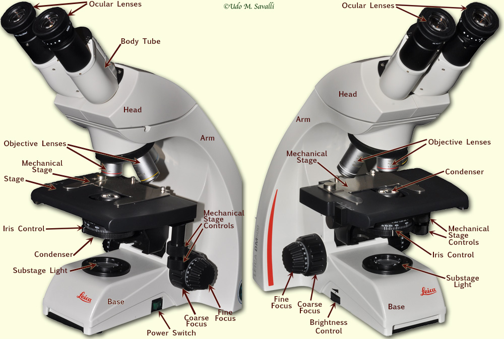
Eyepiece/Ocular Lenses → The user will look through the microscope here. The ocular lenses will magnify the image produced by the objective lenses so the user can see it.
Objective Lenses → Magnifies the specimen and produce a magnified real image. There are three objective lenses: scanning (red) , low (yellow) , high (blue) , and oil immersion (white). The scanning lens as 4x, the low power has 10x, the high power has 40x, and oil immersion has 100x.
Arm → This holds the microscope together. When you are carrying the microscope, always place one hand on the arm.
Nosepiece → Holds & rotates the objective lenses.
Diaphragm → controls & regulates the amount of light.
Body Tube → Located between the eye piece and the nose piece & separates the lenses.
Stage clips → Holds the slide in place.
Stage → This is where the slide is put and studied under.
Light source → This is where the light is coming from, in which the light is used to help us see the specimen.
Base → The bottom of the microscope. When carrying the microscope, put one hand at the bottom of the base.
Course adjustment knob → adjusts the focus of the lens a lot by moving the stage up and down vertically. DO NOT USE WHEN IN HIGH POWER.
Fine adjustment knob → adjusts the focus of the lens a little and allows us to see small details. USE ONLY IN HIGH POWER.
Lenses & Microscopes:
Microscope lenses are biconvex.
The objective lenses create a real image of the object in the tube, and the ocular lens creates an enlarged virtual image of the object.
Changing the curvature of the lens affects the focal length.
As curvature increases, focal length decreases.
As curvature decreases, focal length & magnification increases.
Maximum image magnification happens a smidge before the focal point.
If the object is at the focal point, no image will emerge.
If the object is before the focal point, a virtual enlarged image will be produced.
If the object is between F and 2F, an enlarged inverted image will be produced.
If the object is at 2F, a smaller inverted image will be produced.
Changing the lens is important as it prevents chromatic aberration.
Our eye can change lens to accommodate changes of incident angles caused by the motion of an object.
The farther away from an object, the thinner the lens.
The closer the object, the thicker the lens.
Magnification → ratio of object’s image size to real size.
Resolution → measure of clarity in the image.
It is the minimum distance 2 points can be separated and distinguished as 2 separate points.
Is inversely proportional to wavelength of light used, red light would have the best wavelength.
Higher numerical aperture on lens would have higher resolution.
Contrast → Difference in brightness between light and dark areas of an image.
Staining can increase contrast.
Higher magnification lowers contrast, which enhances resolution.
Using A Microscope - Practicum Prep:
Carry the microscope using one hand on the arm & one hand on the base.
Unwind the cord and plug it in & turn on the power switch.
Adjust the diaphragm:
Put illumination to lowest setting.
Fully open condenser diaphragm
Raise condenser using condenser focus on the side of the stage to the highest.
Set the condenser height.
Remove one of the eyepieces.
Look into the eyepiece into the white circle of light.
Close the condenser and reopen.
Put the eyepiece back.
Adjust the knobs for the eyepiece to 64 on the white line.
Create your slide or use the slide given:
Put the specimen on the center of the slide.
Place a drop of water on the specimen.
Place the coverslip by putting it at an angle and dropping it to prevent airbubbles.
Put the slide on the microscope by moving the slide clips.
Use the coarse adjustment to bring the slide all the way up & use the stage controls to bring the slide in the middle of the condenser lens.
Put the scanning focus in the center by sliding the nosepiece.
Look inside and use coarse and fine adjustments to look at the specimen. Then repeat for low and high power (only use fine for high power).
If the image becomes blurry restart at scanning power.
Draw the specimen.
Draw a circle using the funnel thingy.
Draw the specimen in pencil first and then use colored pencil.
Write a title and create labels with pen.
Write the total magnification (objective lens x 10) in pen.
Use a bracket to estimate the size of the specimen.
When done, put the lens back to scanning power and move the slide holder all of the way to the left.
Estimating size:
Scanning power → total field is 4,000-5,000 um
Low power → Total field is 2000 um
High power → total field is 400-500 um.
Calculating Specimen Size: Field of View/#of Cells
Calculating Magnification of Drawing: Length of drawing/specimen size