W.3.1 Thorax
THORAX VIDEO LECTURE WORKSHEET
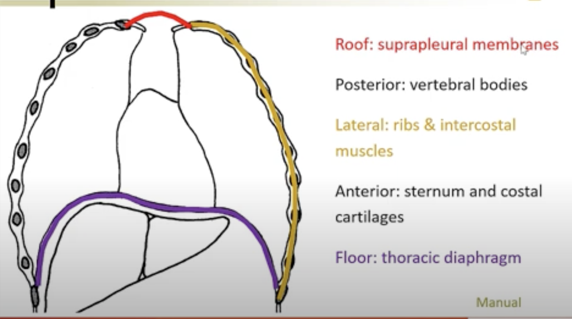
BOUNDARIES OF THE THORAX
Roof: Suprapleural membranes
Posterior: Vertebral bodies
Lateral: Ribs and intercostal muscles
Anterior: Sternum and costal cartilages
Floor: Diaphragm (not explicitly stated, but implied by structure)
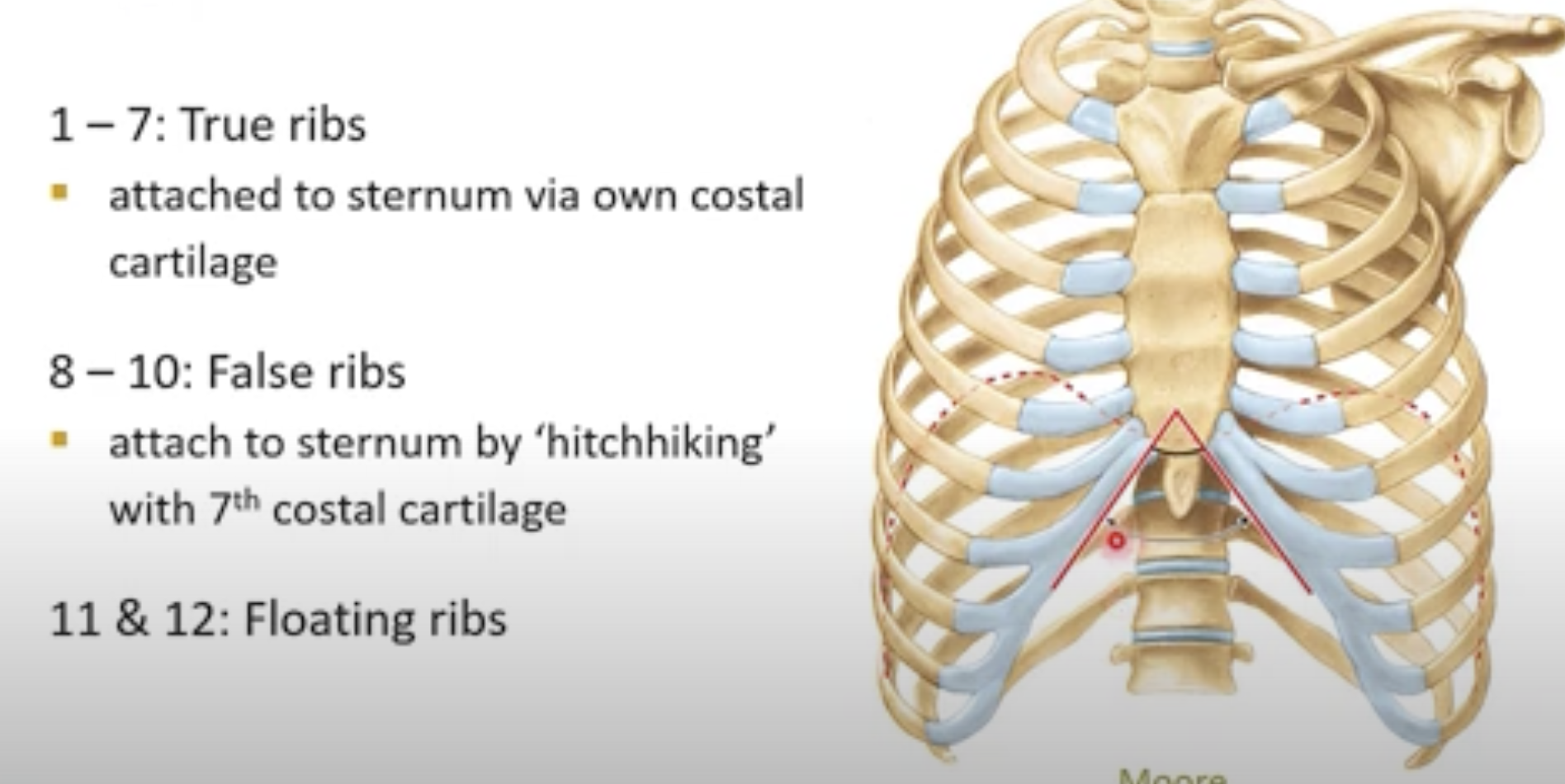
RIB CATEGORIZATION
Ribs 1 – 7
Description: True ribs
Articulation: Each attached to the sternum via its own costal cartilage
Ribs 8 – 10
Description: False ribs
Articulation: Attach to the sternum by "hitchhiking" with the costal cartilage of rib 7
Ribs 11 & 12
Description: Floating ribs
Articulation: No anterior articulation
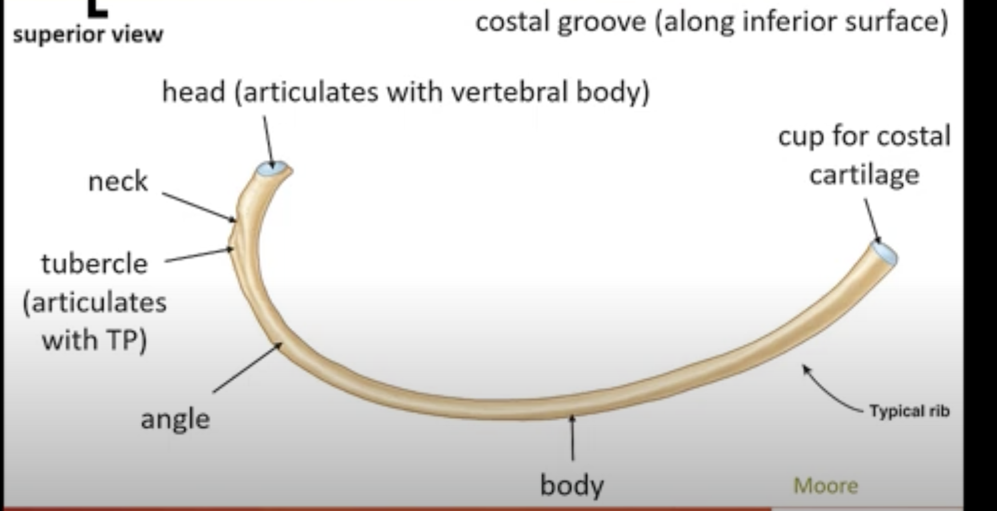
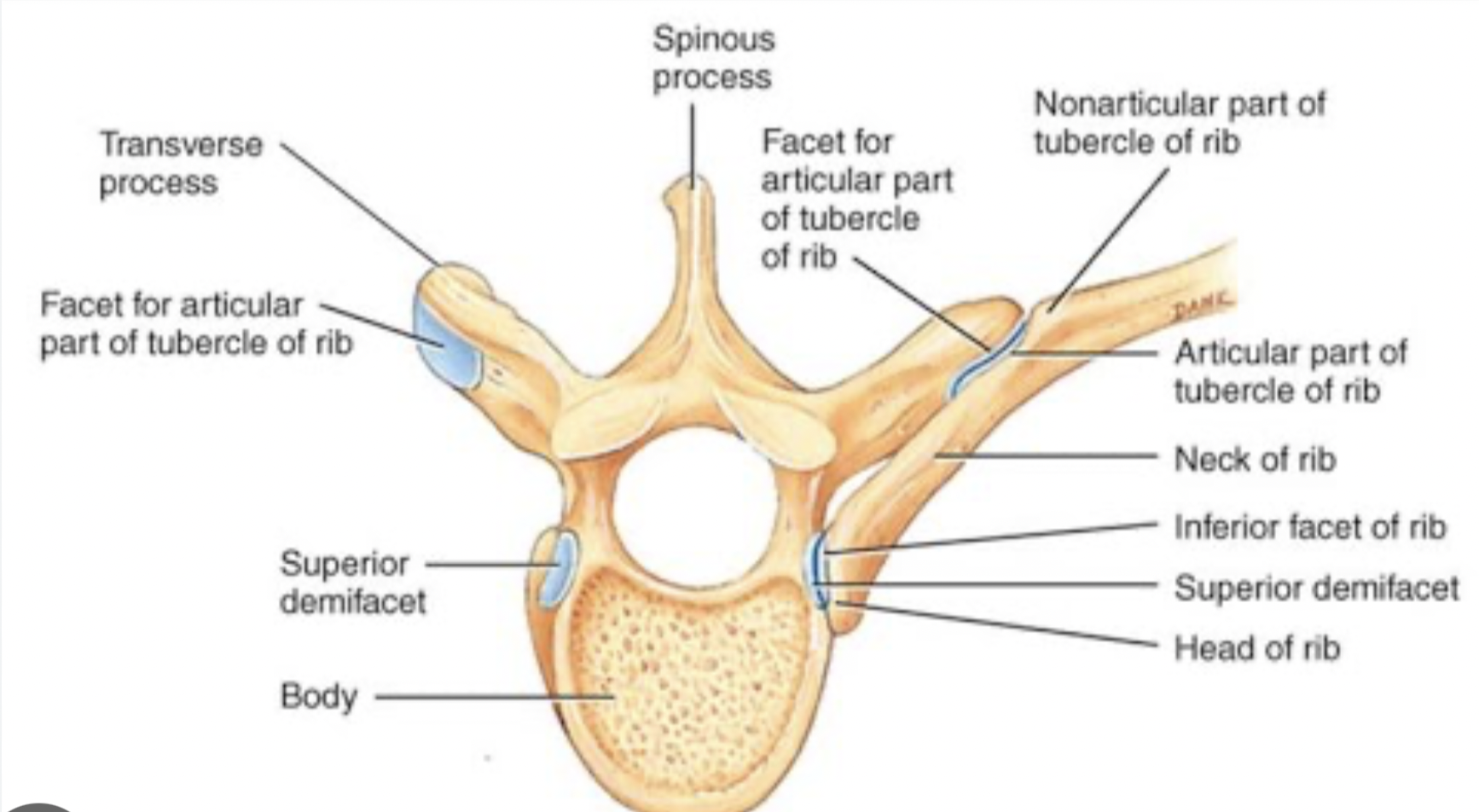
TYPICAL RIBS (3-10)
Head: Articulates with vertebrae
Tubercle: Articulates with transverse process of vertebrae
Body: Main section of the rib
Costal Groove: Along inferior surface for vessels/nerves
Neck: Section just before the head
Angle: Location where rib bends
Cup for Costal Cartilage: Area of attachment to cartilage
ATYPICAL RIBS
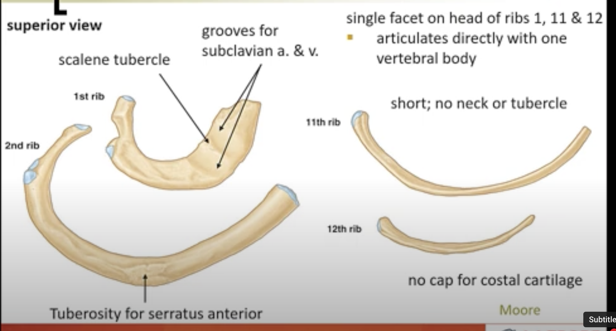
Rib 1:
Grooves for subclavian artery and vein
Tuberosity for serratus anterior
Scalene tubercle
Single facet on head (articulates directly with one vertebral body)
RIB 2
Atypical rib, larger and longer than rib 1
Articulates with the manubrium at the sternomanubrial joint
Has a tuberosity for the serratus anterior muscle
This rib has two facets on the head (articulates with two vertebrae)
Typically has a costal cartilage attachment similar to true ribs
Also provides support for the thoracic wall and plays a role in the mechanics of breathing.
Ribs 11 & 12:
No neck or tubercle
No costal cartilage cup
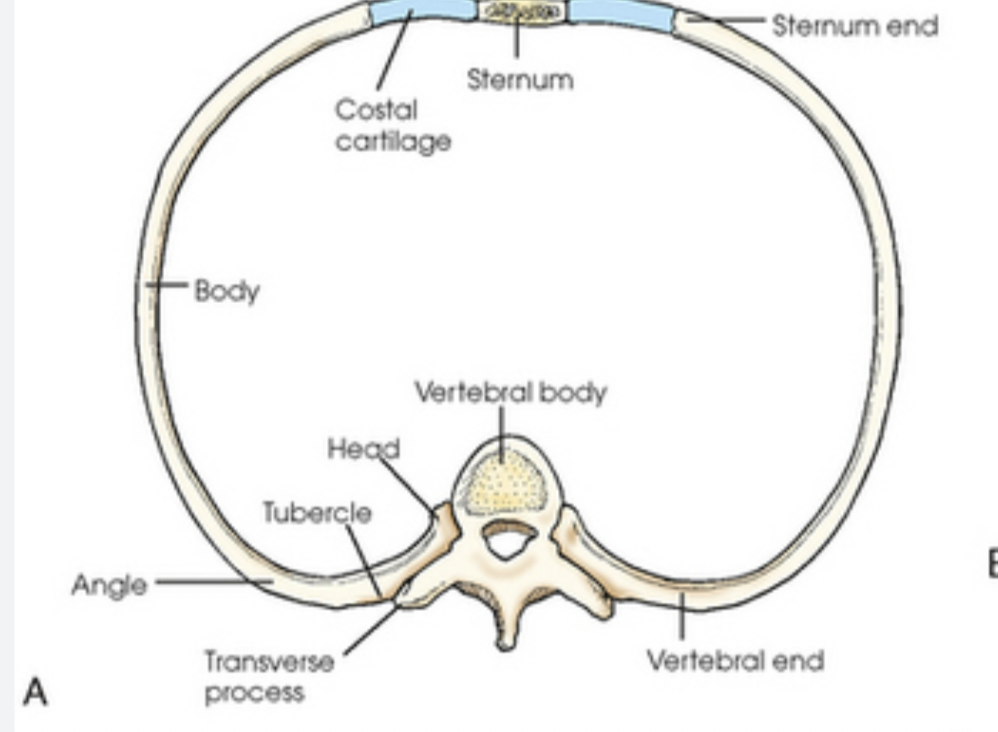
RIBS: ANTERIOR ARTICULATIONS
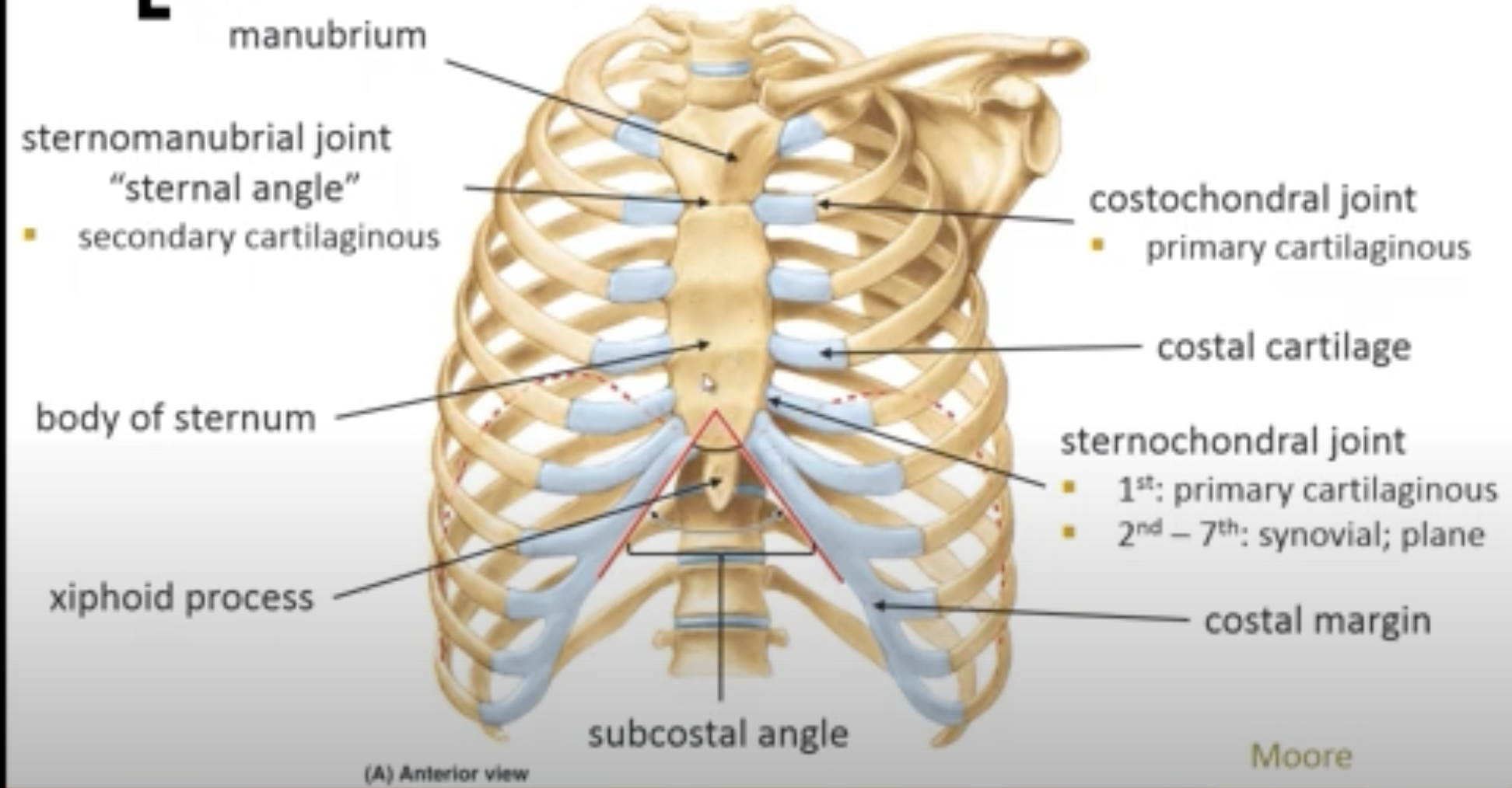
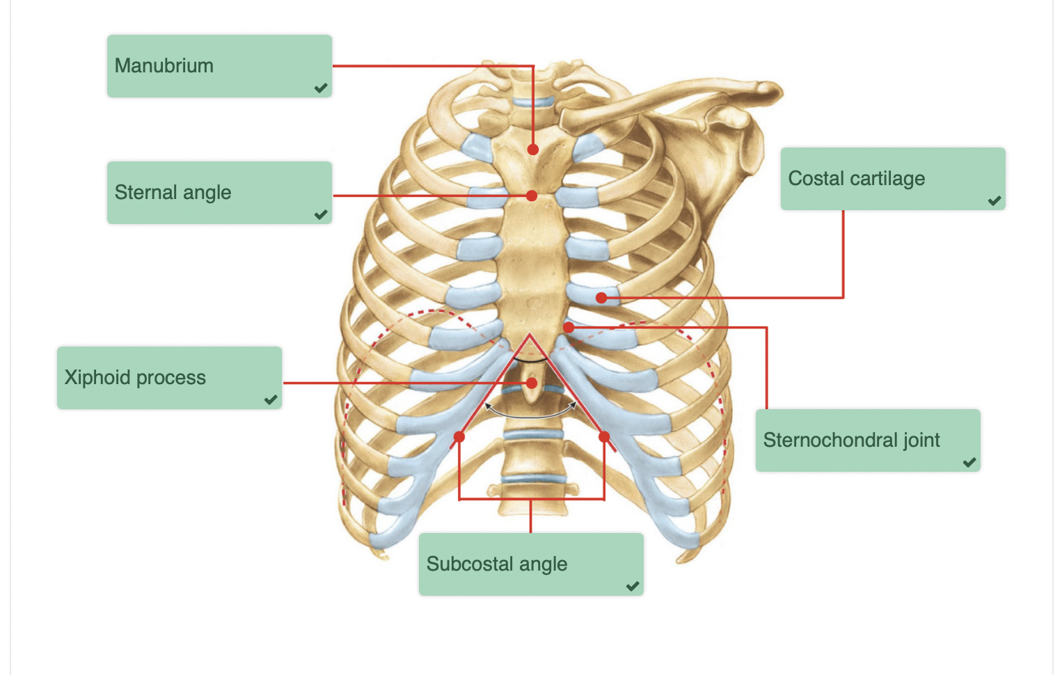
Manubrium: Articulates with first rib
Sternomanubrial Joint: Also known as the "sternal angle"
Body of Sternum: Articulates with ribs 2-7
Xiphoid Process: Terminus of the sternum
Costal Cartilage: Various joints formed (costochondral and sternochondral) for ribs 1-7
costal margin: Inferior border of the thoracic cage, formed by the lower edges of the costal cartilages of ribs 7-10.
RIBS: POSTERIOR ARTICULATIONS
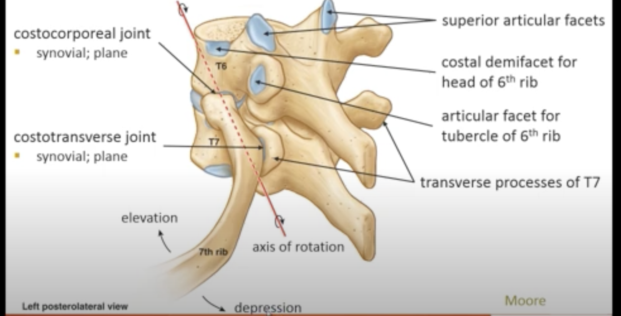
Superior Articular Facets: Articulate with head of ribs
Costocorporeal Joint: For head of the rib articulations
Costotransverse Joint: For tubercle of rib articulation and the transverse process of the vertebrae.
Transverse Processes: Important for rib movement
Elevation and depression axes for rib movement during respiration
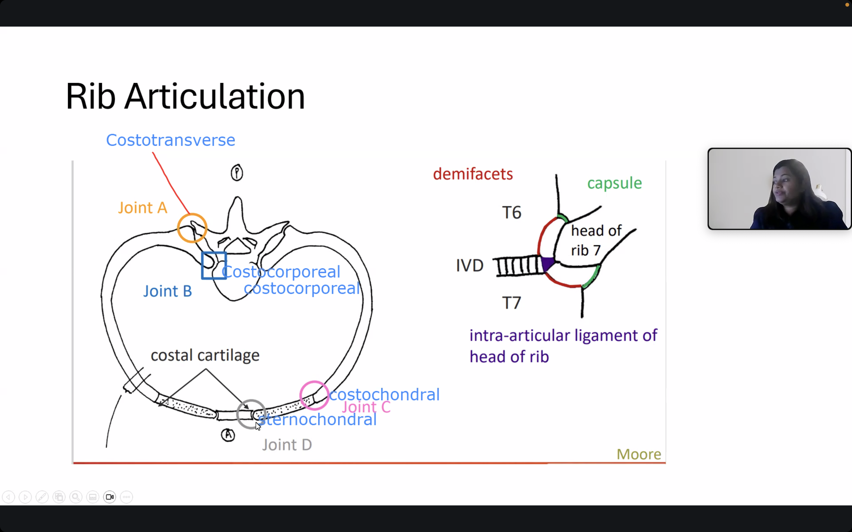
RIB ARTICULATIONS
Costal Cartilage: Connects ribs to sternum and vertebrae
Demifacets & Intervertebral Discs (IVD): Important for rib articulation with vertebrae
Intra-Articular Ligament: Aids in stabilizing the rib heads for movement

list the 4 different joints of rib articulation
Costocorporeal Joint: Articulation between the head of the rib and the vertebrae.
Costotransverse Joint: Articulation between the tubercle of the rib and the transverse process of the vertebrae.
Costochondral Joint: Joint that connects the rib to its costal cartilage.
Sternocostal Joint: Articulation between the costal cartilage of ribs 1-7 and the sternum.
Histological Classification:
Costocorporeal Joint:
synovial joint formed between the head of the rib and the demifacets of adjacent vertebrae.
Costotransverse Joint:
Synovial joint formed between the tubercle of the rib and the transverse process of the corresponding vertebra.
Costochondral Joint:
Primary cartilaginous joint (synchondrosis) connecting the rib to its costal cartilage.
Sternocostal Joint:
Synovial joints (for ribs 2-7) formed between the costal cartilage of the ribs and the sternum. Rib 1 has a non-synovial joint (costal cartilage to manubrium).
Functional Classification:
Costocorporeal Joint:
Function: Facilitates rib movement during respiration, allowing elevation and depression of ribs that modify thoracic volume.
Costotransverse Joint:
Function: Aids rib rotation and movement, contributing to the mechanics of respiration.
Costochondral Joint:
Function: Provides stability and flexibility at the junction where ribs meet costal cartilage, aiding in respiratory movements.
Sternocostal Joint:
Function: Allows the ribs to attach securely to the sternum while permitting limited movement, essential for accommodating thoracic expansion during breathing.
Movements at Rib Articulations
1. Costocorporeal Joint
Movements: Facilitates rib movement during respiration, allowing for elevation and depression of ribs that modify thoracic volume.
2. Costotransverse Joint
Movements: Aids rib rotation and lateral movement, contributing to the mechanics of respiration.
3. Costochondral Joint
Movements: Provides stability and flexibility at the junction where ribs meet costal cartilage, aiding in respiratory movements.
4. Sternocostal Joint
Movements: Allows the ribs (1-7) to attach securely to the sternum while permitting limited movement, essential for accommodating thoracic expansion during breathing.
EMBRYOLOGY
Trilaminar Embryo
Three Layers:
Ectoderm: Surface
Mesoderm: Body wall
Endoderm: Forms gut tube
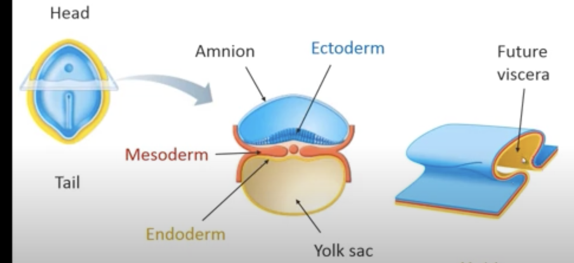
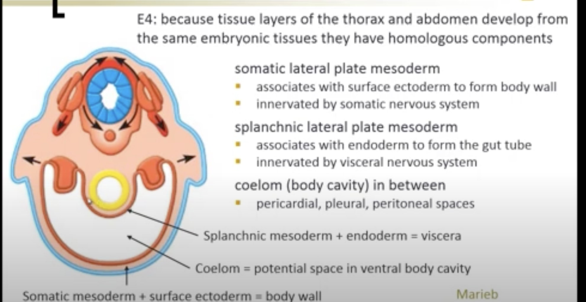
Body Wall vs. Coelomic Cavities
Somatic Lateral Plate Mesoderm: Forms body wall, innervated by:somatic nervous system.
Splanchnic Lateral Plate Mesoderm: Forms gut tube, innervated by: visceral nervous system
Coelom: Potential space in ventral body cavity
pericardial, pleural, peritoneal spaces
Development of thorax and abdomen share common embryonic tissues that have homologous components
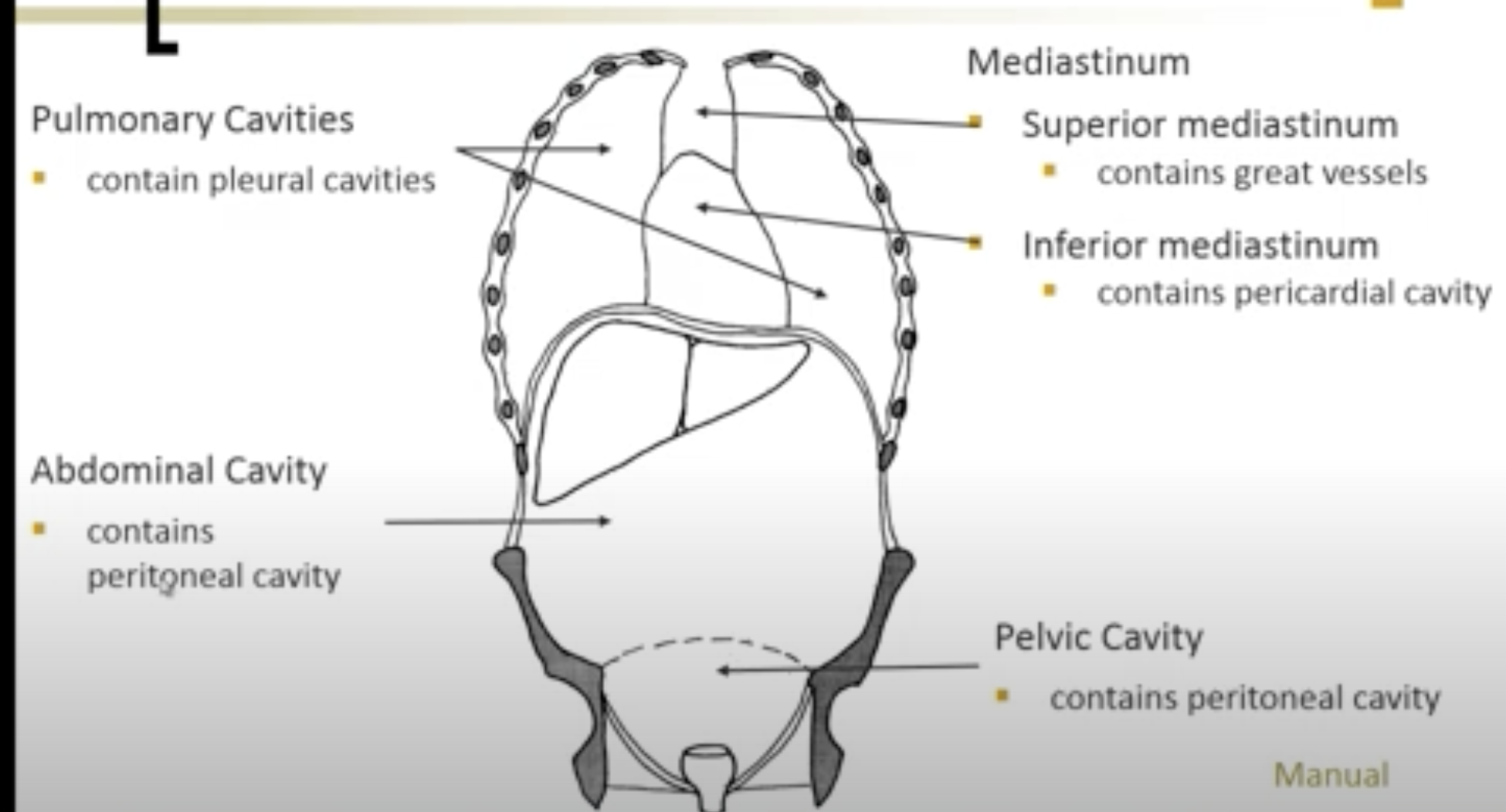
OPEN COELOMIC CAVITIES
Pulmonary Cavities: Surround the lungs
Mediastinum: Contains heart and related structures
Abdominal Cavity: Contains digestive and other organs
Pelvic Cavity: Contains reproductive organs
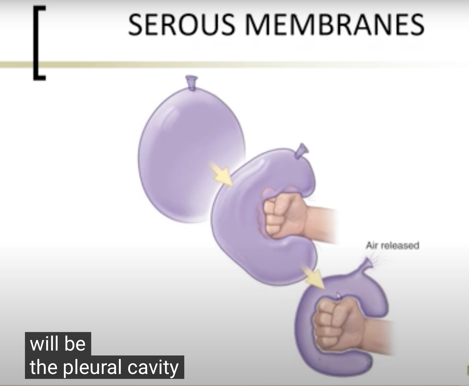
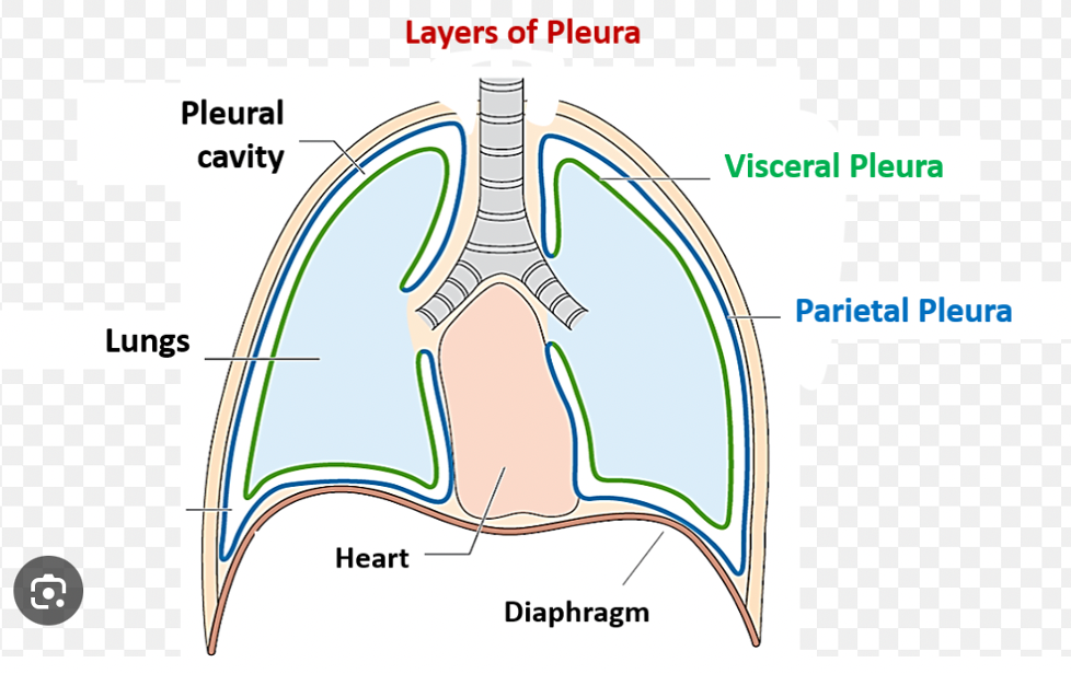
SEROUS MEMBRANES
Contain liquid and provide lubrication to organs in coelomic cavities (specific details not present)
A serous membrane is a thin layer of tissue that lines certain cavities in the body and covers the organs within those cavities. It secretes a lubricating fluid called serous fluid that reduces friction between the organs and the walls of the cavities. For example, serous membranes line the pleural cavities around the lungs, the pericardial cavity around the heart, and the peritoneal cavity in the abdomen.
OPEN vs. POTENTIAL CAVITIES
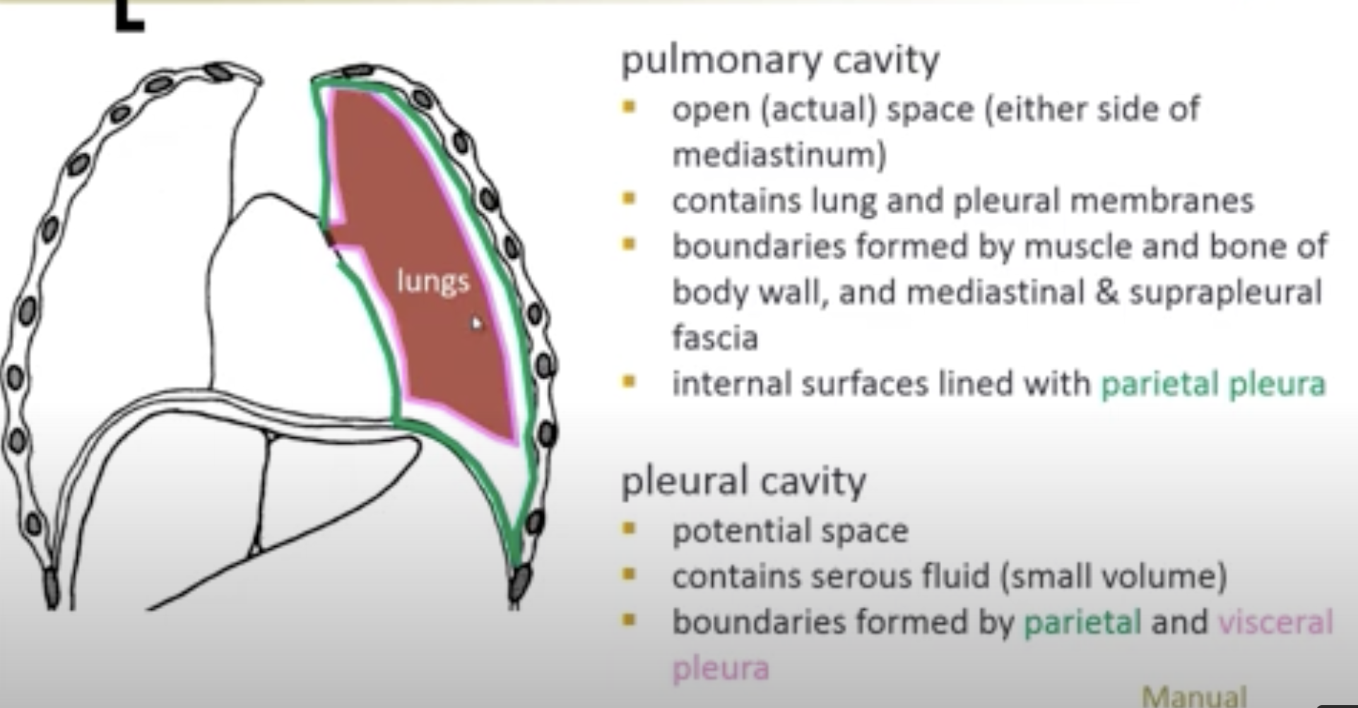
Lungs: Real space (open respiratory pathway)
Contains: Lung tissue
Boundaries: Muscle and bone of body wall, mediastinal and suprapleural fascia
Internal Surfaces: Lined with pleura
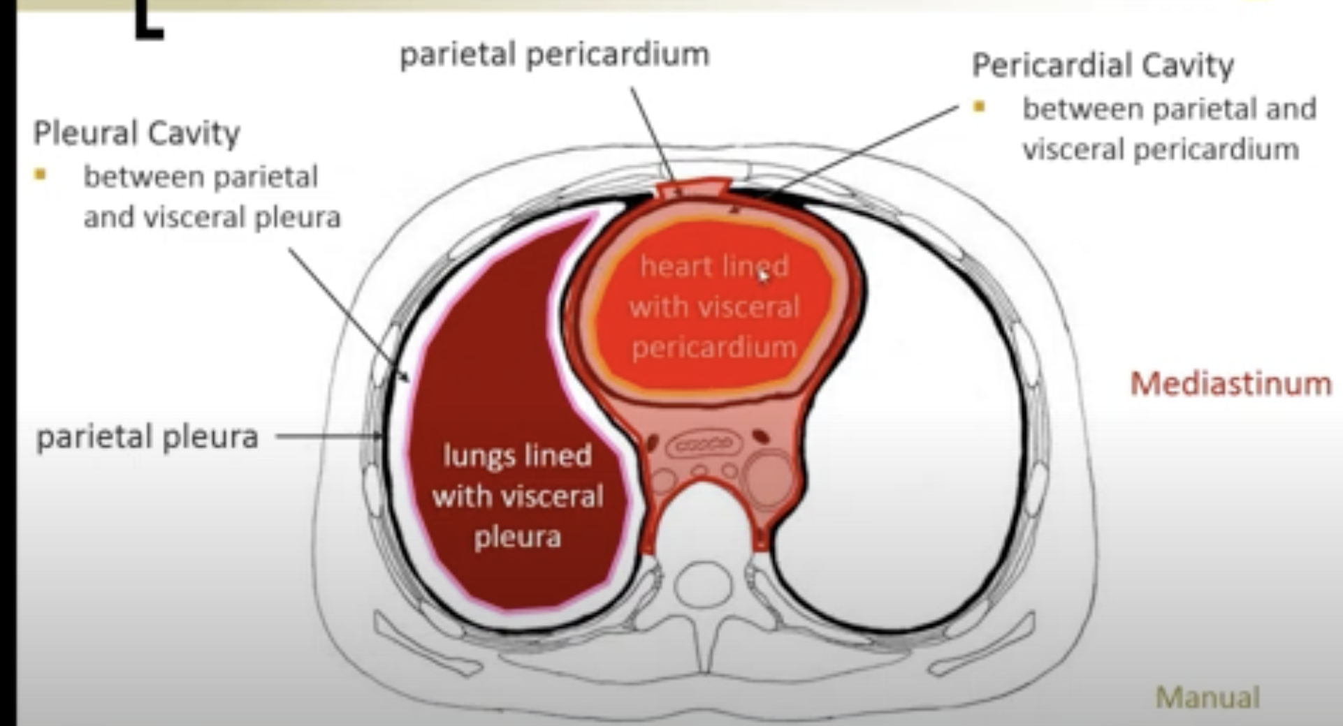
Pleural Cavity:
Potential space, contains serous fluid (small volume)
Boundaries formed by:
PLEURA
Visceral Pleura: Attaches to lungs
Parietal Pleura: Attaches to thoracic cavity wall
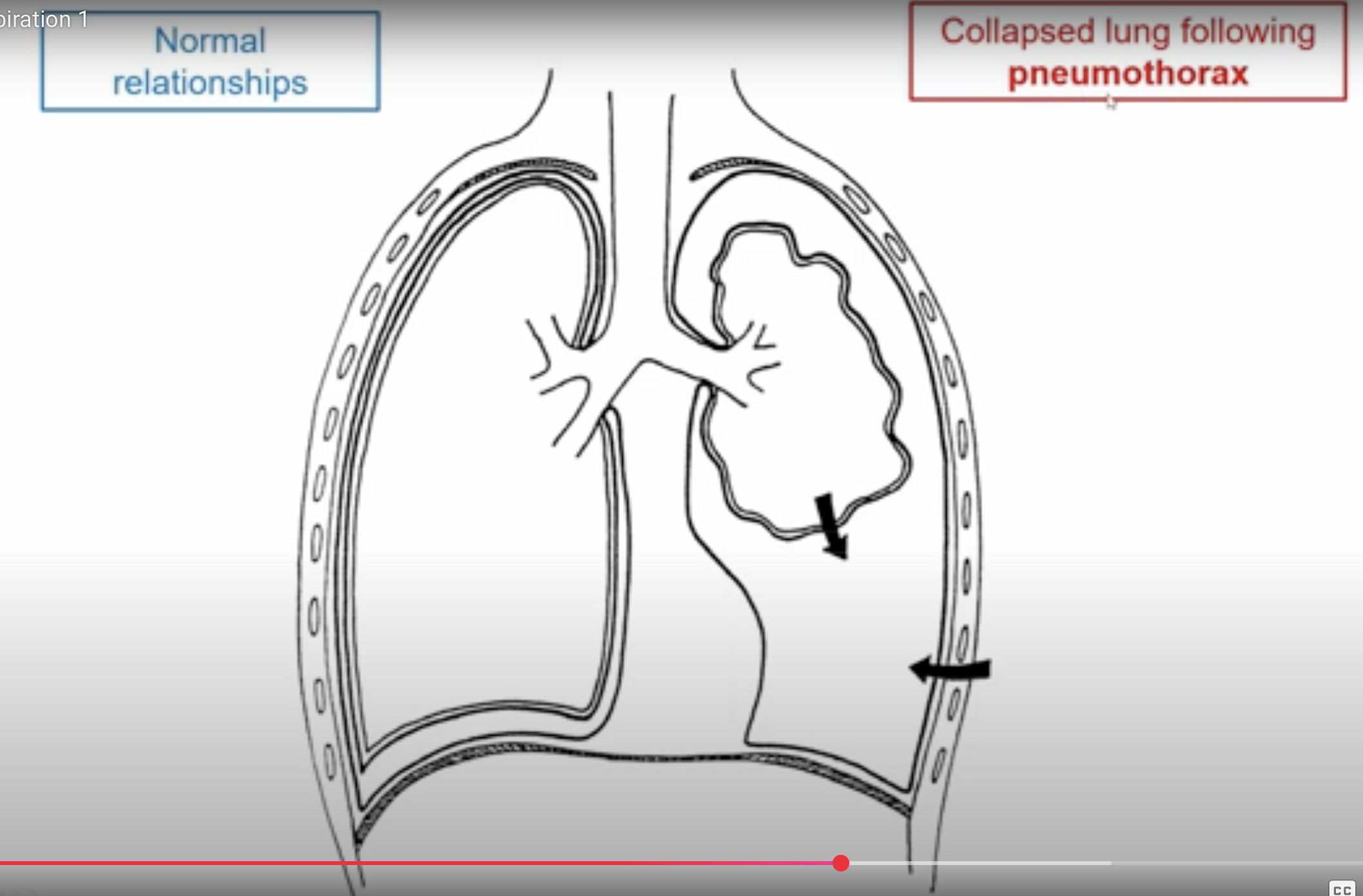
Surface Tension: Provided by pleural fluid, negative pressure (2-8 mm Hg)
Puncture Effect: If punctured, air enters the pleural cavity causing lung deflation
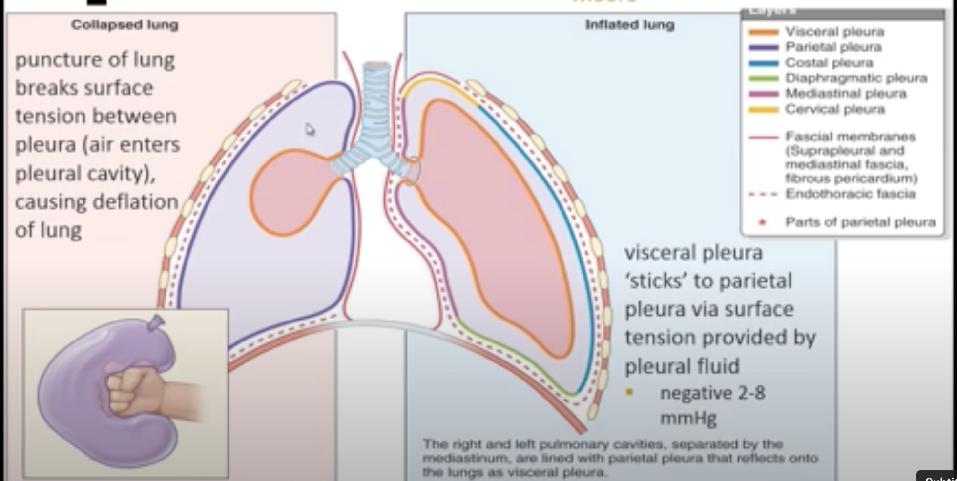
POTENTIAL SPACES
Pleural Cavity: Between visceral and parietal pleura
Mediastinum: Space for heart and vessels
Pericardial Cavity: Between parietal pleura and pericardium (surrounding heart)
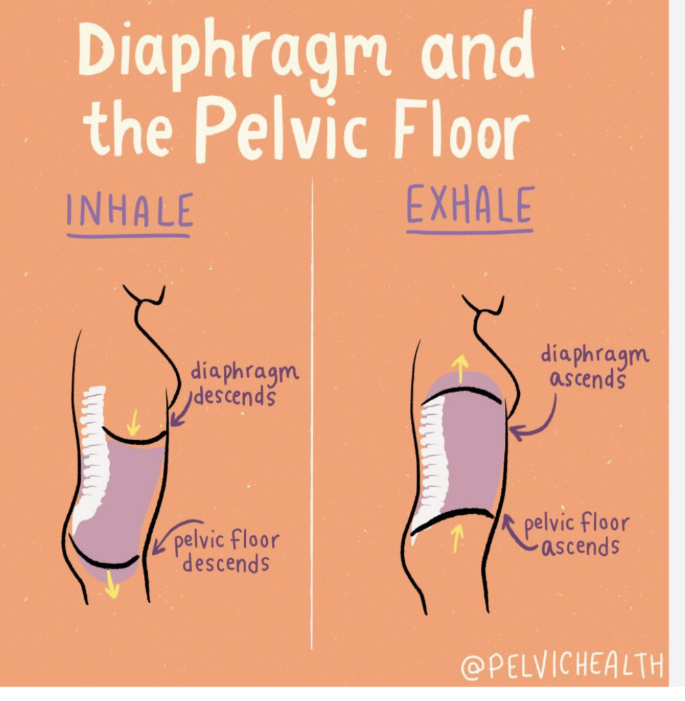
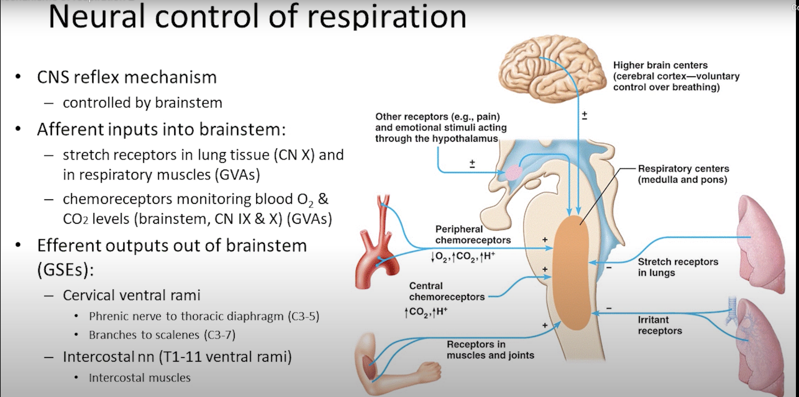
RESPIRATION
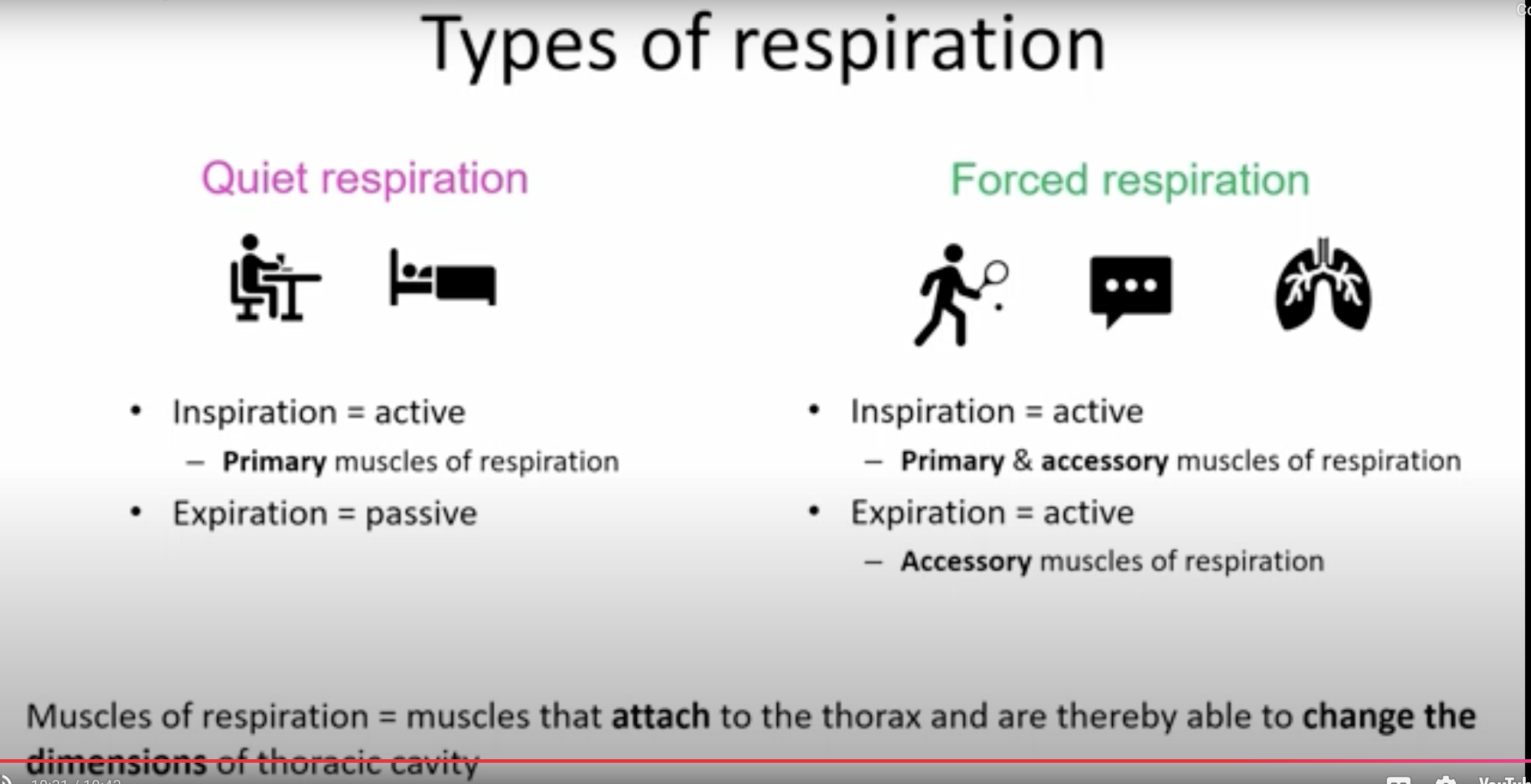
Overview of Respiration
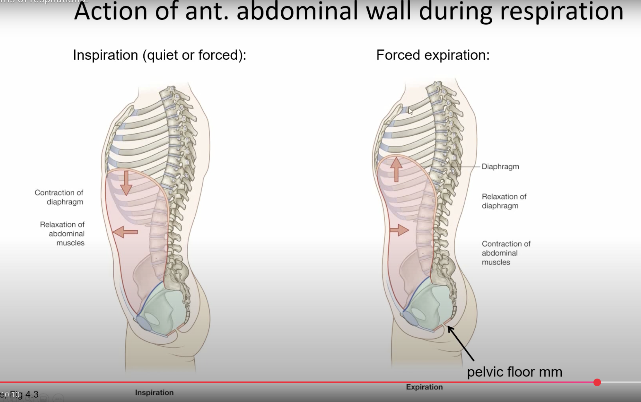
Phase 1: Inspiration (Breathing In)
Drawing air into the lungs
In response to increased thoracic volume
Dependent on muscle activity (diaphragm and accessory muscles of respiration)
Phase 2: Expiration (Breathing Out)
Air passing out of the lungs
In response to decreased thoracic volume
Quiet respiration: Passive process (elastic recoil of diaphragm and lungs)
Forced respiration: Active process (contraction of accessory muscles of respiration)
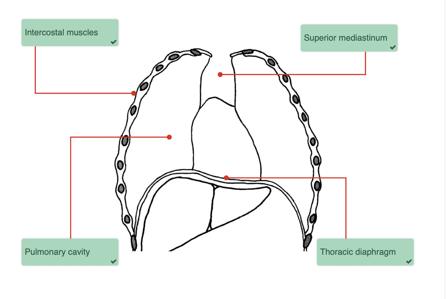
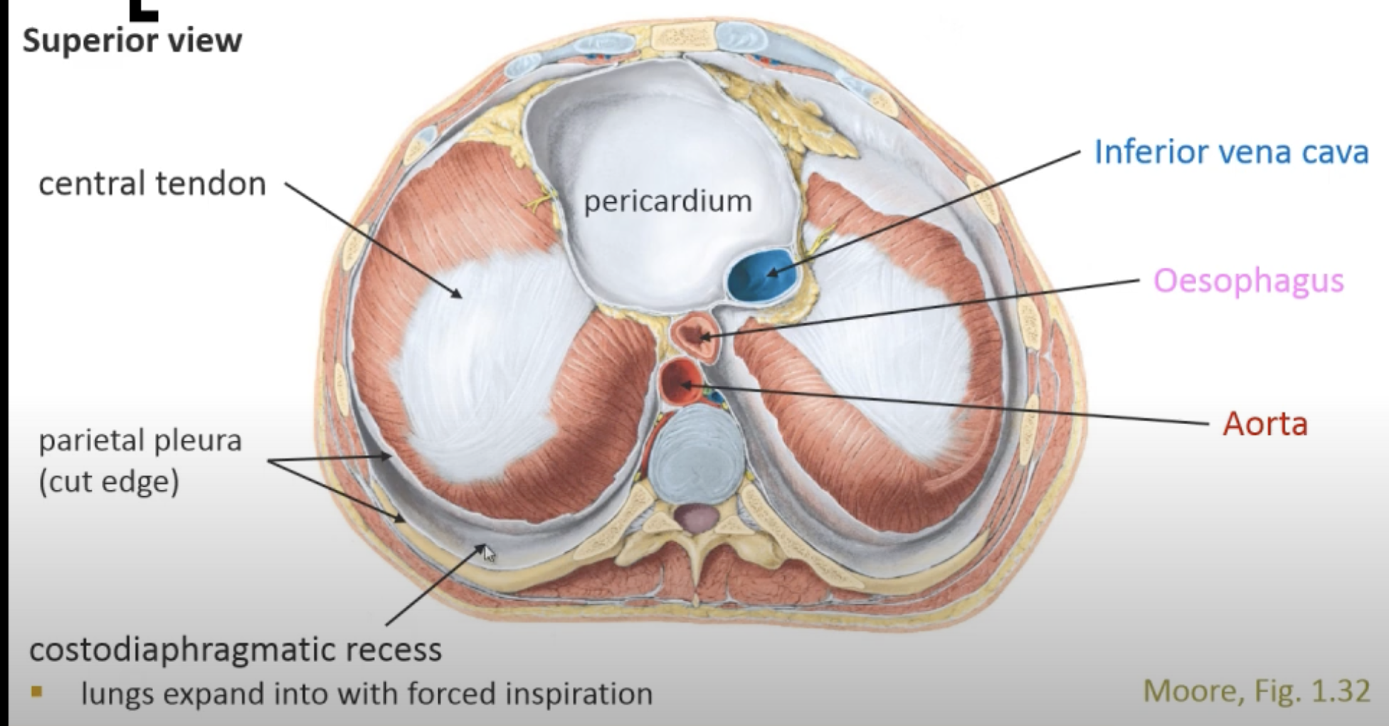
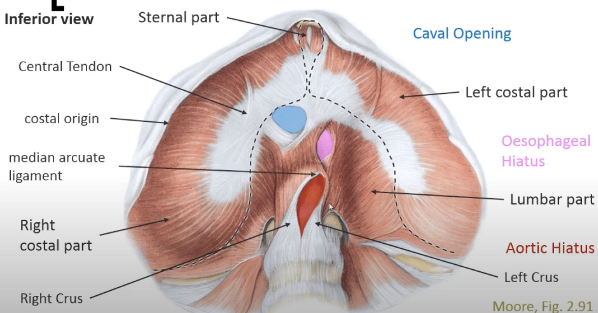
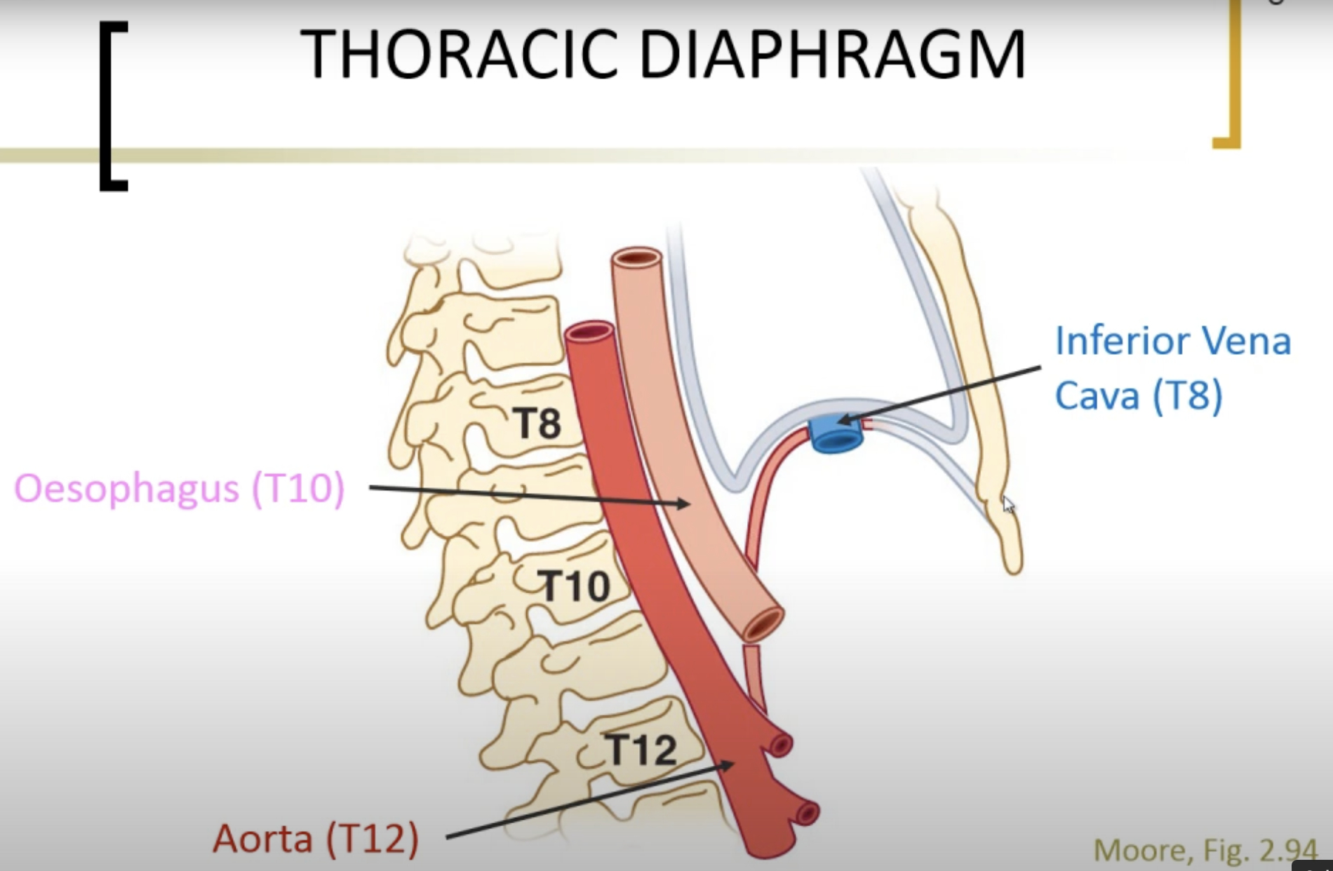
THORACIC DIAPHRAGM
Superior View: Central tendon, pericardium, costodiaphragmatic recess where lungs expand
Inferior View:
Parts include Left Crus, Right Crus, costal origin, Central Tendon, median arcuate ligament, Aortic Hiatus, Oesophageal Hiatus, Caval Opening
Left and Right Crus: Support base of diaphragm
Openings: Aortic hiatus, esophageal hiatus, caval opening (T8, T10, and T12)
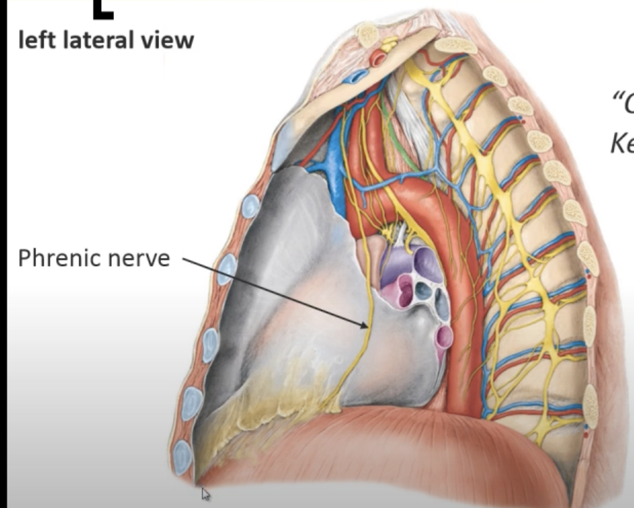
INNERVATION OF DIAPHRAGM
Cervical Roots: "C3, 4, 5: Keep the diaphragm alive."
Outer Edges: Innervated by lower intercostal nerves
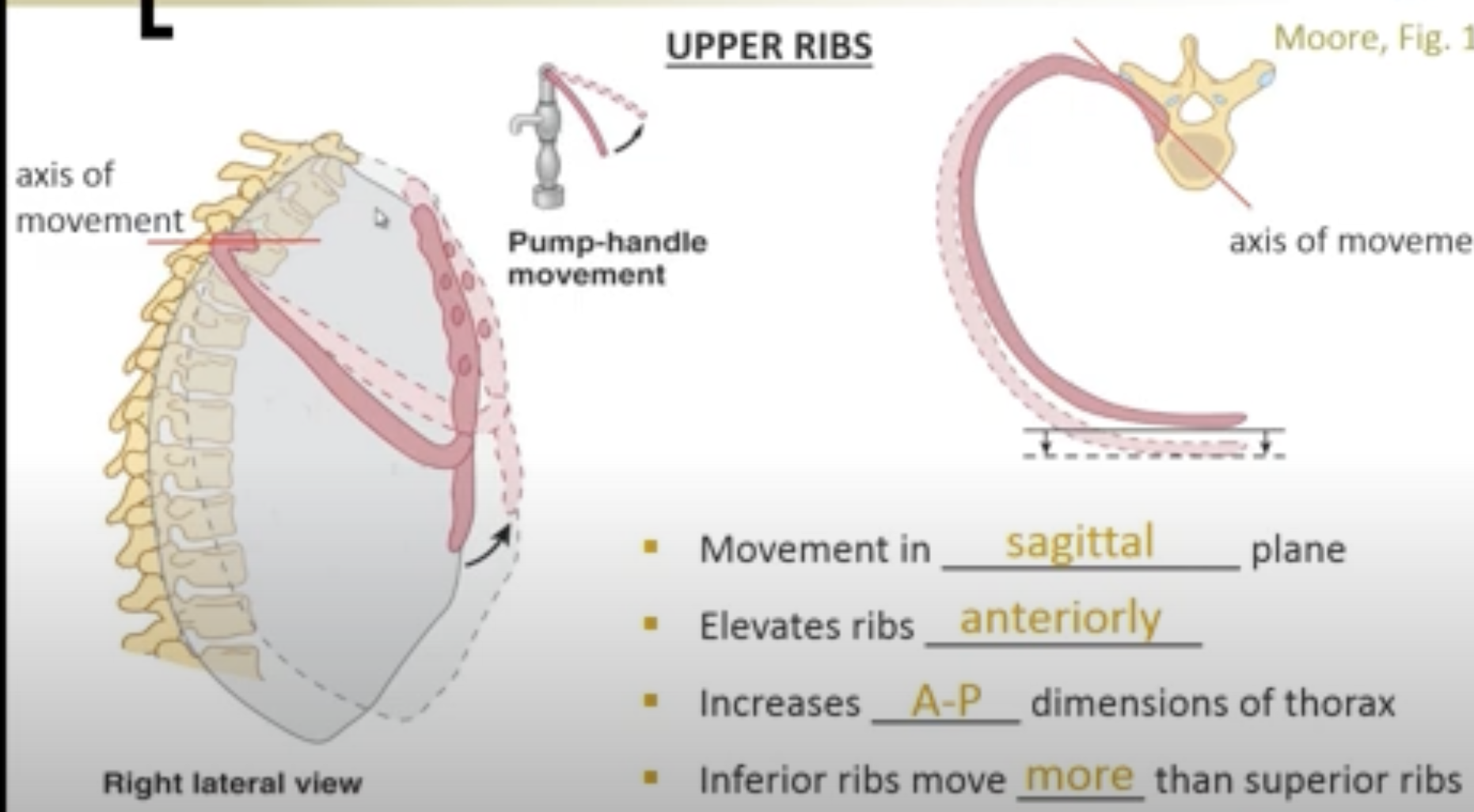
RIB MOVEMENTS
General Rib Movement Mechanics
Movement occurs in a specific plane
Elevation of ribs increases anterior-posterior dimensions of thorax
Inferior ribs move more than superior ribs
Upper Ribs Movement
Movement in sagittal plane
Elevates ribs anterioly
Increases vertical A-P dimensions of thorax- Elevates ribs, increasing dimensions of thorax
Inferior ribs move more than superior ribs
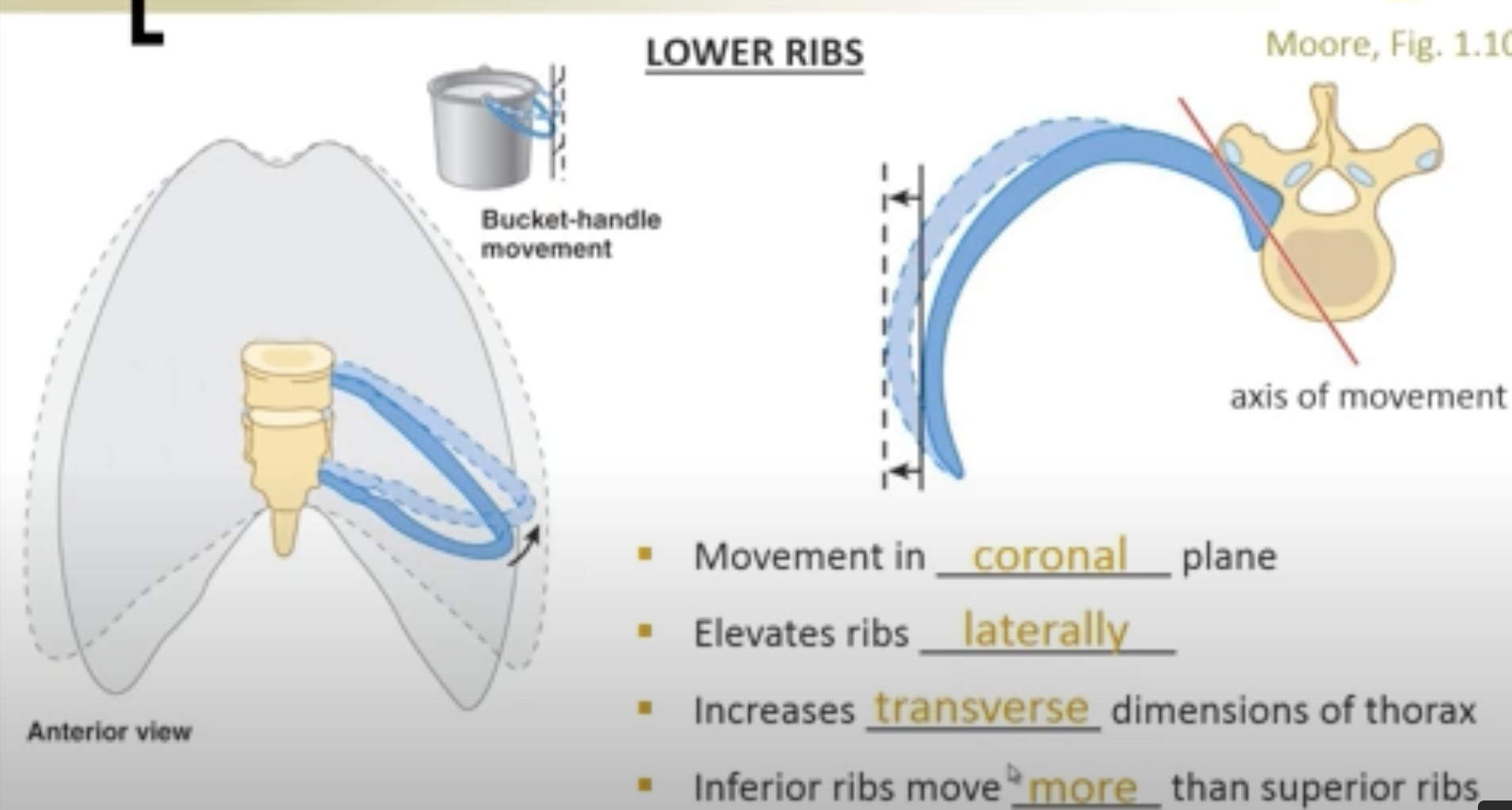
Lower Ribs Movement
Movement in corronal plane
Elevates ribs laterally
Increases transverse dimensions of thorax
Inferior ribs move ___more___ than superior ribs
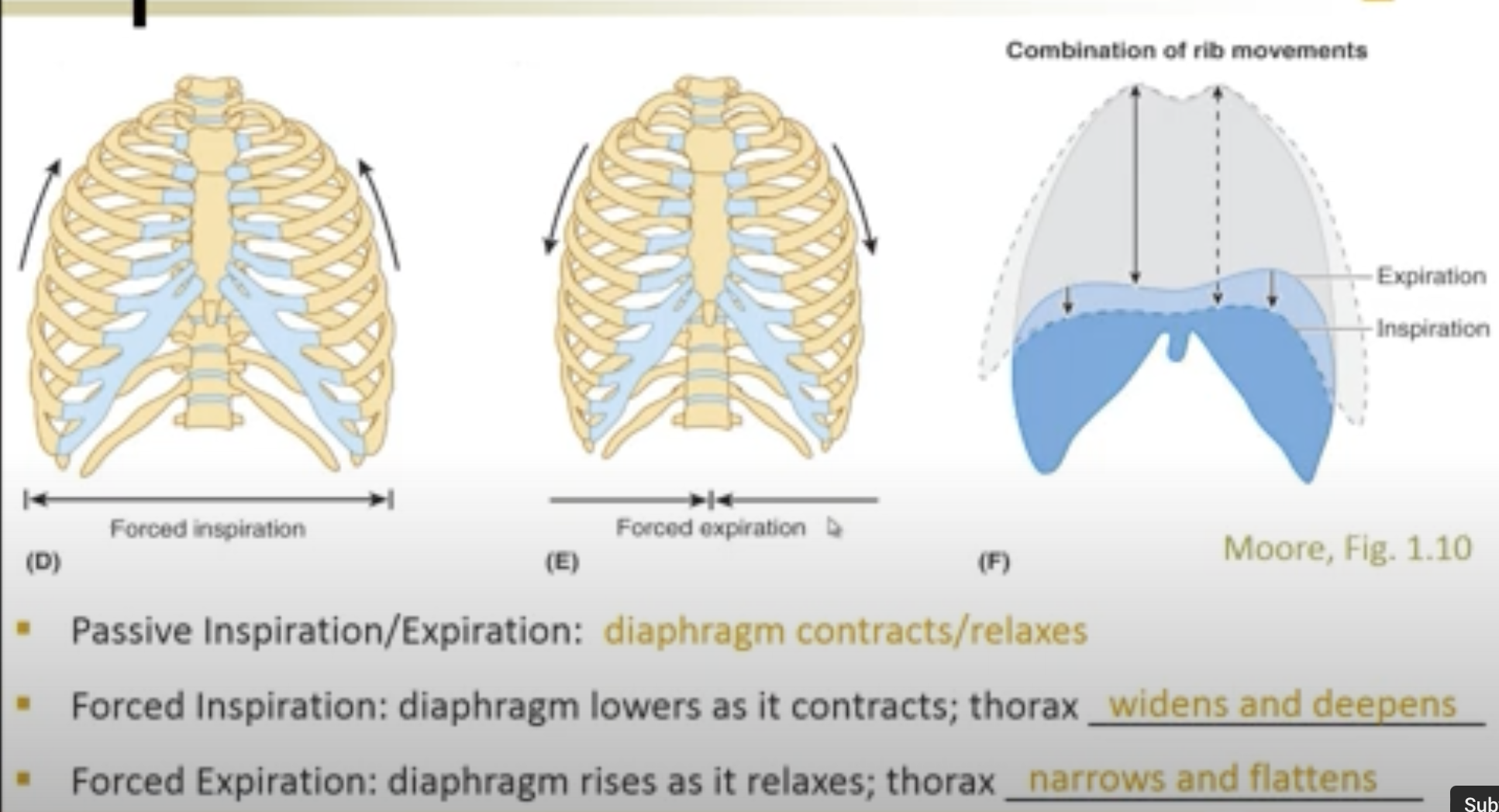
INSPIRATION AND EXPIRATION Modality- RIB MOVEMENTS
Passive vs. Forced Breathing
Passive Inspiration/Expiration: Basic respiratory process
Forced Inspiration:
Diaphragm lowers as it contracts
Thorax expands
Forced Expiration:
Diaphragm rises as it relaxes
Thorax contracts
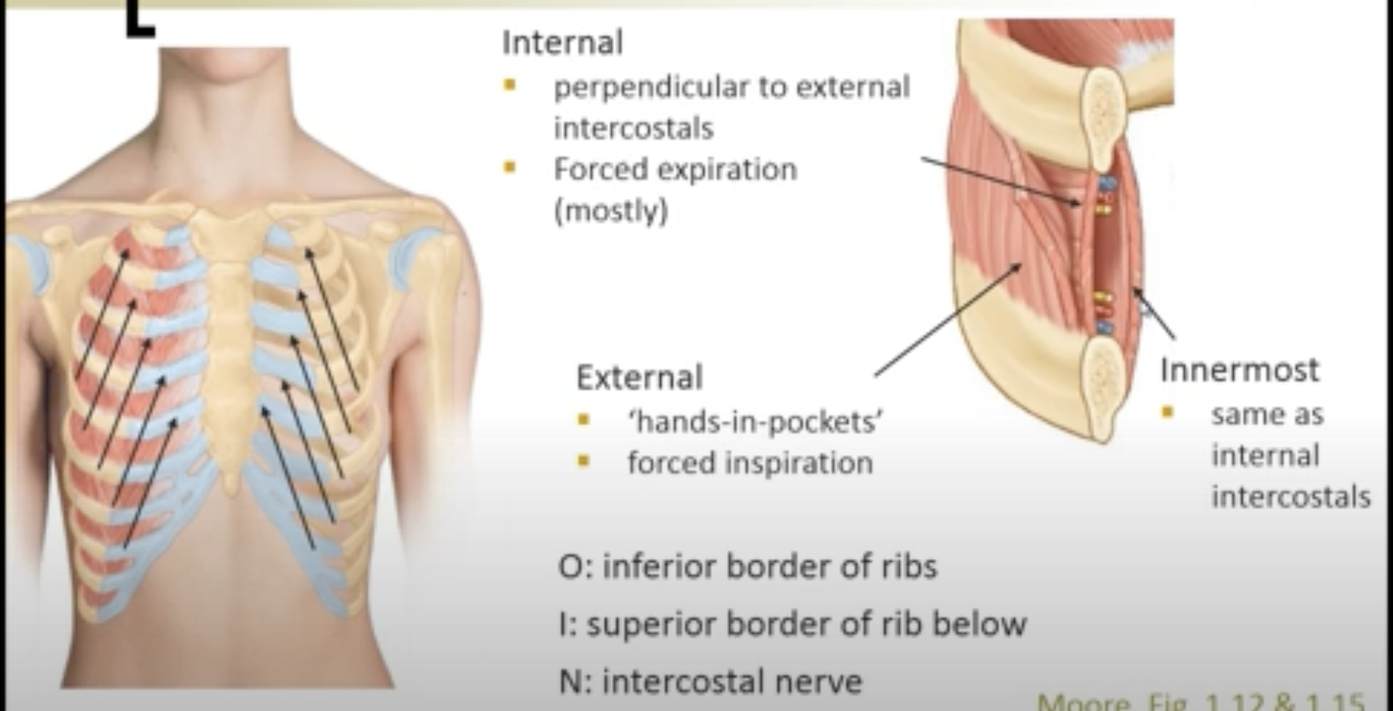
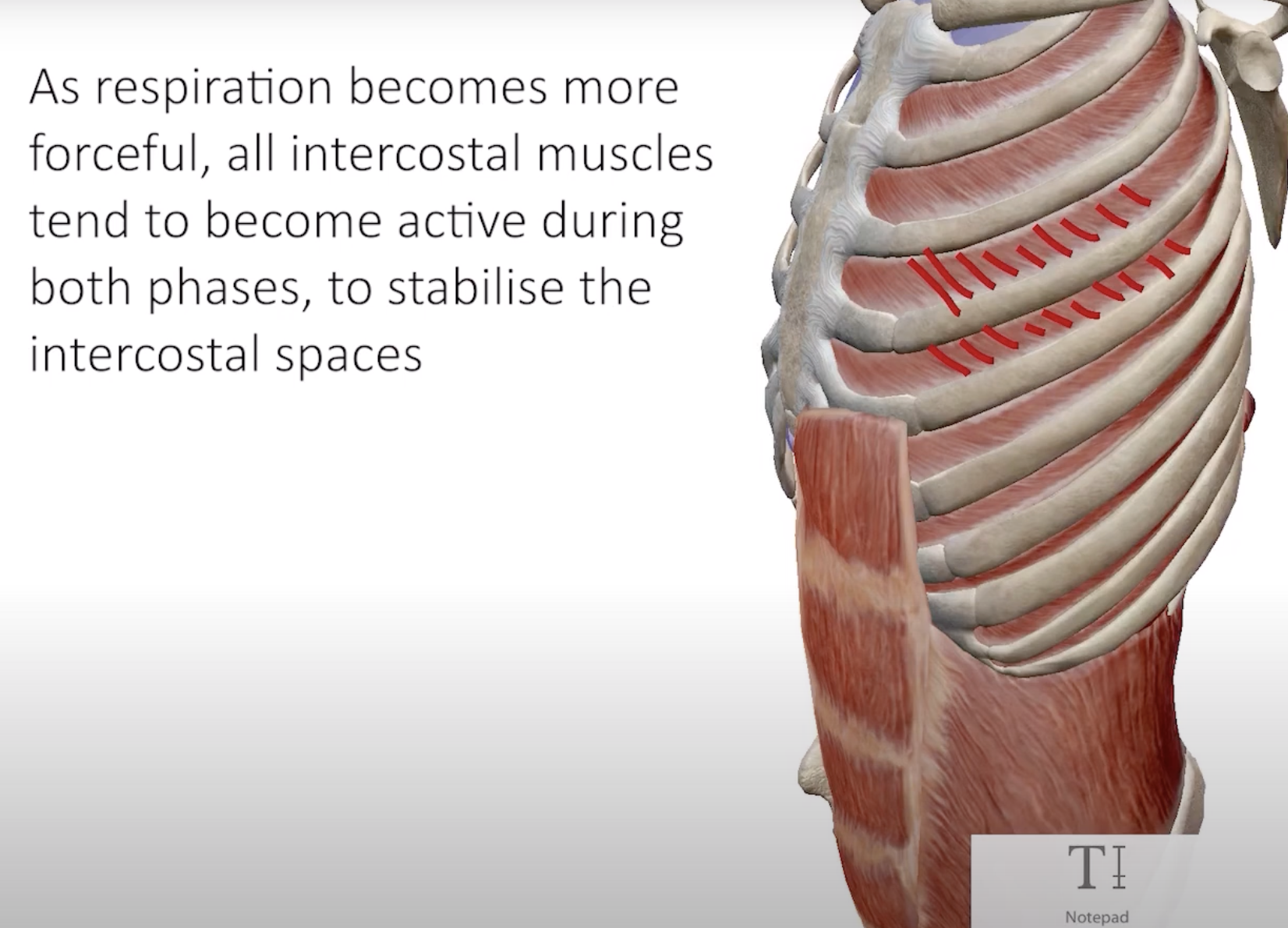
INTERCOSTAL MUSCLES
Origin (O): Inferior border of ribs
Insertion (I): Superior border of rib below
Innervation (N): Intercostal nerve
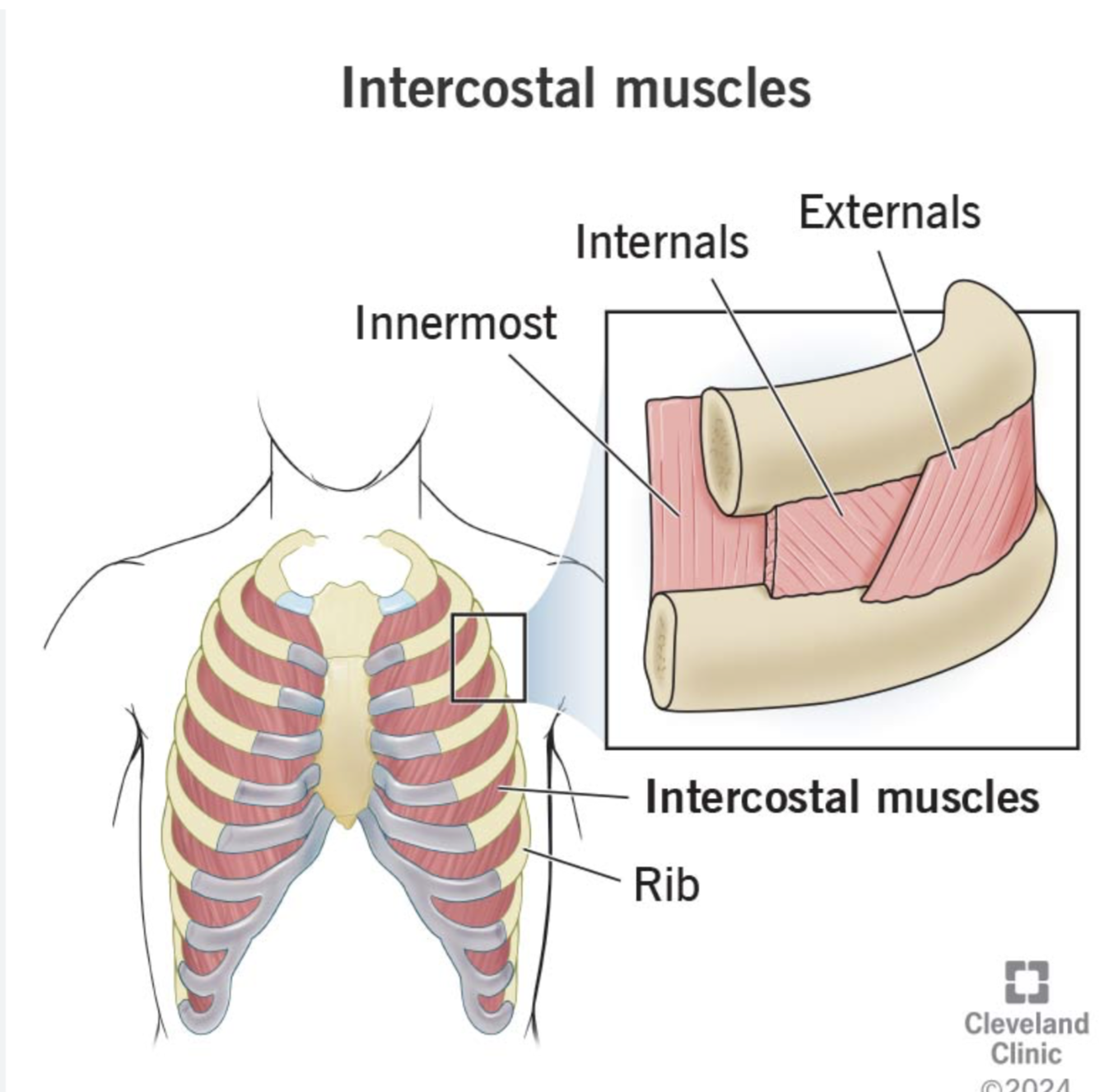
Types of Intercostal Muscles
External Intercostals: Function like "hands in pockets"
Internal Intercostals: Perpendicular to externals, primarily during forced expiration
Innermost Intercostals:
Located deep to the internal intercostals, these muscles assist in the mechanics of breathing by stabilizing the ribcage during contraction.
INTERCOSTAL NEUROVASCULAR BUNDLE
The Intercostal Neurovascular Bundle consists of the intercostal vein, artery, and nerve, located in the subcostal groove of the ribs. It plays a critical role in supplying blood and innervating the intercostal muscles. The clinical relevance of this bundle is its location, which is important for procedures such as needle insertion.
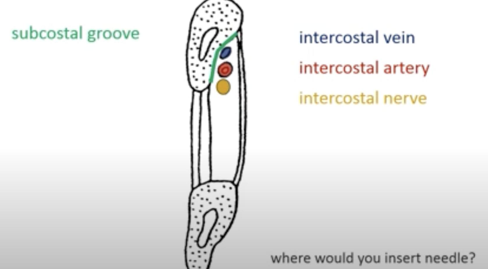
To drain the pleural cavity, a needle is typically inserted in the 5th intercostal space at the anterior axillary line (mid-axillary line) to avoid injury to the intercostal neurovascular bundle. This site is chosen to effectively access the pleural space while minimizing risk of complications.
INTERCOSTAL MUSCLES
Anatomy and Function
External Intercostals
O: Inferior border of ribs
I: Superior border of the rib below
N: Intercostal nerve
Function: Hands-in-pockets movement
Internal Intercostals
Perpendicular to external intercostals
Mainly involved in forced expiration
Innermost Intercostals
Related anatomy of the intercostal neurovascular bundle includes:
Subcostal groove
Intercostal vein
Intercostal artery
Intercostal nerve
Worksheet:
Pleural Linkage: Refers to the connection between the lungs and the thoracic wall.
Surface Tension: Pleural fluid creates surface tension that helps maintain the close apposition of the visceral (lung) and parietal (thoracic wall) pleura.
Negative Pressure: The pleural cavity maintains a negative pressure (2-8 mm Hg) which keeps the lungs inflated by adhering the pleura to the thoracic wall and preventing lung collapse.
serous fluid inbetween parietal and visceral pleura
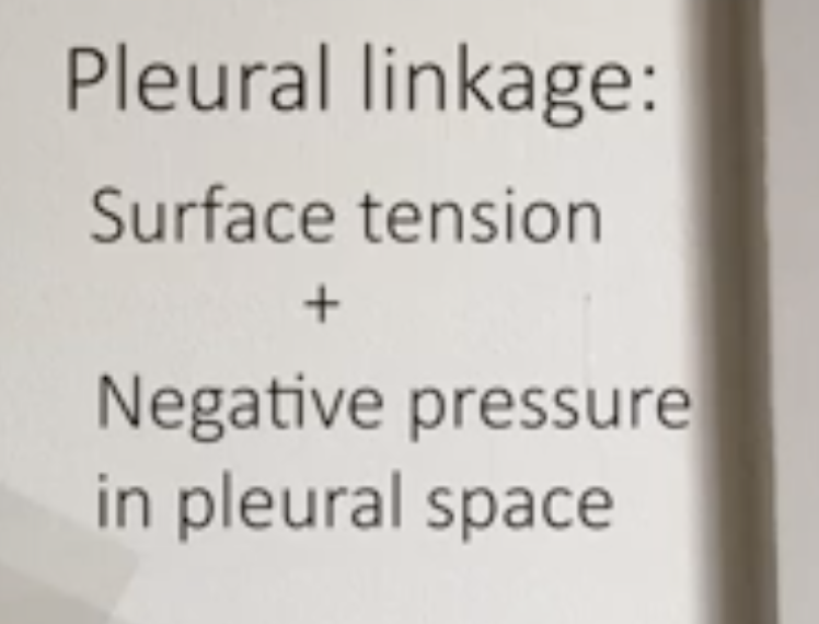
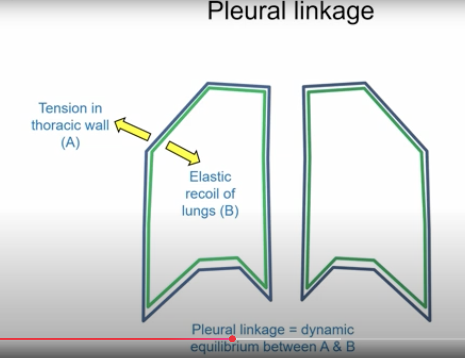
The two mechanisms that contribute to pleural linkage are:
1. Surface Tension: The pleural fluid creates surface tension between the visceral and parietal pleura, which helps in maintaining the close apposition of the two layers.
2. Negative Pressure: The negative pressure within the pleural cavity (ranging between 2-8 mm Hg) plays a crucial role in keeping the lungs inflated by adhering the pleura to the thoracic wall and preventing lung collapse.