Sense Organs
Sense Organs:
- We smell things by our nose, taste by the tongue, hear by ear and see objects by eyes.
- The nose contains ==mucus-coated receptors== which are specialized for receiving the sense of smell and are called ==olfactory receptors.==
- These are made up of olfactory epithelium that consists of three kinds of cells.
- The ==neurons of the olfactory epithelium== extend from the outside environment directly into a pair of broad bean-sized organs, called the olfactory bulbs, which are extensions of the ==brain’s limbic system.==
- Both nose and tongue detect dissolved chemicals.
- The chemical senses of ==gustation (taste) and olfactory (smell)== are functionally similar and interrelated.
- The tongue detects tastes through taste buds, containing gustatory receptors.
- With each taste of food or sip of the drink, the brain integrates the differential input from the taste buds and a complex flavor is perceived.
Eye:
- Our paired eyes are located in sockets of the skull called orbits
Parts of an Eye:
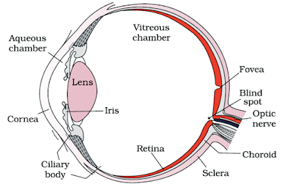
- The adult human eyeball is nearly a spherical structure.
- The wall of the eyeball is composed of three layers.
- The external layer is composed of ==dense connective tissue and is called the sclera.==
- The anterior portion of this layer is called the ==cornea.==
- The ==choroid layer== is thin over the posterior two-thirds of the eyeball, but it becomes thick in the anterior part to form the ciliary body.
- The middle layer, the choroid, contains ==many blood vessels and looks bluish in color.==
- The ciliary body itself continues forward to form a ==pigmented and opaque structure called the iris== which is the ==visible colored portion== of the eye.
- The eyeball contains a ==transparent crystalline lens== that is held in place by ==ligaments== attached to the ciliary body.
- In front of the lens, the aperture surrounded by the iris is called the ==pupil.==
- The diameter of the pupil is regulated by the ==muscle fibers of the iris.==
- The inner layer is the retina and it contains three layers of neural cells – from inside to outside –
- Ganglion cells
- Bipolar cells
- Photoreceptor cells.
- There are two types of photoreceptor cells, namely, rods and cones.
- These cells contain ==light-sensitive proteins called photopigments.==
- The ==daylight (photopic) vision and color vision are functions of cones and the twilight (scotopic) vision is the function of the rods.==
- The rods contain a ==purplish-red protein called rhodopsin== or visual purple, which contains a derivative of ==Vitamin A.==
- In the human eye, there are three types of cones that possess their own characteristic photopigments that respond to red, green, and blue lights.
- The sensations of different colors are produced by various combinations of these cones and their ==photopigments.==
- When these cones are stimulated equally, a sensation of white light is produced.
- The optic nerves leave the eye and the retinal blood vessels enter it at a point medial to and slightly above the posterior pole of the eyeball.
- Photoreceptor cells are not present in that region and hence it is called the ==blind spot.==
- At the posterior pole of the eye lateral to the blind spot, there is a yellowish pigmented spot called ==macula lutea with a central pit called the fovea.==
- The fovea is a thinned-out portion of the retina where ==only the cones are densely packed.==
- It is the point where the ==visual acuity (resolution) is the greatest.==
- The space between the cornea and the lens is called the ==aqueous chamber and contains a thin watery fluid called aqueous humor.==
- The space between the lens and the retina is called the ==vitreous chamber and is filled with a transparent gel called the vitreous humor.==
Mechanism of Vision:
- The light rays in visible wavelength focussed on the retina through the cornea and lens generate potentials (impulses) in rods and cones.
- As mentioned earlier, the photosensitive compounds (photopigments) in the human eyes are composed of ==opsin (a protein) and retinal (an aldehyde of vitamin A).==
- Light induces dissociation of the retinal from opsin resulting in changes in the structure of the opsin.
- This causes membrane permeability changes.
- As a result, potential differences are generated in the photoreceptor cells.
- This produces a signal that generates action potentials in the ganglion cells through the bipolar cells.
- These action potentials (impulses) are ==transmitted by the optic nerves== to the visual ==cortex area of the brain==, where the neural impulses are analyzed and the image formed on the retina is recognized based on earlier ==memory and experience.==
The Ear:
- The ears perform two sensory functions, ==hearing and maintenance of body balance.==
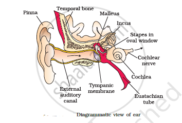
- Anatomically, the ear can be divided into three major sections called
- The outer ear:
- The outer ear consists of the pinna and ==external auditory meatus== (canal).
- The pinna collects the ==vibrations== in the air which produce sound.
- The external auditory meatus leads inwards and extends up to the ==tympanic membrane== (the ear drum).
- There are very fine hairs and ==wax-secreting glands== in the skin of the pinna and the meatus.
- The tympanic membrane is composed of ==connective tissues== covered with skin outside and with mucus membrane inside.
- The middle ear:
- The middle ear contains three ossicles called the malleus, incus, and stapes which are attached to one another in a ==chain-like fashion.==
- The malleus is attached to the ==tympanic membrane and the stapes== are attached to the ==oval window== of the cochlea.
- The ear ossicles increase the ==efficiency of transmission of sound waves== to the inner ear.
- The inner ear:
- A ==Eustachian tube connects the middle ear cavity with the pharynx.==
- The Eustachian tube helps in ==equalizing the pressures on either side== of the eardrum.
- The fluid-filled inner ear called labyrinth consists of two parts, the bony and the membranous labyrinths.
- The bony labyrinth is a series of channels.
- Inside these channels lies the membranous labyrinth, ==which is surrounded by a fluid called perilymph.==
- The ==membranous labyrinth is filled with a fluid called endolymph.==
- The coiled portion of the labyrinth is called the ==cochlea.==
- The membranes constituting ==cochlea, the reissner’s and basilar==, divide the surrounding perilymph-filled bony labyrinth into an ==upper scala vestibuli and a lower scala tympani.==
- The space within the cochlea called scala media is filled with ==endolymph.==
- At the base of the cochlea, the scala vestibule ends at the oval window, while the scala tympani terminates at the round window which opens to the middle ear.
- The organ of Corti is a structure located on the ==basilar membrane which contains hair cells that act as auditory receptors.==
- The hair cells are present in rows on the internal side of the organ of Corti.
- The basal end of the hair cell is in close contact with the afferent nerve fibers.
- A large number of processes called ==stereocilia== are projected from the apical part of each hair cell.
- Above the rows of the hair cells is a thin elastic membrane called a ==tectorial membrane.==
- The inner ear also contains a complex system called ==vestibular apparatus, located above the cochlea.==
- The vestibular apparatus is composed of ==three semi-circular canals and the otolith== (the macula is the sensory part of the saccule and utricle).
- Each semi-circular canal lies in a different plane at right angles to each other.
- The membranous canals are suspended in the perilymph of the bony canals.
- The base of the canals is swollen and is called the ampulla, which contains a projecting ridge called ==crista ampullaris== which has hair cells.
- The ==saccule and utricle contain a projecting ridge called macula.==
- The crista and macula are the specific receptors of the vestibular apparatus responsible for the ==maintenance of the balance of the body and posture.==
Mechanism of Hearing:
- The external ear receives sound waves and directs them to the ear drum.
- The ==eardrum vibrates== in response to the sound waves and these vibrations are transmitted through the ==ear ossicles== (malleus, incus, and stapes) ==to the oval window.==
- The vibrations are passed through the oval window ==onto the fluid of the cochlea, where they generate waves in the lymph.==
- The waves in the lymph induce a ==ripple in the basilar membrane.==
- These movements of the basilar membrane bend the hair cells, pressing them ==against the tectorial membrane.==
- As a result, ==nerve impulses== are generated in the associated afferent neurons.
- These impulses are transmitted by the ==afferent fibers via auditory nerves== to the ==auditory cortex of the brain==, where the impulses are analyzed and the sound is recognized.