TV4101 - Diagnostic Tests in Cardiology
Thoracic radiographs can give an idea of cardiac size using the vertebral heart score
Can also have idea if LA is enlarged
Most important - only widelh avail modality that you have cardiogenic oedema which you generally need to have CHF
Always take an Orthogonal view?
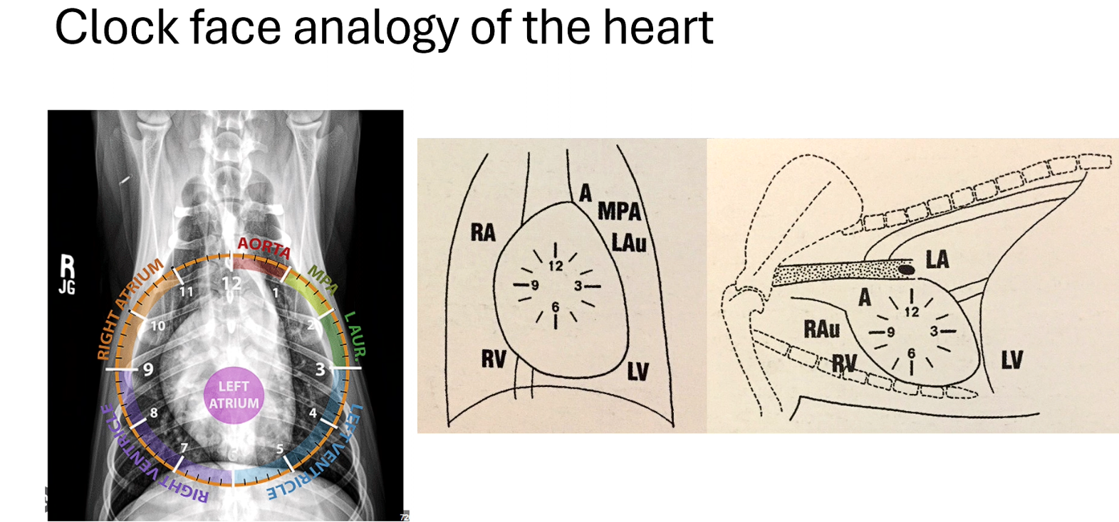
Clock allwos us to see which chamber is enlarged
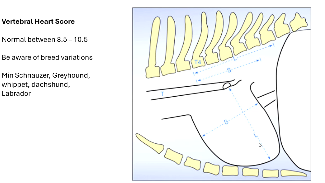
Measuring from carina down to apex of heart then a perpendicular line at widest point of heart
Then measure each line to see how many vertebrae they cover starting from T4 and that is you VHS
VHS another disadvantage
Diseases can cause enlarged hearts e.g. Hypertrophy or pericardial effusion
As such VHS is more a cardiac silhouette if the pericardium is full of fluid VHS will be larger than normal but we can’t diff btw chambers with radiograph
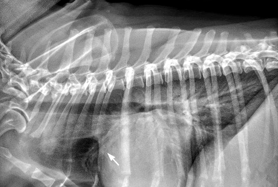
Arrow - Pulmonary vein larger than Pulmonary artery
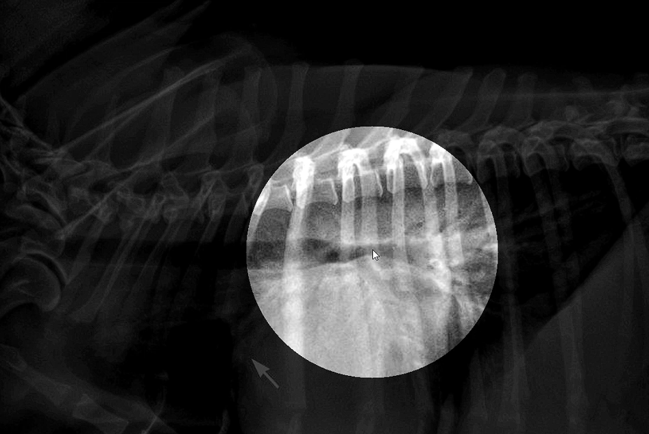
Left Atrial bulge
Both of these signs = Left atrial enlargement with congestive heart failure
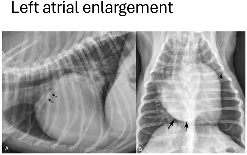
Can also see dorsal elevation of trachea
VHS is 14
This is a globoid cardiac silhouette
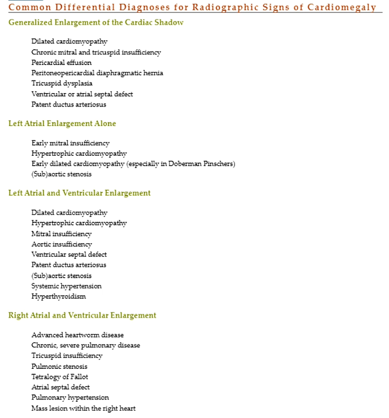
DDX?
Peric eff
Dilated cardiomyopathy
Severe valvular disease if you have severe remodelling
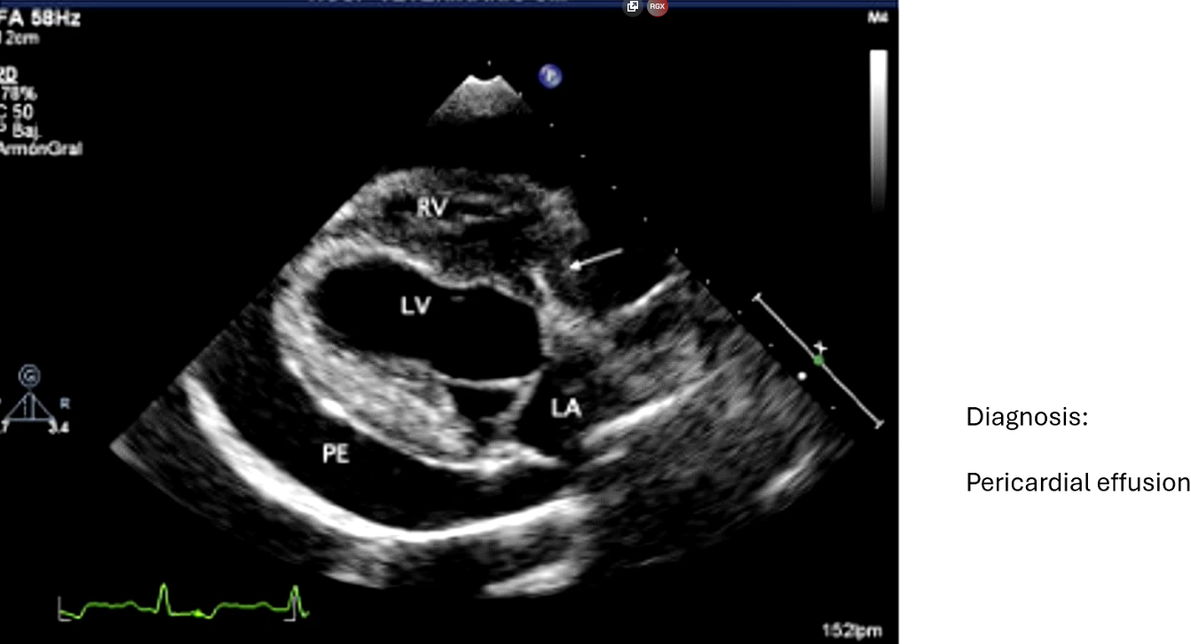

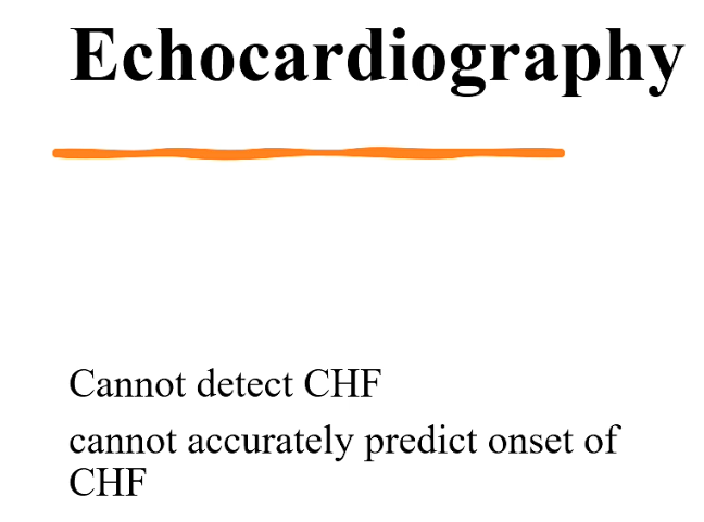
Echocardiography
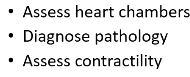
Assess chambers - shape, thickness of wall
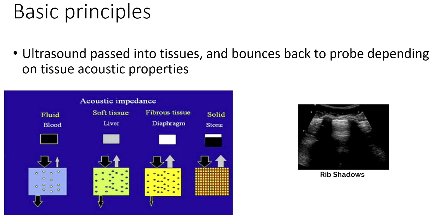
Fluid takes up no sound so is black
Bone is solid and bounces sound back so quite white
Does make an acoustic shadow underneath it
Another acoustic barrier is lung surface - fluid air surface
Seen as a bunch of lines going back and forth
No tissue detail due to bouncing
Liver absorb some sound
Echo sees only muscle blood and fluid

Phased Array chad probe
Small footprint - emits sound from 1-2 crystals so a small precise beam/point
Can do both kinds of doppler
Doopler types
Physics tool to measure blood flow
Bottom left pic shows colour flow doppler
Spectral doopler - diff way to plot flow (middle photo) - depicted as a graph
Anything floweing towards probe is a blood flow spike above baseline
Regurgitation is seen as little troughs
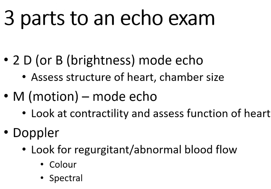
B mode / brightness mode
See a 2d image
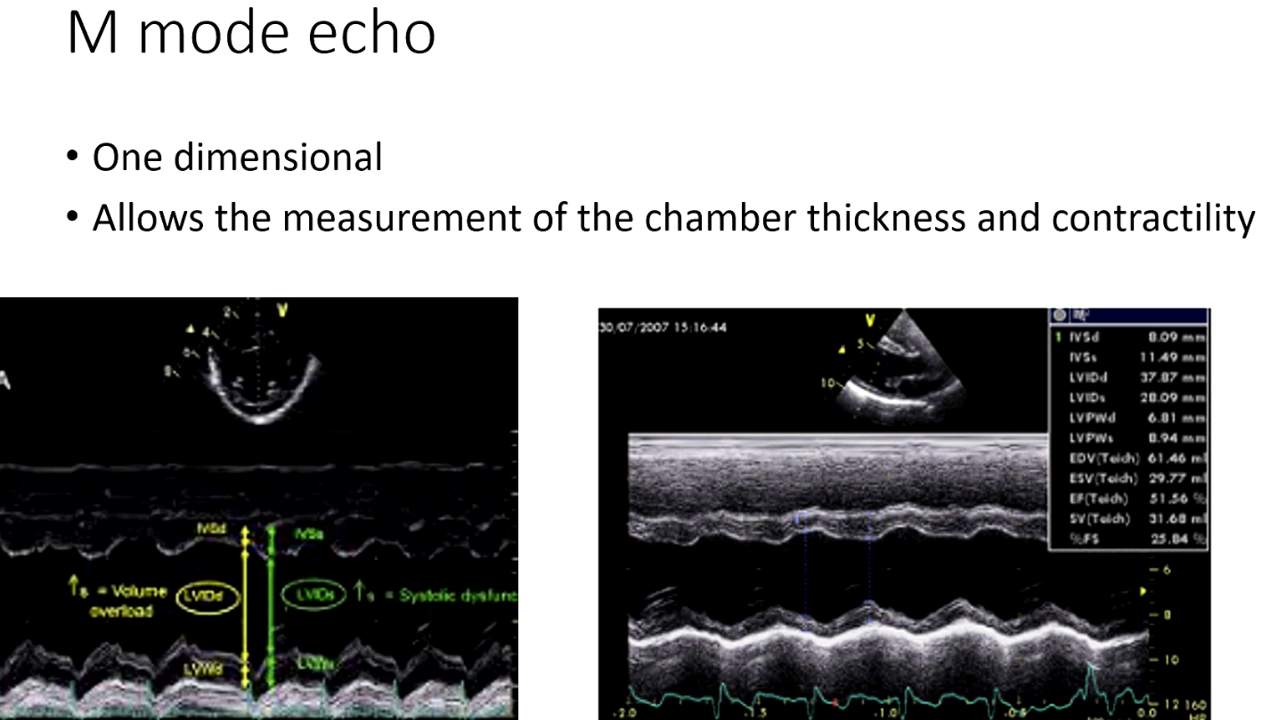
M mode
1 dimensional
Left pics
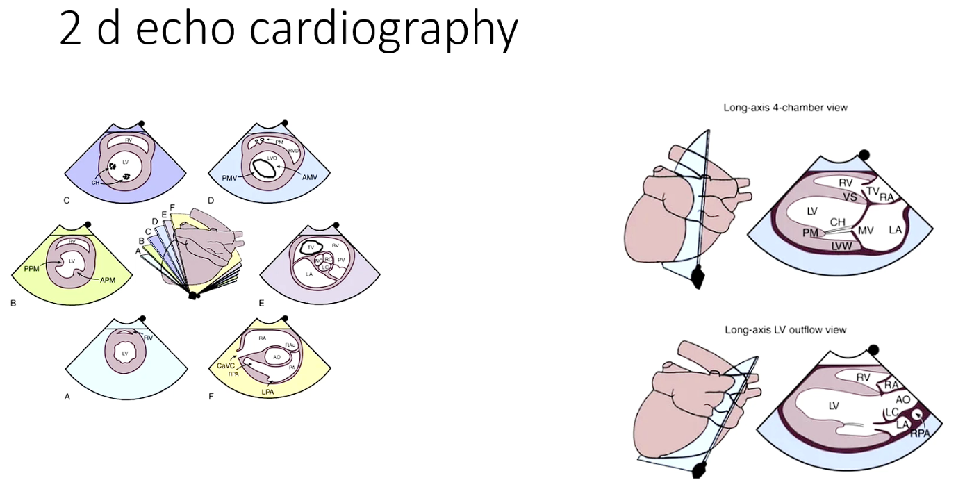
Transverse plane
Moving probe up higher towards aota and will be able to see more chambers (E)
Ratio of LA to aorta to determine if its is enlarged
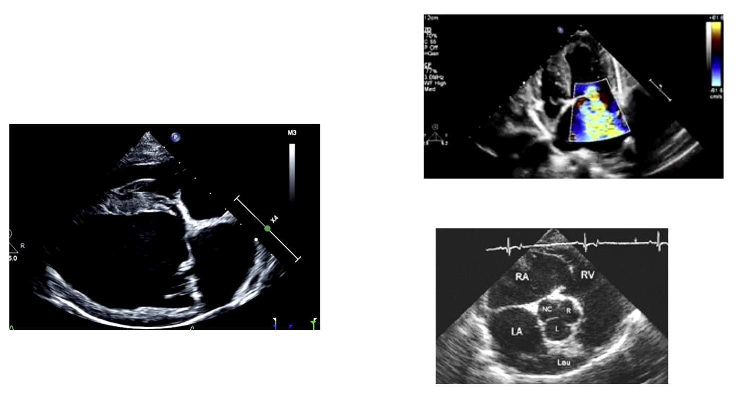
Left pic - Dog with DCM
Enlarged chamber
Valve is open so issue there
Top right
Colour flow doppler see the triangle of regurgitation
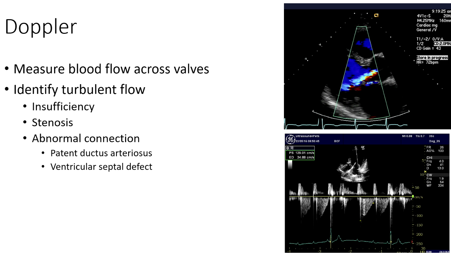
Turbulent flow
Vale insufficiency
Valve stenosis
Radiograph limit - csn’t assess chamber size
Thoracic X-ray advantage - can detect oedema and potential dx chf
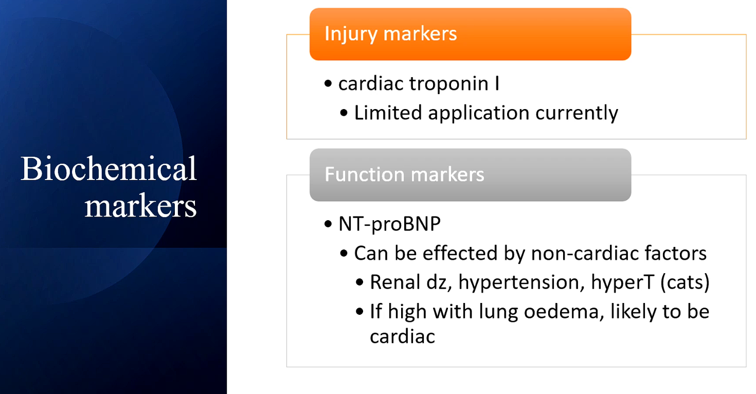
Trponin might have in horse with ionophore poisoning
Function marker
NT-proBNP = N terminal protein (hasn’t been coverted into active form yet) Brain naturalistic peptide
Actually comes from LA wtf?
When LA is stretched
Released to offest toe actions of rrare? rest? system and sympath nervous system - but itsn’t strong enough to beat them so no therapetuic value
It is a surrogate for neuro hormonal activaion in CHF (diagnostic value)
Also affected by Cushing’s
Or on fluids