Strength and Conditioning Exam 1
Chapter 1: Structure and Function of the Body
Skeletal System
Composed of 206 bones in the adult body
Provides leverage, support, and protection
Pulled on by muscles, ligaments, bones, and tendons.
Axial Versus Appendicular Skeleton
Axial: Skull, Vertebral Column (C1-Coxyx), Ribs and Sternum
Appendicular: Everything else
Types of Joints
Joint: Point at which bones meet
Fibrous: Allow virtually no movement
–Example: Sutures of the skull
Cartilaginous: Allow limited movement
–Example: Intervertebral
Synovial: Allow considerable movement
–Example: Elbow, knees, shoulder
Uniaxial: Operate as a hinge, rotate about one axis
–Example: Elbow, Knee
Biaxial: Operate in two perpendicular axes
–Example: Ankle
Multiaxial: Allow movement in all three axes
—Example: Shoulder, hip
Vertebral Column
Vertebral bones separated by flexible disks that allow for movement
–Cervical vertebrae (neck region): 7 (C1, C2)
–Thoracic vertebrae (upper back): 12
–Lumbar vertebrae (lower back): 5 (L4-L5)
–Sacral vertebrae (make up rear of pelvis): 5
–Coccygeal vertebrae (form vestigial tail extending down from the pelvis): 3-5
Muscular System
Macrostructure and microstructure
–Each skeletal muscle is an organ that contains muscle tissue, connective tissue, nerves, and blood vessels.
–Fibrous connective tissue, or epimysium, covers the body’s more than 430 skeletal muscles
A motor unit consists of a motor neuron and the muscle fibers it innervates.
There are typically several hundred muscle fibers in a single motor unit.
Epimysium (the outer layer)
Perimysium (surrounding each fasciculus, or group of fibers)
Endomysium (surrounding individual fibers
The arrangement of myosin (thick) and actin (thin) filaments gives skeletal muscle its striated appearance.
•The discharge of an Action Potential from a motor nerve signals the release of Ca2+ from the Sarcoplasmic Reticulum into the myofibril, causing tension development in muscle.
In stretched muscle the I-bands and H-zone are elongated, and there is low force potential due to reduced crossbridge–actin alignment. When muscle contracts (here partially), the
I-bands and H-zone are shortened
Sliding-filament theory of muscular contraction IN ORDER
–Resting phase
–Excitation–contraction coupling phase
–Contraction phase
–Recharge phase
–Relaxation phase
The number of crossbridges that are formed between actin and myosin at any instant in time dictates the force production of a muscle.
Calcium and ATP are necessary for crossbridge cycling with actin and myosin filaments
Neuromuscular System
Activation of muscles
–The extent of control of a muscle depends on the number of muscle fibers within motor unit
– Muscles that function with great precision may have as few as 1 junction.
– Muscles that require less precision may have several 100’s or junctions.
all-or-none principle: All of the muscle fibers in the motor unit contract and develop force at the same time. There is no evidence that a motor neuron stimulus causes only some of the fibers to contract. Similarly, a stronger action potential cannot produce a stronger contraction
Unfused Tetanus: When after exercise a muscle twitches slightly for a brief period of time
Fused Tetnaus: When a muscles locks up, ex: a cramp
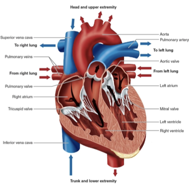
Cardiovascular System
Heart
–Valves
Tricuspid valve and mitral (bicuspid) valve
Aortic valve and pulmonary valve
–Conduction system
-Sa Node: Natural pacemaker
Av Node: Send the signals to the rest the the body
Cardiac Conduction
Rhythmicity and conduction properties of myocardium
–Influenced by cardiovascular center of medulla
–Signals transmitted through sympathetic and parasympathetic nervous systems
–Bradycardia: Less than 60 BPM
–Tachycardia: More than 100 BPM
Cardiovascular System
Heart
–Electrocardiogram records all heart electrical activity at the surface of the body. (EKG/ECG)
Blood vessels
–Blood vessels are close circuit systems (if u get cut in a bv you will bleed out)
–The Arterial system: carries oxygenated blood away from the heart to the body.
–The Venous system: returns deoxygenated blood to the heart
–Arteries
–Capillaries
–Veins
Blood
– Hemoglobin: transports oxygen in the blood and serves as an acid–base buffer. These are inside RBC.
– Red Blood Cells: facilitate carbon dioxide removal
Key Point
The Cardiovascular System transports nutrients and removes waste products while helping to maintain the environment for all the body’s functions. Blood transports O2 from lungs to the tissues for use in cellular metabolism; and it transports the CO2 back to the lungs, where it is removed from the body.
Respiratory System
The exchange of O2 and CO2 is the primary function of the respiratory system.
Exchange of air
–The amount and movement of air and expired gases in and out of the lungs are controlled by the expansion and recoil of the lungs. This is controlled by the diaphragm.
Chapter 2: Biomechanics of Resistance Exercise
Key Terms
Biomechanics: The mechanisms through which components(muscles, tendons, ligaments) interact to create movement
Agonist : The muscle most directly involved in bringing about a movement; also called the prime mover.
Antagonist : A muscle that can slow down or stop the movement.
Synergist : A muscle that assists indirectly in a movement
Mechanical advantage: The ratio of the moment arm through which an applied force acts to that through which a resistive force acts.
Concentric muscle action: Full range of motion where the muscle I shortened thorough the motion (Positive) The forces generated within the muscle and acting to shorten it are greater than the external forces acting at its tendons to stretch it.
Eccentric muscle action: In a tension extension state lowering back down (negative) . The forces generated within the muscle and acting to shorten it are less than the external forces acting at its tendons to stretch it.
Isometric Contraction: A muscle action in which the muscle length does not change, because the contractile force is equal to the resistive force. The forces generated within the muscle and acting to shorten it are equal to the external forces acting at its tendons to stretch it.
Isokinetic Contraction: A muscle action in which the muscle contracts at a constant velocity. Specialized equipment is typically required to maintain this constant speed throughout the range of motion.
Isotonic Contraction: A muscle action in which the muscle tension remains constant throughout the range of motion, while the muscle shortens or lengthens. This term often refers to dynamic movements where the load is relatively constant, although true constant tension is rare in most exercises.
Skeletal Musculature
Skeletal musculature: A system of muscles enables the skeleton to move.
Origin = Proximal (toward the center of the body) attachment.
Insertion = Distal (away from the center of the body) attachment.
First-class lever: the muscle force and resistive force act on opposite sides of the fulcrum
Second-class lever: the muscle force and resistive force act on the same side of the fulcrum,
third-class lever: the muscle force and resistive force act on the same side of the fulcrum, with the muscle force acting through a moment arm shorter than that through which the resistive force acts.
Anatomical Planes and Major Body Movements
Anatomical Reference point: The body is erect, the arms are down at the sides, and the palms face forward, this is called
The Sagittal plane slices the body into left–right sections.
The Frontal plane slices the body into front–back sections.
The Transverse/Longitudinal plane slices the body into upper–lower sections.
Human Strength and Power
Strength: The capacity to exert force at any given speed .
Acceleration: The change in velocity per unit of time. This is associated with resistive force by Newton’s second law (Force = Mass × Acceleration
Power: Outside of the scientific realm, power is loosely defined as “ Explosive strength .” Also defined as the time rate of doing work. Is found by Work / Time
Positive Work: Force exerted on an object and the distance it travels. Is found by Force × Displacement
Negative Work: Occurs during an eccentric contraction
Biomechanical factors in human strength: Neural control the force output. More neuron junctions the more you can control the muscle
Sources of Resistance to Muscle Contraction
When the weight is horizontally closer to the joint, there are less resistance forces (making it easier)
When the weight is horizontally farther from a joint, there are more resistance forces (making it harder)
Fluid resistance is the resistive force encountered by an object moving through a fluid (liquid or gas), or by a fluid moving past or around an object or through an opening.
The more an elastic component is stretched, the more it'll come back
The “fluid ball” resulting from contraction of the deep abdominal muscles and the diaphragm.
Valsalva Maneuver: The glottis is closed, thus keeping air from escaping the lungs, and the muscles of the abdomen and rib cage contract, creating rigid compartments of liquid in the lower torso and air in the upper torso. “Holding your breath to protect your organs.
The shoulder is prone to injury during weight training because of its structure and the forces to which it is subjected.Warm up with relatively light weights.Follow a program that exercises the shoulders in a balanced way. Exercise at a controlled speed.
The knee is prone to injury because of its location between two long levers.
Elbows and wrists: The primary concern involves overhead lifts. However, the most common source of injury to these areas is from overhead sports such as throwing events or the tennis serve.
Chapter 3: Bioenergetics of Exercise and Training
Key Terms
bioenergetics: The flow of energy in a biological system; the conversion of macronutrients into biologically usable forms of energy. (eating)
catabolism: The breakdown of large molecules into smaller molecules, associated with the release of energy. (breakdown of large molecules to small molecules)
anabolism: The synthesis of larger molecules from smaller molecules; can be accomplished using the energy released from catabolic reactions. (Taking small molecules and building it up into large molecules)
exergonic reactions: Energy-releasing reactions that are generally catabolic. (breakin down)
endergonic reactions: Require energy and include anabolic processes and the contraction of muscle. (building up)
metabolism: The total of all the catabolic or exergonic and anabolic or endergonic reactions in a biological system.
adenosine triphosphate (ATP): Allows the transfer of energy from exergonic to endergonic reactions. (the basis of all energy in our body)
lactate threshold (LT): The exercise intensity or relative intensity at which blood lactate begins an abrupt increase above the baseline concentration.
Biological Energy Systems
Three basic energy systems/metabolic pathways exist in muscle cells to replenish ATP:
–Phosphagen system
–Glycolysis
—Oxidative system
Phosphagen system
–Provides ATP primarily for short-term, high-intensity activities (e.g., resistance training and sprinting) and is active at the start of all exercise regardless of intensity (all exercise starts in this system, can also be called the ATP-PCR system)
–Creatine kinase catalyzes the synthesis of ATP from CP and ADP
The body does not store enough ATP for exercise. It replenishes after exercise ends.
Some ATP is needed for basic cellular function.
The phosphagen system uses the creatine kinase
reaction to maintain the concentration of ATP.The phosphagen system replenishes ATP rapidly
Glycolysis: The breakdown of carbohydrates—either glycogen stored in the muscle or glucose delivered in the blood—to resynthesize ATP
ADP: adenosine diphosphate
ATP = adenosine triphosphate
NAD+, NADH = nicotinamide adenine dinucleotide
Krebbs Cycle: A central metabolic pathway in the mitochondria that breaks down acetyl-Coenzyme A (derived from carbohydrates, fats, and proteins) to produce electron carriers (NADHNADH and FADH2FADH2) for subsequent ATPATP generation in the electron transport chain, releasing CO2CO2 as a byproduct.
The endT result of glycolysis is pyruvate may proceed in one of two directions: either Pyruvate can be converted to lactate. or ATP resynthesis occurs at a faster rate but is limited in duration.
—This process is sometimes called anaerobic glycolysis (or fast glycolysis).
Energy yield of glycolysis:
•Glycolysis from one molecule of blood glucose yields a net of two ATP molecules.
•Glycolysis from muscle glycogen yields a net of three ATP molecules.
The oxidative (aerobic) system: Primary source of ATP at rest and during low-intensity activities. Uses primarily carbohydrates and fats as substrates (EX: walking)
Fat oxidation: Triglycerides stored in fat cells can be broken down by hormone-sensitive lipase. This releases free fatty acids from the fat cells into the blood, where they can circulate and enter muscle fibers. Some free fatty acids come from muscles
Protein oxidation: Protein is not a significant source of energy for most activities.
Substrate Depletion and Repletion
Phosphagens
–Creatine phosphate can decrease markedly
(50-70%) during the first stage (5-30 seconds) of high-intensity exercise and can be almost eliminated as a result of very intense exercise to exhaustion.–Postexercise phosphagen repletion can occur in a relatively short period; complete resynthesis of ATP appears to occur within 3 to 5 minutes, and complete creatine phosphate resynthesis can occur within 8 minutes.
Glycogen
–The rate of glycogen depletion is related to exercise intensity.
At relative intensities of exercise above 60% of maximal oxygen uptake, muscle glycogen becomes an increasingly important energy substrate; the entire glycogen content of some muscle cells can become depleted during exercise
Glycogen is the most important energy substrate
–Repletion of muscle glycogen during recovery is related to postexercise carbohydrate ingestion.
•Repletion appears to be optimal if 0.7 to 3.0 g of carbohydrate per kilogram of body weight is ingested every 2 hours following exercise.
excess postexercise oxygen consumption (EPOC): Oxygen uptake above resting values used to restore the body to the preexercise condition; also called postexercise oxygen uptake, oxygen debt, or recovery O2.
VO2max = maximal oxygen uptake
The appropriate exercise intensities and rest intervals allows for the “selection” of specific energy systems during training and results in more efficient and productive regimens for specific athletic events with various metabolic demands.
Metabolic Specificity of Training
Interval training: Much more training can be accomplished at higher intensities. Difficult to establish definitive guidelines for choosing specific work-to-rest ratios
High-intensity interval training (HIIT): Brief repeated bouts of high-intensity exercise with intermittent recovery periods to elicit cardiopulmonary, metabolic, and neuromuscular adaptations.Suggested work-to-rest ratios >1:1
Combination training: Adds aerobic endurance training to the training of anaerobic athletes in order to enhance recovery (because recovery relies primarily on aerobic mechanisms). May reduce anaerobic performance capabilities, particularly high-strength, high-power performance