Topic 5_Anatomy of the Muscular System
Muscle System Overview
Chapter 9: p. 281-282
Chapter 10: p. 323-384 (Sections 10.1 – 10.5, omit p. 325-327)
Content focus: only the muscles included in the PowerPoint presentation.
Usage Rights
Educational Only: This PowerPoint presentation is for educational purposes only.
Do not post or distribute without permission from the author.
Affiliation: Université d’Ottawa | University of Ottawa
Skeletal Muscles Overview
Learning Objectives
Describe the functions of skeletal muscles.
Describe characteristics of muscle tissue.
Understand the gross structure of skeletal muscles, including membranes and tendons/aponeuroses.
Functions of Skeletal Muscles
Produce movement
Maintain posture and body position
Stabilize joints
generate heat- shivering
Additionally:
protects organs forms valves controls pupil size
support soft tissue
guard entracnes and exit
store nutritional reserves.
Characteristics of muscle tissue
excitability (responsiveness)
Contractility (the ability to shorten and generate force) is another key characteristic, allowing muscles to perform work and produce movement.
Extensibility: ability to be stretched
Elasticity: ability to recoil or resume resting length after being stretched
Fascicle Arrangements
Identify different types of fascicle arrangements in skeletal muscles.
Define:
Agonist: muscle that provides major force for a movement.
Antagonist: opposes/reverses a particular movement.
Synergist: helps the prime mover and reduces undesirable movement.
Fixator: a type of synergist that immoblizes bone.
Origin: attachment of muscle that remains fixed.
Insertion: attachment that moves during contraction.
Muscle Nomenclature
Explain the general nomenclature of skeletal muscles.
Discuss origin, insertion, and action of:
Muscles of facial expression.
Muscles that move the eyeballs, mandible, and tongue.
Muscles of the neck.
Muscles moving the vertebral column.
Respiratory muscles.
Muscles of the pelvic floor and perineum.
Muscles that move the pectoral girdle.
Muscles of the arm, forearm, thigh, leg, hand, and foot.
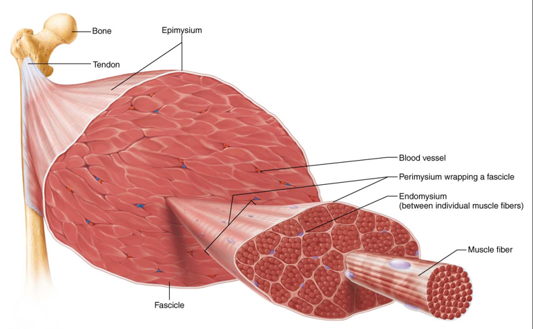
Connective Tissue Sheaths
Types of Connective Tissue Wrappings
Endomysium: fine areolar CT surrounding each muscle fiber.
Perimysium: dense irregular CT around fascicles (groups of muscle fibers).
Epimysium: dense, irregular CT surrounding the entire muscle.
Deep Fascia: coarser layer binding muscles into functional groups.
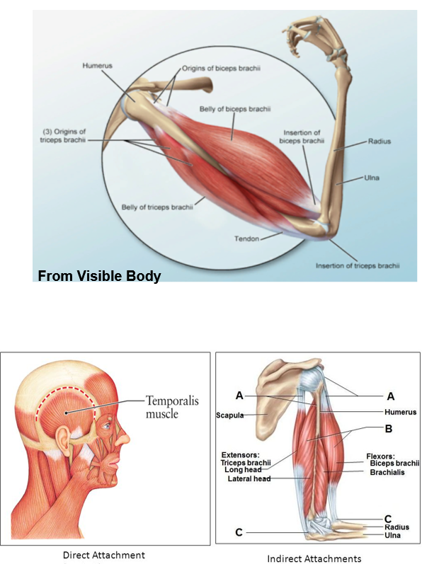
Muscle Attachments
Direct Attachment: epimysium fused to the periosteum of bone or perichondrium of cartilage.
Indirect Attachment: most muscles attach via tendons or aponeuroses, spanning joints and attaching to bones at two points.
Muscle Actions and Interactions
muscles can only pull;never push
muscles can work together or in opposition
3 functional groups: prime movers, antagonists, synergists
muscles can change their role depending on the movement
Functional Groups
Prime Mover (Agonist): major force for a specific movement.
e.g. brachialis in elbow flexion
Antagonist: opposes the action of prime movers.
e.g. triceps in elbow flexion
Synergist: assists the prime mover to create the same movement.
add force
reduce unecessary movements
holding a bone steady: fixators
All muscles can change roles depending on the activity (ex: triceps brachii).
Fascicle Arrangement
Types of Fascicle Arrangements
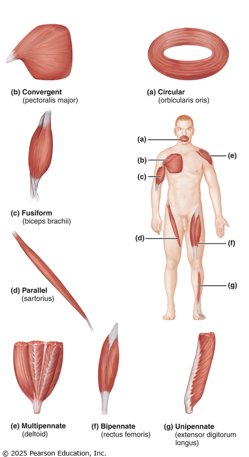
Circular: concentric rings around openings (e.g., orbicularis oculi).
Parallel: fascicles parallel to long axis (e.g., sartorius, biceps brachii).
Pennate: short fascicles attaching obliquely to a central tendon (unipennate, bipennate, multipennate).
Convergent: fascicles converge towards a single tendon (e.g., pectoralis major).
Naming Skeletal Muscles
Criteria for Naming
Location: associated with a bone or body region (e.g., intercostals).
Shape: named based on shape (e.g., deltoid = triangle).
Size: descriptors (e.g., maximus for largest).
Direction: fiber orientation (e.g., rectus = straight).
Number of Origins: e.g., biceps (two heads).
Attachments: named for points of origin and insertion.
extensor carpi radialis longus
muscle that extends the wrist and is located along the radial side of the forearm.
when it gets this specific, usually means there is a carpi ulnaris, otherwise it would just be extensor carpi longus.
Muscles of Facial Expression
Key Muscles
Epicranius (Frontal and Occipital Bellies): raises eyebrows and fixes the aponeurosis.
Platysma: tenses neck, depresses the mandible.
Orbicularis Oculi: protects eyes, enables blinking.
Zygomaticus: raises corners of the mouth.
Buccinator: involved in whistling and chewing.
Muscles of Mastication
Key Muscles
Masseter: primary jaw closure muscle.
Temporalis: elevates and retracts mandible.
Innervated by mandibular division of cranial nerve V.
Eye Movement Muscles
Key Muscles and Functions
Lateral Rectus: moves the eye laterally (innervated by CN VI).
Medial Rectus: moves the eye medially (innervated by CN III).
Superior Rectus: elevates the eye (innervated by CN III).
Inferior Rectus: depresses the eye (innervated by CN III).
Superior Oblique: depresses and turns the eye laterally (innervated by CN IV).
Inferior Oblique: elevates and turns the eye laterally (innervated by CN III).
Muscles of the Neck
Key Muscles
Sternocleidomastoid: prime mover for head flexion.
Scalenes: elevates first two ribs and aids in neck rotation.
Back and Vertebral Muscles
Key Muscles
Erector Spinae: prime mover of back extension.
Quadratus Lumborum: helps lateral flexion of the vertebral column.
Respiratory Muscles
Key Muscles
External Intercostals: elevate the rib cage during inspiration.
Internal Intercostals: depress the rib cage during expiration.
Diaphragm: prime mover of inspiration, flattening during inhalation.
Abdominal Wall Muscles
Key Muscles
Rectus Abdominis: flexes and stabilizes the pelvis.
External Obliques: aid in trunk rotation.
Transversus Abdominis: compresses abdominal contents.
Muscles of the Pelvic Floor
Key Functions
Levator Ani and Coccygeus: support pelvic organs, resist intra-abdominal pressure.
Muscles of the Upper Limb
Shoulder Joint Muscles
Pectoralis Major: prime mover of arm flexion, adduction, and medial rotation.
Latissimus Dorsi: prime mover of arm extension and adducts the arm.
Deltoid: prime mover of arm abduction.
Elbow Joint Muscles
Biceps Brachii: flexes elbow and supinates forearm.
Triceps Brachii: powerful forearm extensor.
Muscles of the Lower Limb
Key Groups
Iliopsoas: major hip flexor.
Quadriceps Femoris: prime mover of knee extension.
Hamstrings: prime movers of thigh extension and knee flexion.
Leg and Ankle Muscles
Gastrocnemius: plantar flexes the foot.
Soleus: assists in plantar flexion.
Tibialis Anterior: prime mover of dorsiflexion.
Intrinsic Muscles of the Hand and Foot
Control precise movements of fingers and toes; not required to memorize specific muscles.