Cytoskeleton, Extracellular Components, & Infectious Diseases Review
Cytoskeleton:
Network of fibers stretching through the cytoplasm.
Plays a major role in organizing the structures & activities of the cell.
Can also serve as tracks transporting stuff from one place to another.
Cytoskeleton Functions
Gives mechanical support to the cell and maintain its shape.
This is essential for the animal cells since they don’t have cell walls.
Stabilized by a balance of opposing forces exerted by its elements.
Provides anchorage in organelles and helps them stay in place.
It can dismantle itself and can reassemble itself in a new place in the cell, which helps change the shape of the cell.
Interacts with motor proteins that leads to cell motility (includes both changes in cell location and the movements of the parts in the cell).
They work together with plasma membrane molecules to allow cells to move along fibers.
The cytoskeleton acts as a track on which motor proteins “walk” across that carry vesicles or organelles.
EX. vesicles carrying neurotransmitter molecules use motor proteins to “walk” across the cytoskeleton track to the tips of axons.
Manipulates the plasma membrane by bending it forward to form food vacuoles or vesicles via phagocytosis.
 Components of the Cytoskeleton:
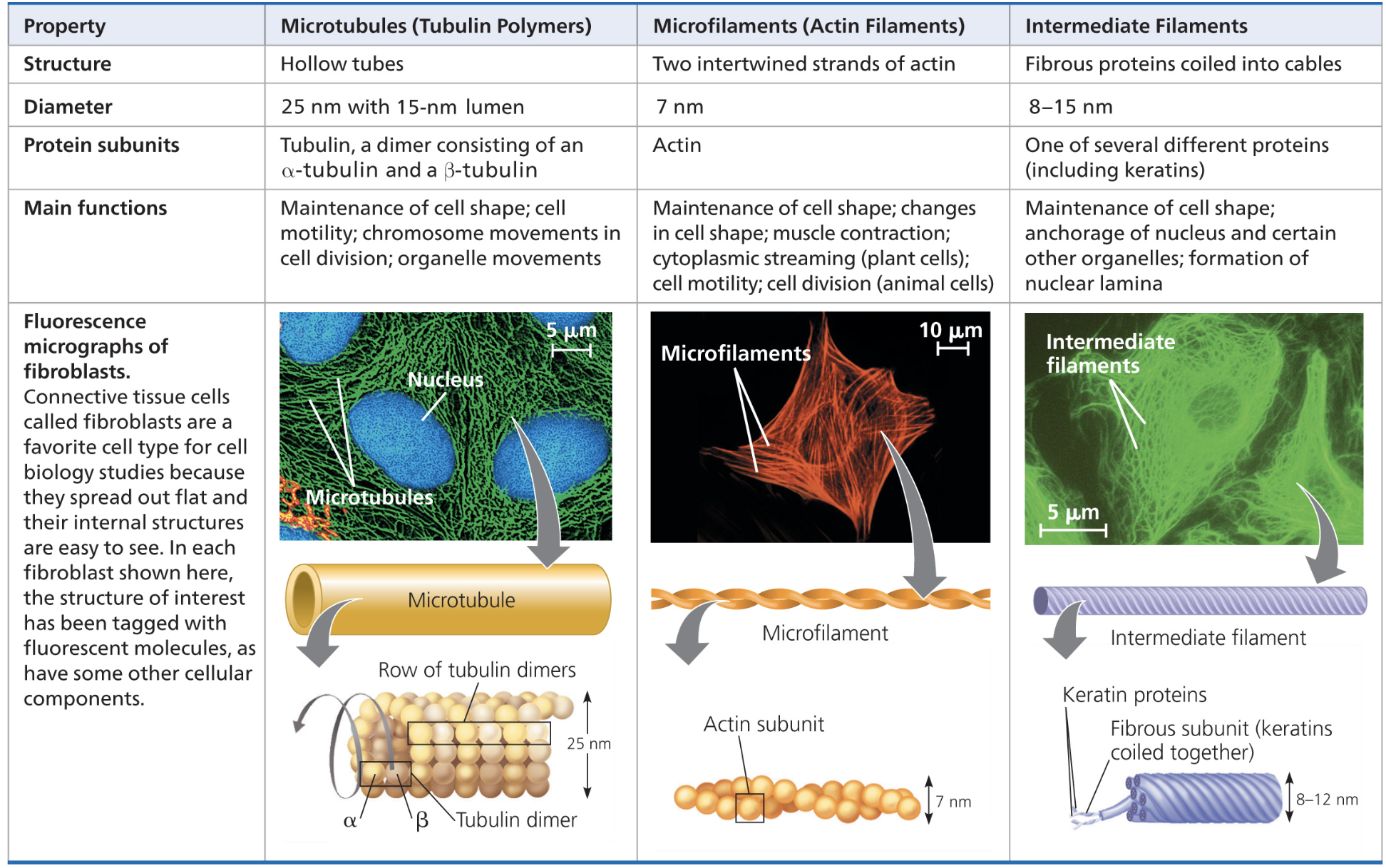
Microtubules:
Hollow rods made from tubulin, all eukaryotic cells have microtubules.
Tubulin is a dimer → a molecule with 2 components.
Has alpha tubulin and beta tubulin.
Grow by adding tubulin dimers; can be disassembled & reassembled by removing & attaching tubulin dimers.
The ends of microtubules are slightly different; one end (called the plus side) can accumulate & release tubulin dimers much more quicker than the other → grows & shrinks more significantly during cellular activities.
Shape & support cells, act as tracks where organelles w/ motor proteins can move on.
Can guide vesicles from the ER to the Golgi apparatus and from the Golgi to the plasma membrane.
Involved in the separation of chromosomes during cell division.
Centrioles & Centrosomes (IN ANIMAL CELLS):
Microtubules grow out of the centrosome → region that is located near the nucleus.
Pair of centrioles found in the centrosome
Cylinder of 9 sets of triplet microtubules.
Helps with mitosis.
Cilia & Flagella
Composed of microtubules, their unique arrangement is responsible for the beating of the structure.
Have a group of microtubules in an extension of the plasma membrane (called the basal body) in a 9 + 2 pattern (9 pairs of microtubules in a ring with 2 in the middle)
Nonmotile cilia have a 9 + 0 pattern.
Motor proteins called dyneins are attached in between each other MT doublet for bending.
Cilia → specialized for locomotion, short appendages, occurs in large numbers on cell surface.
Alternating strokes, like the oars of a boat.
Can act as an antenna for nonmotile cells, transmits molecular signals from the cell’s environment to its interior which can lead to changes in cell activity.
Is crucial for brain function & embryonic development.
Flagella → specialized for locomotion, long appendages, usually only one or two per cell.
Undulating motion, tail of a fish.
Unicellular protists & bacteria use cilia & flagella that act as locomotor appendages.
Can move fluid over the surface of the tissue if it is attached to the end of cell that is attached tightly.
EX. Cilia on the windpipe sweeps mucus out of the lungs.
EX. Cilia on the oviducts helps move the egg to the uterus in the female reproductive system.

Microfilaments:
Thin solid rods made from actin, is a twisted double chain of actin proteins.
Bears tension to keep the cell together.
Creates a 3D network in the inside of the plasma membrane to support cell shape.
Gives the outer cytoplasmic layer, cortex, a solid consistency.
Are the core of microvilli in ANIMAL CELLS → increase the cell’s surface area.
Interact w/ filaments made from myosin to cause contraction of muscle cells.
Creates localized contractions & motility in amoebas by extending pseudopodia & moving towards them.
Actin - protein interactions lead to cytoplasmic streaming in PLANT CELLS → circular flow of cytoplasm.
Intermediate Filaments (ONLY IN ANIMAL CELLS):
Made from molecular subunits of a certain family of proteins that include keratin.
Are more permanent fixtures of cells.
Reinforce the shape of the cell & fixes the position of certain organelles.
Nucleus sits in a cage made of intermediate filaments.
Extracellular Components:
Cell Wall (PLANTS) 🌱 🪴
Protects the plant cell, maintains the plant cell shape, and prevents excessive uptake of water.
Holds the plant up against gravity.
Are much thicker than plasma membranes
Made up of cellulose with a combination of other polysaccharides and proteins.
Young plant cells first have a primary cell wall
Primary cell walls of adjacent plant cells are connected withe middle lamella, which acts as a glue layer of pectin.
When the plant cell stops growing, the primary cell wall is either hardened or a secondary cell wall is added.
Extracellular Matrix (ANIMALS)
Made up of glycoproteins such as collagen, which are embedded with proteoglycans.
Proteoglycan → small core protein w/ many carbohydrate chains covalently attached to it.
Some cells are attached to the ECM by ECM glycoproteins like fibronectin.
Bind to cell surface receptor proteins called integrins .
Regulates cell behavior
EX. cells in developing embryos migrate by matching its orientation of microfilaments with the orientation of the ECM.
Can influence gene activity in the nucleus.
ECM information reaches the nucleus via mechanical & chemical signaling (includes microfilaments) Changes in cytoskeleton influences this signaling, which leads to changes in protein and cell function.
Plasmodesmata (PLANTS) 🌱 🪴
Found in plant cell walls, are channels that connect plant cells together.
Unify the plant into one living continuum.
Water & small solutes travel in plasmodesmata from cell to another cell, even protein & RNA can do this too.
They travel to the plasmodesmata via cytoskeleton fibers.
Junctions in ANIMAL cells:
Three types of junctions, mainly found in epithelial tissue
Tight Junctions
Desmosomes
Gap Junctions
Tight junctions:
PM of neighboring cells are tightly pressed against each other
Form continuous seals that prevent leakage of extracellular fluid
Desmosomes:
Function like rivets & fasten cells together into strong sheets
Intermediate filaments anchor desmosomes in the cytoplasm
Attach muscle cells together.
Gap Junctions:
Provide cytoplasmic channels between cells, similar to plasmodesmata.
Consists of membrane proteins that extend from the membranes of the two adjacent cells.
Create pores where ions, sugars, amino acids, and small molecules can pass through from cell to cell.
Required for cell to cell communication in heart and muscle tissue.
Bacteria Morphology:
The average size of bacteria is 2 micrometers w/ a diameter of 0.5 micrometers.
There are 3 major shapes of bacteria:
Spherical (cocci)
Rod shaped (bacilli)
Spiral
Spherical shaped (cocci)
Can occur singularly or in groups resembling chains or clusters.
Monococcus → single spherical shaped bacteria
Diplococcus → pair of spherical shaped bacteria.
Streptococcus → chain of multiple spherical shaped bacteria, think a necklace.
Staphylococcus → cluster of multiple spherical shaped bacteria, like a cluster of grapes
Rod shaped (bacilli)
Rod shaped, can occur singularly or in chains
Bacillus → single unattached bacterial cell.
Diplobacillus → pair of two rod shaped bacteria
Streptobacillus → chain of rod shaped bacteria.
Found in many types of bacteria.
Spiral shaped
Spirochetes → more tightly wound & are cork screw shaped
Can resemble coils or commas, such as vibrio.
Gram Staining:
It is a technique to differentiate between gram positive and gram negative bacteria.
Gram positive bacteria have a thick layer of peptidoglycan in their cell walls and are more simpler.
Gram negative bacteria don’t have much peptidoglycan in their cell walls and are more complex.
Gram staining has three processes:
1) Staining with crystal violet and gram iodine (primary stain):
The bacteria sample is stained purple with crystal violet. Gram iodine acts as a complex and fixes the dye to the bacteria to prevent easy removal of the dye.
2) Rinsing with ethanol (decolorization):
Ethanol dehydrates the peptidoglycan layer, so it shrinks and tightens around the cell wall. Because of this, the crystal violet in gram positive bacteria cannot be washed away from the ethanol. However, since there is less peptidoglycan in gram negative bacteria, the crystal violet is able to be washed away, so purple color is lost.
At this step, gram positive bacteria still look purple and gram negative bacteria look colorless.
3) Staining with safranin (secondary stain):
The bacteria sample is stained with safranin, which appears pink. In gram positive bacteria, safranin has no effect on the purple stain as it is a lighter and weaker stain than the crystal violet. In gram negative bacteria, the safranin is retained and the gram negative bacteria appear pink.
Gram staining can be used to determine if infections are caused by gram positive or gram negative bacteria.
This makes a big difference; gram negative bacteria can cause fever or shock since they have toxic lipid components in their cell wall and are more resistant to antibiotics.
However, gram positive bacteria can have virulent strains that can be antibiotic resistant.
Viruses 🦠 :
Viruses are nonliving:
Cannot carry out metabolic activity outside of a host cell.
Cannot be killed using antibiotics
Don’t have cell walls.
However, viruses have the following characteristics:
They are acellular
Parasitic
Can have either a RNA or a DNA genome
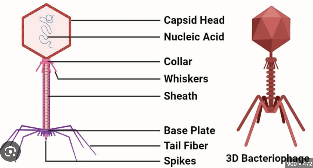
Has a protein capsid
The most common form of a viral structure is the bacteriophage.
Viruses can’t make proteins because they simply don’t have ribosomes.
They are obligate intracellular parasites and can only reproduce in host cells.
Viruses can only affect a certain number of host species, this is called their host range.
For example, viruses with broad host ranges like West Nile virus can infect mosquitos, birds, and humans. Measles have a very narrow host range, since they can only infect humans.
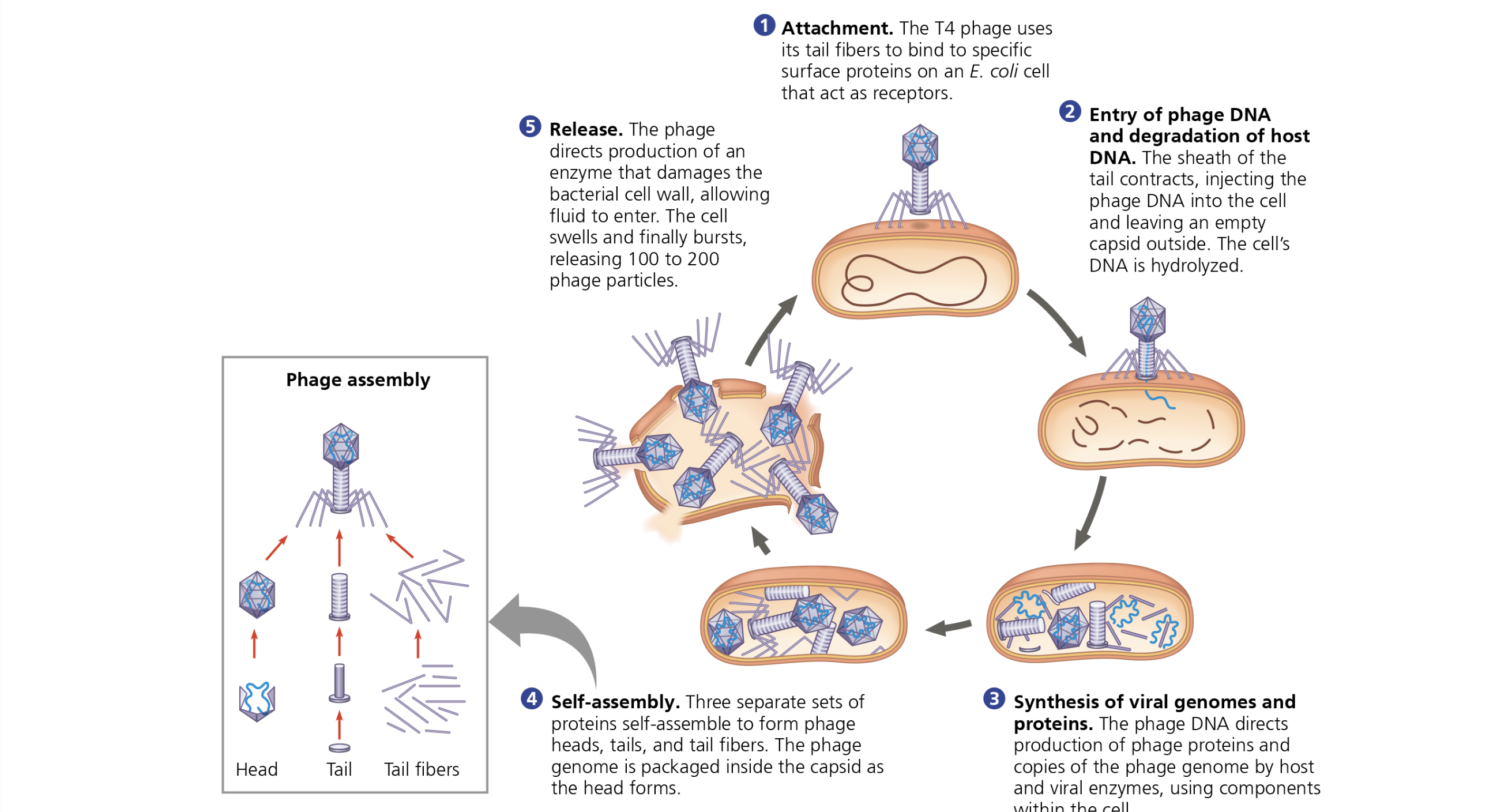
The Lytic Reproductive Cycle:
This cycle results in the death of the host cell.
A bacteriophage binds to surface proteins of a bacterium
The phage injects its genetic material into the bacterium and the host cell’s DNA is hydrolyzed.
The phage’s DNA with the host cell’s DNA is used to create copies of the phage genome and phage proteins.
The phage proteins and genomes self assemble to create bacteriophages inside the cell.
When the host cell becomes full of the viruses, it lyses/breaks open and releases the bacteriophages into the environment.
The Lysogenic Cycle
Does not kill the host cell and allows replication of the phage genomes
The phage binds to the surface proteins of a host cell.
The bacteriophage injects its reproductive material into the cell.
The phage reproductive genomes is incorporated into the host genome, creating a prophage
Whenever the host cell reproduces, the prophage is also reproduced along with the host genome.
Infectious Diseases: