Primary Tooth Trauma
Dental Trauma
• TDIs are common. The mouth is the second most common area to injure.
• 22% of children 0-6 years have sustained TDI
• Injury related to the teeth and/or periodontium (Gingiva, periodontal ligament or alveolar bone) • Many involve soft tissue injury (lips, tongue etc)
• Most common injury in primary teeth: injury to periodontal ligament resulting in luxation
• Non-accidental injuries (50% of children who have suffered physical abuse will have an oro-facial injury)
Aetiology Primary Dentition
• Boys: 30-40%
• Girls: 15-30%
Peak age 2-4 years
Trauma history
• Who are they with? Who has PR?
• Where did the injury occur? Contamination. Legal issues
• How did the injury occur? Impact zone
• When did the injury occur? Essential for appropriate management
• Any other injuries?
• Was there a period of unconsciousness? If so, for how long? Amnesia, nausea and vomiting are all signs of brain damage and require medical attention.
• Is there any disturbance in the bite? An affirmative answer may indicate a luxation injury with displacement, an alveolar or jaw fracture or a fracture of the condylar region.
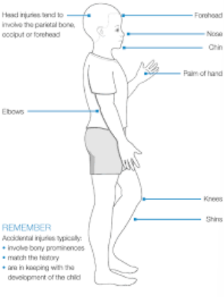
Non- accidental injury?
• Does the history fit with the injury?
ALWAYS CONSIDER NAI
• 50% of children at risk will have oro-facial injuries
• Repeated episodes of trauma
• Delayed presentation
• Failure to follow up
Medical history
• Full medical history…
Things to particularly consider:
• Condition where there is an infection risk
• Conditions affecting compliance
• Bleeding disorders
• Allergies
• Tetanus status
Examination
• Clean the face and the oral cavity with water or saline. This cleaning will make the patient feel more comfortable and facilitate extraoral and oral examination.
• Photographs are recommended
• Extraoral wounds (bruises or lacerations)
• Palpation of Facial Skeleton
• Palpation of TMJ
• Observation of Mandibular Opening and Closing
IO – Soft Tissue Examination
• Soft Tissue injury
• Missing tooth/fragment in lip?
• Examine and note: Gingiva, fraenum, palate, lips, tongues, cheeks
IO – Hard Tissue Examination
• fractures
• pulpal exposures
• colour changes
• displacement
• disturbance in occlusion
• mobility of teeth
• mobility of segment
• palpation of alveolar process
Dental Trauma Chart
Mobility
Colour
TTP (manual)
Sinus pathology
Vitality test
Percussion sound
Position of displacement
Sensibility tests are unreliable in primary teeth and therefore not recommended.
Classification of Trauma to Primary Teeth
Essential reading
• IADT 2020
• Trauma guidelines
Enamel Fracture
• No radiograph required
• Smooth sharp edges
• Encourage good OH to prevent plaque accumulation
• Encourage a return to a normal diet as soon as possible.
Enamel/Dentine Fracture
• No pulp exposure – uncomplicated
• Location of missing fragment
• Soft tissue x-ray may be required
• Cover exposed dentine with composite (or GIC)
• Encourage good OH to prevent plaque accumulation
• Care to prevent further trauma, but to encourage a return to a normal diet
Enamel/Dentine/Pulp Fracture
• Pulp involvement – complicated
• Location of missing fragment
• IOPA and soft tissue x-ray may be required
• Pulpotomy or Extraction
• Encourage good OH to prevent plaque accumulation
• Care to prevent further trauma, but to encourage a return to a normal diet
Crown-root Fracture
• With or without pulp involvement
• LA removal of loose fragments to assess restorability – restore or extract
• Possible rapid referral to the Paeds team
• Encourage good OH to prevent plaque accumulation
• Care to prevent further trauma, but to encourage a return to a normal diet
Root Fracture
• Coronal fragments may be extruded and mobile
• There may be premature occlusal contact
• Xray required (most commonly apical or mid-third)
• If the coronal fragment is not displaced – no treatment
• If displaced and not excessively mobile, even with occlusal interference can monitor to spontaneously reposition
• If excessively mobile – extract the coronal fragment
• Encourage good OH to prevent plaque accumulation
• Care to prevent further trauma, but to encourage a return to a normal diet
Alveolar Fracture
• Mobility and dislocation of segment
• Occlusal interference
• Reposition under LA
• Stabilise and splint for 4 weeks
Concussion
• Tender to touch but has not been displaced. Normal mobility
• Observation
• Encourage good OH to prevent plaque accumulation
• Care to prevent further trauma, but to encourage a return to a normal diet
Subluxation
• Tender to touch, increased mobility but has not been displaced.
• May have bleeding from gingiva
• Observation
• Encourage good OH to prevent plaque accumulation
• Care to prevent further trauma, but to encourage a return to a normal diet
Extrusive luxation
• Partial displacement of tooth out of the socket
• Mobile. Occlusal interference may be present.
• IOPA
• If no occlusal interference then allow to spontaneously reposition
• Interference/Excessive mobility/Extruded >3mm extract under LA
Lateral luxation
• Displaced in a palatal/lingual or labial direction
• Immobile
• May have occlusal interference
• No interference – allow to spontaneously reposition (6 months)
• If interference – extraction
• Encourage good OH to prevent plaque accumulation
• Care to prevent further trauma, but to encourage a return to a normal diet
Intrusive luxation
• Displaced through labial bone, can impinge on developing successor
• Allow to spontaneously reposition (6 months – 1 year)
• Encourage good OH to prevent plaque accumulation
• Care to prevent further trauma, but to encourage a return to a normal diet
Avulsion
• Tooth is lost from socket
• Locate missing tooth – environment/soft tissues/ingested/aspiration
• Do NOT reimplant primary teeth
• Encourage good OH to prevent plaque accumulation
• Care to prevent further trauma, but to encourage a return to a normal diet
Follow up
• Simplified follow up:
• Long-term arrangements need to be in place
• 2 weeks, 4 weeks, 3 months, 6 months, yearly to 5 years and beyond
Core Outcome Set (COS)
To be recorded at each review appointment (+/-):
– PDL healing
– Pulp Space healing
– Pain
– Discolouration
– Tooth loss
– Quality of Life
– Aesthetics (Pt perception)
– Trauma-related dental anxiety
– No. of clinical visits
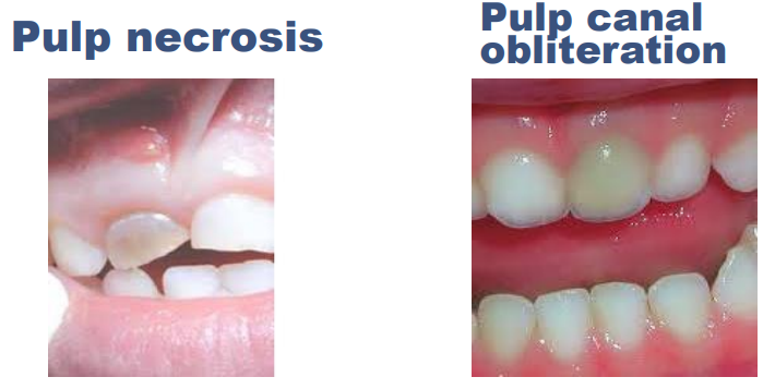
Pulp necrosis: lost vitality, sinus involvement (infection), extract
Advice for parents/guardians
Risk to the primary tooth. The tooth may lose vitality (die)
Look out for:
• Discolouration
• Swelling/lumps on the gum
• Mobility
• Tenderness/pain
Risk of damage to the permanent tooth
Complications to the permanent tooth
• Opacity
• Hypoplasia
• Dilaceration
• Root angulation alteration
• Arrest of root development
• Delayed eruption due to granulation tissue (up to a year)
• Ectopic eruption (lost guidance)
• Impaction to malformation