Anatomy & Physiology
Circulatory (Class 3/17/25)
Arteries away, veins back,
Nutrients and waste exchange occurs in the capillaries, where the thin walls allow for the transfer of oxygen, carbon dioxide, and other substances between blood and tissues.
What is the function of the mammalian circulatory system?
transport things around your body (nutrients and waste)
Clotting, circulates platelets
Defense, it circulates leukocytes
Heat, it circulates heat through your body to help maintain a stable internal temperature and support metabolic processes.
Describe the structure of the mammalian heart in detail, including all chambers, major vessels in and out, and valves.
4 Chambers
2 Types
2 Atriums and 2 Ventricles
Atria are small and have little muscle, most of the work is done with gravity
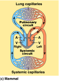
Ventricles are larger and more muscular, RIght goes to lungs and left goes to body
The right atrium receives deoxygenated blood from the body through the superior and inferior vena cavae.
The left atrium receives oxygenated blood from the lungs via the pulmonary veins.
The right ventricle pumps deoxygenated blood to the lungs through the pulmonary artery.
The left ventricle pumps oxygenated blood to the rest of the body through the aorta.
The 4 chambers are entirely separate the oxygenated and deoxygenated blood should not be mixed!
4 Valves
Tricuspid Valve: The valve located between the right atrium and the right ventricle, ensuring that blood flows in one direction and preventing backflow during contraction.
Pulmonary Valve:
Aortic Valve - located between the left ventricle and the aorta, it prevents backflow of blood into the ventricle after contraction.
7 Vessels
Aorta: The largest artery that carries oxygenated blood from the heart to the body.
Pulmonary arteries: Two arteries that carry deoxygenated blood from the right ventricle to the lungs.
Pulmonary veins: Four veins that return oxygenated blood from the lungs to the left atrium.
Superior vena cava: A large vein that carries deoxygenated blood from the upper body to the right atrium.
Inferior vena cava: A large vein that carries deoxygenated blood from the lower body to the right atrium.
Coronary arteries: Arteries that supply blood to the heart muscle itself.
Coronary veins: Veins that drain deoxygenated blood from the heart muscle back to the right atrium.
Notes:
Myocardium: heart muscle
Pericardium: FLuid filled sac around heart
Endothelium- epithelial cells that line the chambers and vessels

Trace the pathway a red blood cell would follow as it enters the heart from the body, out to the lungs, back to the heart, and back to the body. Describe the change in oxygen content as it males this journey.
We have a closed system of blood vessels or the heart
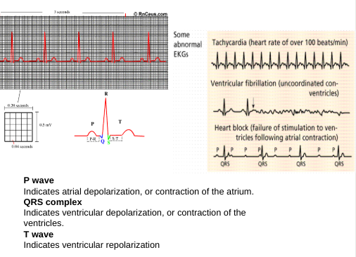
How is the heartbeat regulated? Describe the function of the AV and SA nodes. Describe how an action potential travels around the heart.
heart’s natural pace maker, located in the right atrium made of pace maker cells send signals to av node and acts as a relay
when the sa fires the atrium contracts and then the av node receive this and sends a signal to the ventricles
The brief pause is vital to make sure the chambers can fill
Circulatory (Class 3/18/25)
Compare arteries, veins, capillaries in terms of function and general structure.
Discuss the genetic and environmental factors that affect blood pressure. Explain what the top and bottom numbers mean in a typical reading.
What materials are exchanged between the capillaries and body cells? Between the body cells and the lungs? What process allows this exchange to occur?
Describe the composition of the blood in detail. Include in your answer the function of plasma proteins, erythrocytes, leukocytes, and platelets. What substances does the blood transport?
Arteries
Function: Carry oxygenated blood away from the heart (except pulmonary arteries which carry deoxygenated blood to the lungs).
Structure: Thick muscular walls to withstand high pressure; narrow lumen.
Veins
Function: Carry deoxygenated blood back to the heart (except pulmonary veins which carry oxygenated blood from the lungs).
Structure: Thinner walls than arteries; larger lumen; contain valves to prevent backflow.
Capillaries
Function: Site of exchange between blood and tissues; transfer of oxygen, carbon dioxide, nutrients, and waste occurs here.
Structure: Very thin walls (one cell thick) allowing for efficient diffusion; smallest blood vessels.
Factors Affecting Blood Pressure
Genetic Factors
Hereditary: Family history can play a significant role in predisposition to hypertension (high blood pressure).
Genetics: Certain genes may contribute to the regulation of blood pressure and salt sensitivity.
Environmental Factors
Diet: High salt intake can elevate blood pressure; diets rich in fruits, vegetables, and whole grains are beneficial.
Physical Activity: Regular exercise can help maintain healthy blood pressure levels.
Weight: Obesity increases the risk of hypertension.
Stress: Chronic stress may lead to temporary spikes in blood pressure.
Alcohol and Smoking: Excessive alcohol consumption and smoking can raise blood pressure.
Understanding Blood Pressure Readings
Typical Reading: Blood pressure is measured in millimeters of mercury (mmHg) and presented as two numbers.
Systolic Pressure (Top Number): The pressure in the arteries when the heart beats and pumps blood.
Diastolic Pressure (Bottom Number): The pressure in the arteries when the heart is resting between beats.
Normal Range: A typical reading is around 120/80 mmHg. Elevated readings may indicate prehypertension or hypertension, warranting further assessment and management.
Materials Exchanged Between Capillaries and Body Cells
Oxygen: Diffuses from the capillaries into body cells for cellular respiration.
Carbon Dioxide: Produced by body cells during metabolism, it diffuses into the capillaries to be transported to the lungs.
Nutrients: Glucose, amino acids, and fatty acids diffuse from the capillaries into body cells to provide energy and building materials.
Waste Products: Urea and other metabolic wastes move from body cells into capillaries for removal.
Materials Exchanged Between Body Cells and Lungs
Oxygen: Diffuses from the alveoli of the lungs into the blood in the pulmonary capillaries.
Carbon Dioxide: Moves from the blood in the pulmonary capillaries into the alveoli to be exhaled.
Process Allowing Exchange
Diffusion: The primary process for exchange occurs through diffusion, where substances move from areas of higher concentration to areas of lower concentration across thin capillary walls.
Gas Exchange in the Respiratory System
Between Alveoli and Blood
Location: Gas exchange occurs in the alveoli, which are tiny air sacs in the lungs.
Process: Oxygen from inhaled air diffuses across the alveolar walls into the blood in the surrounding pulmonary capillaries due to a concentration gradient (higher concentration in alveoli, lower in blood).
Carbon Dioxide: Conversely, carbon dioxide in the blood (higher concentration) diffuses into the alveoli (lower concentration) to be exhaled.
Between Blood and Body Cells
Location: Gas exchange between blood and body cells occurs in systemic capillaries.
Process: Oxygen carried by hemoglobin in red blood cells diffuses from the blood (higher concentration) into body cells (lower concentration) for cellular respiration.
Carbon Dioxide: As cells metabolize nutrients, they produce carbon dioxide, which diffuses from the cells (higher concentration) into the blood (lower concentration) to be transported back to the lungs for exhalation.
Overall, gas exchange relies on diffusion driven by concentration gradients, ensuring that oxygen is delivered to tissues and carbon dioxide is removed efficiently.
Composition of Blood
Blood is a vital fluid responsible for transporting various substances throughout the body. It consists of several components:
1. Plasma
Description: It is the liquid portion of blood, making up about 55% of its volume.
Functions:
Transports nutrients, hormones, and proteins to the parts of the body that need it.
Contains plasma proteins such as:
Albumin: Helps maintain osmotic pressure and transport substances.
Globulins: Function in immune response and transport.
Fibrinogen: Crucial for blood clotting.
2. Erythrocytes (Red Blood Cells)
Description: The most abundant cells in blood, typically shaped like biconcave discs.
Functions:
Transport oxygen from the lungs to the body tissues.
Carry carbon dioxide from the tissues back to the lungs for exhalation.
Contain hemoglobin, a protein that binds to oxygen and carbon dioxide.
3. Leukocytes (White Blood Cells)
Description: A diverse group of cells that form part of the immune system.
Functions:
Body’s defense mechanism against infections and foreign invaders (bacteria, viruses, parasites).
Various types include lymphocytes, monocytes, neutrophils, eosinophils, and basophils, each with specific roles.
4. Platelets (Thrombocytes)
Description: Small cell fragments that are essential for blood clotting.
Functions:
Participate in hemostasis by causing the blood to clot at the site of injury, preventing excessive bleeding.
Substances Transported by Blood
Oxygen: From the lungs to body tissues.
Carbon Dioxide: From body tissues to lungs for exhalation.
Nutrients: Such as glucose, amino acids, and fatty acids from the digestive tract to cells.
Waste Products: Such as urea and creatinine to the kidneys for excretion.
Hormones: Secreted by endocrine glands and transported to target tissues.
Summary
The composition of blood is essential for various bodily functions including transportation, immune response, and blood clotting. Understanding these components highlights the importance of blood in maintaining homeostasis and health.
The Four Valves of the Heart
Tricuspid Valve
Location: Between the right atrium and the right ventricle.
Function: Ensures one-way blood flow from the right atrium to the right ventricle, preventing backflow during contraction.
Pulmonary Valve
Location: Between the right ventricle and the pulmonary artery.
Function: Prevents backflow of blood into the right ventricle after it has pumped deoxygenated blood to the lungs.
Mitral Valve (Bicuspid Valve)
Location: Between the left atrium and the left ventricle.
Function: Allows one-way blood flow from the left atrium to the left ventricle, preventing backflow during contraction.
Aortic Valve
Location: Between the left ventricle and the aorta.
Function: Prevents backflow of blood into the left ventricle after it has been pumped into the aorta to supply the body with oxygenated blood.
EMO: stimulates the production of red blood cells, when they are low it will increase erythropoietin (EPO) levels, prompting the bone marrow to enhance red blood cell production to maintain adequate oxygen transport in the body.
Respiratory
What is the function of the respiratory system?
The respiratory system is responsible for facilitating the exchange of oxygen and carbon dioxide between the body and the environment, enabling cellular respiration and maintaining homeostasis.
Trace a molecule of oxygen from the nose to a cell in the toe. Trace a molecule of CO2 from a cell in the toe to the external environment.
Dissolves into plasma —> binds to hemoglobin within red blood cells —> converted into carbonic acid
Describe the structure of the alveoli, and their relationship with the circulatory system. How does the structure of the alveoli allow them to carry out their function?
Describe how gas exchange happens between the alveoli and the blood, and between the blood and body cells.
Describe the structure and function of hemoglobin.
tetrameric quaternary structure, it can carry 4 O2 Molecules
Oxygen Distoriation:
Endotherms “Warm blood”: maintain a constant body temperature
We burn calories to keep ourselves warm
Higher metabolic rate, and require more oxygen
Small endotherms need more oxygen
Metabolic rate is inversely proportional to body size
Ectotherms "Cold blooded", cannot maintain a constant body temperature and will fluctuate with the environment: organisms rely on external sources of heat to regulate their body temperature, which affects their metabolic processes and overall energy levels.
They rely on the external environment
Digestive
A complete system, the alimentary canal = 2 holes
Travels one way, and two opening the motuh and anuc
Ingestions: Taking food in
Digestion: mechanical (chewing, churning, crushing) or chemical (enzymes that break down each other the 4 specific macromolecule), there are specialized areas for each Macromolecule
absorption: MOSTLY the first part of the small intestine (a little bit of the stomach)
Elimination: removal of the excess substances
monomers are absorbed into blood
What is the function of the digestive system?
Breaks down food chemically & mechanically
Be able to trace, in detail the digestion of a hamburger, including all necessary organs, enzymes, and cells.
What is peristalsis? A bolus? Emulsification? Sphincters?
Keeps the food moving, a series of contractions in the smooth muscles that moves food throughout the system
Bolus: a mass of food that has been chewed and mixed with saliva,
Describe the swallowing reflex. What is the purpose of this reflex?
What are the digestive functions of the pancreas, liver, gall bladder, and duodenum?
the are accessory organs, meaning foood never actually travels through them and are connected to the main system via ducts that
Pancreas: Secretes enzymes into the sm. int and helps to regulate blood sugar levels by producing insulin and glucagon as needed.
What are the functions of the jejunum and ileum?
Describe the major function of the colon. What is the function of the abundant bacteria found here?
Explain the relationship between the digestive and circulatory systems.
Amylase: an enzyme that catalyzes the hydrolysis of starch into sugars, playing a crucial role in the digestive process.
Lipase: an enzyme responsible for breaking down fats into fatty acids and glycerol, which is essential for the absorption of dietary lipids.
Bile: a digestive fluid produced by the liver and stored in the gallbladder, that aids in the emulsification of fats, making them more accessible for lipase action during digestion.
Pepsin: an enzyme secreted by the stomach that breaks down proteins into smaller peptides, initiating the process of protein digestion.
mastication: the process of chewing food in the mouth, which mechanically breaks it down into smaller pieces and mixes it with saliva, making it easier for enzymes to further digest the food in the gastrointestinal tract.
peristalsis: the series of wave-like muscle contractions that move food through the digestive tract, playing a crucial role in ensuring that the food is efficiently propelled from the esophagus to the stomach and through the intestines.
deglutition: the process of swallowing, which involves the coordinated actions of the tongue, soft palate, and muscles of the throat to move food from the mouth to the esophagus.
Excretory
What is the function of the excretory system? How is it different from the digestive system?
Filters blood, removes metabolic waste (waste that comes from chemical reactions) and external toxins, maintains blood pH, maintains levels of electrolytes and nutrients
Helps to measure BP
Secretes hormone to help regulate
Helps to regulate red blood cell production
Balances water, glucose, ions
Identify all organs of this system and briefly explain their function.
Kidneys → Ureters →Bladder
Kidneys: Filter blood to remove waste and excess substances, producing urine for excretion.
What happens if they don’t work? = If the kidneys fail to function properly, waste products and excess fluids can accumulate in the body, leading to conditions such as edema, hypertension, and even kidney disease.
Ureters: Transport urine from the kidneys to the bladder.
What happens if they don’t work? = If the ureters become blocked or damaged, urine may back up into the kidneys, which can cause kidney damage, infection, and severe pain.
Bladder: Stores urine until it is ready to be expelled from the body.
What happens if it doesn't work? = If the bladder fails to function properly, issues such as urinary incontinence, frequent urination, or urinary retention can occur, greatly affecting the quality of life.
Urethra: The tube that carries urine from the bladder out of the body.
The renal artery goes into the kidneys and branches into a shit ton of capillaries
The renal vein comes from the kidneys to the superior vena cava
Identify at least 3 substances/chemicals/molecules that are regulated by the kidney:
Blood pressure hormones (e.g., renin)
How? Hormonal regulation occurs through the kidneys' ability to produce and secrete hormones, which help maintain various physiological functions. For instance, erythropoietin stimulates red blood cell production; calcitriol regulates calcium and phosphate balance; and aldosterone influences sodium retention and potassium excretion.
Electrolytes (e.g., sodium, potassium) calcium, magnesium)
How?
Acid-base balance components (e.g., bicarbonate) play a crucial role in maintaining the body's pH levels and overall homeostasis.
Differentiate between filtration, reabsorption, secretion, & excretion.
What is the relationship of nephron to kidney?
In each kidney there are about 1 Million that filter the blood
What is the relationship between the excretory and circulatory systems? Between the excretory and digestive?
file:///media/fuse/drivefs-9db870e809258606c799aec315125a40/root/Record%20(online-voice-recorder.com).mp3
Urea: the urea is less toxic than ammonia and is more soluble in water, allowing it to be transported easily in the bloodstream to the kidneys for excretion. more common in mammals
“All you wanna do is Eat and Kill people”
Reproductive
Explain how each of the following contributes to male fertility: testes, seminiferous tubules, leydig (interstitial)cells, sertoli (sustentacular)cells.
cells, scrotum, epididymis, vas deferens, seminal vesicles, prostate gland, bulbourethral gland, erectile tissue.
Function and locations of the bulbourethral gland: The bulbourethral gland, also known as Cowper's gland, is located beneath the prostate gland and within the urogenital diaphragm; its primary function is to produce a pre-ejaculatory fluid that lubricates the urethra and neutralizes any acidity, thus creating an optimal environment for sperm during ejaculation.
Function of the testis: The testis produces sperm and hormones, particularly testosterone, which are crucial for male fertility. The seminiferous tubules, located within the testes, are specifically responsible for the production of sperm through the process of spermatogenesis. Leydig cells produce testosterone, which is essential for the development of male reproductive tissues and the maintenance of libido. Sertoli cells, on the other hand, support and nourish developing sperm cells, providing the necessary environment for spermatogenesis to occur. In addition, the testis also plays a role in the regulation of other hormones, such as inhibin, which helps to control the production of sperm by providing feedback to the anterior pituitary gland.
Function of the Vas Deferens: The vas deferens is a muscular tube that transports sperm from the epididymis to the ejaculatory duct, playing a vital role in the male reproductive system. It is part of the spermatic cord and helps propel sperm during ejaculation through peristaltic contractions. Additionally, the vas deferens serves as a storage site for sperm, allowing for maturation and concentration before they join with seminal fluid to form semen. Overall, its function is essential for facilitating successful reproduction by ensuring that sperm are effectively delivered to the urethra.
Function of the Prostate Gland: The prostate gland is a walnut-sized organ located below the bladder and surrounds the urethra. It produces seminal fluid, which nourishes and transports sperm during ejaculation. This fluid constitutes a significant portion of semen, contributing to its volume and acting as a medium for sperm motility. Furthermore, the prostate gland plays a role in sperm activation and increases the chances of successful fertilization. Its proper functioning is crucial for overall male reproductive health.
Function of the Urethra in Men: The urethra in men serves as a conduit for both urine and semen, extending from the bladder through the penis. It is divided into three sections: the prostatic urethra, which runs through the prostate; the membranous urethra, which is surrounded by the pelvic muscles; and the spongy urethra, which runs through the penis. During urination, the urethra allows for the expulsion of urine, while during ejaculation, it facilitates the discharge of semen. This dual function is essential for male reproductive and urinary health, and any obstruction or dysfunction can lead to various medical issues.
Main hormones in male reproductive system:
Testosterone: The primary male sex hormone, responsible for the development of male characteristics, spermatogenesis, and libido.
Luteinizing hormone (LH): Stimulates testosterone production from the Leydig cells in the testes.
Follicle-stimulating hormone (FSH): Aids in the production of sperm by promoting the function of Sertoli cells in the testes.
Explain how each of the following contributes to female fertility: ovaries oviduct, uterus, endometrium, bartholin’s gland, vagina.
Describe the hormonal changes that occur during the human female reproductive (menstrual) cycle. What effect do these changes have on the endometrium? What effect do these changes have on the ovaries & follicles?There are 5 hormones involved, Describe the effects of each one, and how each fluctuates during the cycle.
The 5 hormones in the female anatomy:
Estrogen: Produced primarily by the ovaries, estrogen levels rise during the follicular phase, promoting endometrial growth and maturation of follicles.
Progesterone: Secreted by the corpus luteum after ovulation, progesterone stabilizes the endometrium for potential implantation of an embryo.
Luteinizing hormone (LH): Responsible for triggering ovulation, LH levels surge mid-cycle, leading to the release of a mature egg from the ovary.
Follicle-stimulating hormone (FSH): Stimulates follicle development and estrogen secretion during the follicular phase, with levels decreasing after ovulation.
Gonadotropin-releasing hormone (GnRH): Secreted by the hypothalamus, GnRH regulates the release of FSH and LH from the pituitary gland, with secretion patterns altering during different phases of the menstrual cycle.
The Testicles are a gland and are under the regulation of the hypothalamus, the GnRH will tell the pituitary to secrete FSH and LH
Both cause the production of testosterone- this causes the sperm formation
In females, the hormonal regulation initiates with the hypothalamus releasing GnRH, which stimulates the anterior pituitary gland to produce FSH and LH.
We fucking crazy….
Day one of period (ever) tells the hypothalamus to release GnRH which stimulates the release of FSH and LH from the pituitary and causes the follicle to mature.
FSH (Follicle-Stimulating Hormone): Promotes the growth and maturation of ovarian follicles, stimulating estrogen production. Levels rise in the early follicular phase.
LH (Luteinizing Hormone): Triggers ovulation and the formation of the corpus luteum, surging mid-cycle.
Estrogen: Produced by growing follicles, this hormone prepares the endometrium for implantation and also regulates FSH and LH levels. Builds the uterine lining. The increase in the FSH and LH causes the ovulation, which is the release of a mature egg from the dominant follicle, typically occurring around the midpoint of the menstrual cycle.
Progesterone: Secreted by the corpus luteum after ovulation, it maintains the endometrium for potential embryo implantation and inhibits further ovulation. Keeps the Uterine lining from building
Inhibin: Produced by the follicles and corpus luteum, it suppresses FSH production once sufficient follicles mature.
These hormonal fluctuations ensure proper development within the ovaries and coordinated preparation of the endometrium throughout the menstrual cycle.
Describe the hormonal cycle that regulates reproduction in human males.
Describe the processes of spermatogenesis & oogenesis. What are the main differences between the 2?
Spermatogenesis is the process of sperm cell development that occurs continuously in the testes, resulting in millions of sperm being produced daily, while oogenesis is the formation of present at birth and undergo maturation monthly during the menstrual cycle.
In spermatogenesis, the process includes spermatogonia developing into primary spermatocytes, then secondary spermatocytes, and finally into spermatids before maturing into spermatozoa, while in oogenesis, the primary oocytes undergo meiosis and complete their division only during ovulation, producing one mature ovum and polar bodies from each cycle.
Additionally, spermatogenesis takes about 64 days to complete, whereas oogenesis can take many years, as the primary oocytes are arrested in prophase I until ovulation occurs.
What is menopause? Menopause is the time in a woman's life, typically occurring between the ages of 45 and 55, when the menstrual cycle ends, signaling the cessation of ovarian function and a significant decline in hormone production, particularly estrogen and progesterone, leading to various physical and psychological symptoms. During this phase, women may experience symptoms such as hot flashes, night sweats, mood changes, and decreased libido, indicating the profound impact of hormonal changes on overall health.