Topic 3_The Human Skeleton I
Topic 3B: The Skeleton I
Introduction
Overview of bones, cartilage, and ligaments in the human skeleton.
Reference: Chapter 7, page 19-245 from a specific source.
Section Objectives
Skull Structure
Cranial and Facial Bones: Identify, describe, and mark notable features.
Sutures & Sinuses: Understand connections and anatomical structure.
Bony Boundaries: Describe orbits and nasal cavity structures.
Vertebral Column
Recognize regions and normal curves, ligaments, and intervertebral discs.
Discuss structure of cervical, thoracic, and lumbar vertebrae.
Note the anatomy of sacrum and coccyx.
Thoracic Cage
Detailed anatomy of the sternum and ribs.
Distinguish between true and false ribs.
Skeleton Overview
Major Divisions
Axial Skeleton:
Comprises bones of the skull, vertebral column, and rib cage.
Appendicular Skeleton:
Comprises bones of the upper and lower limbs; includes pectoral and pelvic girdles to attach limbs to the axial skeleton.
Human Skeleton Statistics
Total Bones: 206 bones; contributes to approximately 20% of body weight.
The Skull
Structure
Comprised of 22 Bones total:
Cranial Bones: Enclose the brain, attach to muscles.
Facial Bones: Provide framework for face, cavities for senses, openings for air and food passage, anchor teeth and facial expressions.
Most skull bones are flat, except for the mandible, which is not.
Sections of the Skull
Cranial Vault: Upper parts including forehead.
Cranial Base: Contains bony ridges dividing into anterior, middle, and posterior fossae.
Contains various cavities, such as nasal and ear cavities.
Named Openings: 85 openings for major vessels and nerves including foramina and canals.
Cranial Bones
Types of Bones:
Parietal (2)
Temporal (2)
Frontal
Occipital
Sphenoid
Ethmoid

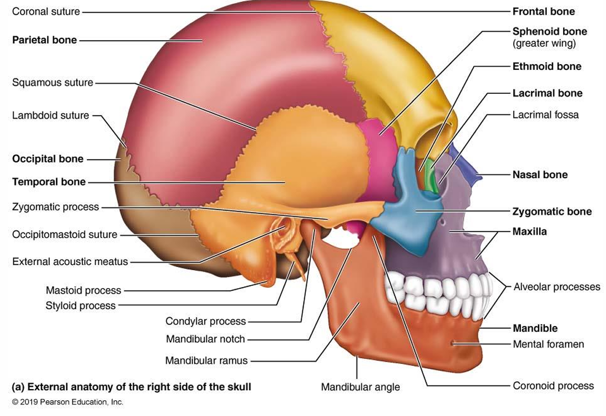
Frontal Bone
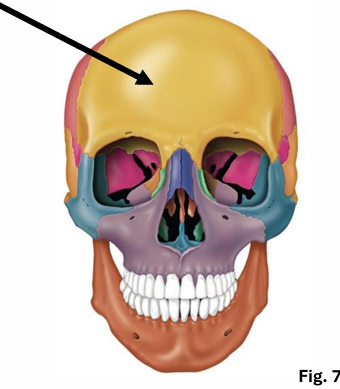
Features
Forms the anterior portion of the cranium and roof of orbits.
Contains frontal sinuses, located to the sides of glabella.
Vertical part: forehead, called squamous region
note:
supraorbital margin
supraorbital foramen
glabella area
the smooth area located between the eyebrows, just above the nasal bridge.
Parietal Bones
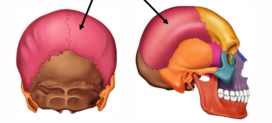
Features
Form superior and lateral aspects of the skull.
Connected through sutures to adjacent bones.
Major sutures:
Coronal: Between parietal and frontal bones.
Sagittal: Divides parietal bones.
Lambdoid: Between parietal and occipital.
Squamous: Between parietal and temporal.
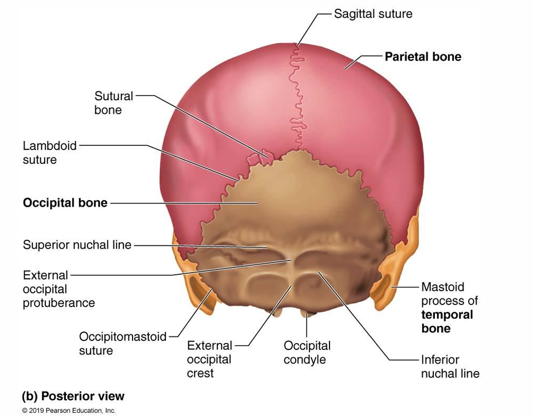
Occipital Bone
Features
Forms posterior skull artwork.
Connects with parietal and temporal bones.
Contains foramen magnum for spinal cord passage.
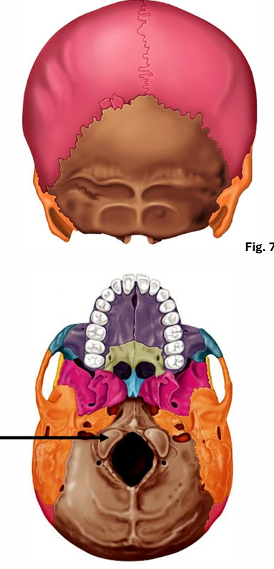
Foramen magnum: hole through which brain connects with spinal chord
Occipital condyles: site of articulation with first cervical vertevrae
condyle: rounded part of bone usually for articulation
fossa: depression in bone
External occipital crest: ride down middle of occipial
External occipital protuberance: bump as bone comes toward spine
Nuchal lines: ridges on the external occipital bone that provide attachment for muscles and ligaments associated with the neck.
Temporal Bones
Structure
Covers inferior and lateral surfaces of the skull.
Positions: Squamous, tympanic, and petrous regions.
Features include: zygomatic process, external auditory meatus, and mastoid process.
Squamous:
flattened
zygomatic process of the temporal bone
mandibular fossa: recieves condyle of mandible to form jaw joint
Tympanic:
Tympanic membrane surrounds external acoustic meatus:serves to transmit sound vibrations to the middle ear.
Petrous:
bump behind ear is called a mastoid process.
Mastoid process: major attachment area for some neck muscles
Styloid process: muscles for tongue and some neck
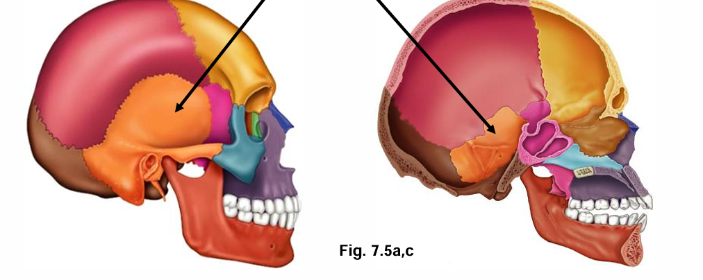
Sphenoid Bone
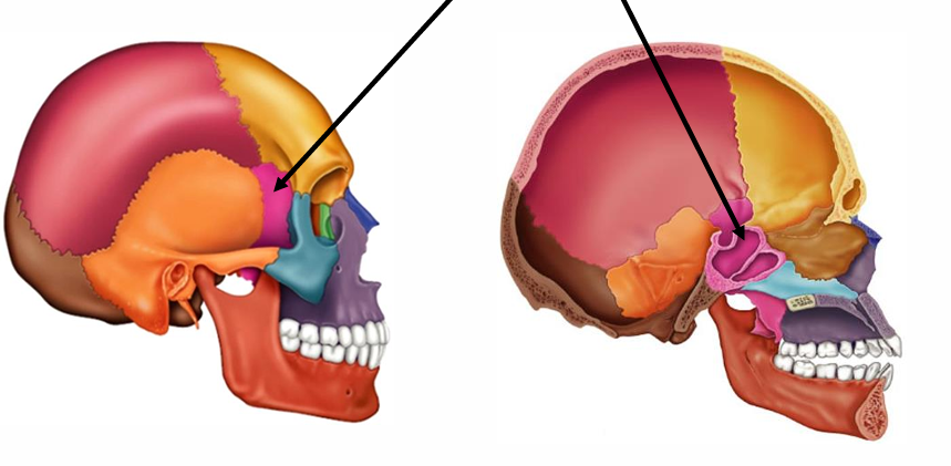
Characteristics
A complex bat-shaped bone contributing to the cranial fossa.
Key Features: Optic foramina, superior orbital fissure, and sella turcica for pituitary gland.
keystone bone: because it connects to all the other cranium bones.
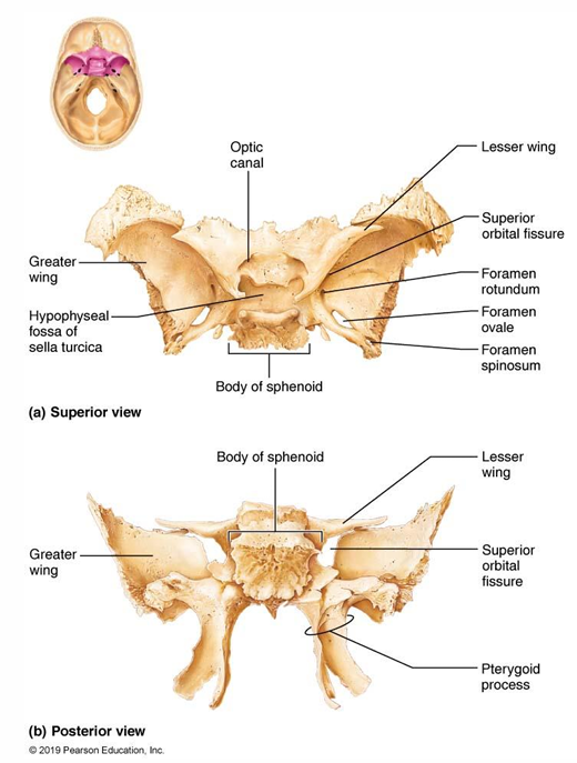
Optic foramina
Superior orbital fissure
Foramen rotundum and ovale
Foramen spinosum
sella turcica
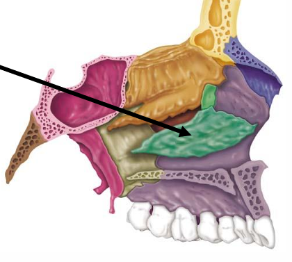
Ethmoid Bone
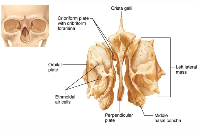
Anatomy
Forms part of the nasal cavity and orbits.
Contains cribriform plate for olfactory nerves and contributes to nasal septum.
cribriform plate
roof of nasal; cavity, floor of anterior cranial fossa
tiny holes (olfactory foramina) allow transmit info to olfactory bulb
perpendicular plate
projects inferiorly to contribute to the nasal septum
crista galli
anchor dura mater of brain
lateral amsses contain ethmoid sinuses
mediallly are superior and middle nasal conchae, which are thin, scroll-like bones that help to filter and humidify the air we breathe.
Major Sutures of the Skull
Connect cranial bones through the identified sutures: Coronal, Squamous, Sagittal, and Lambdoid.
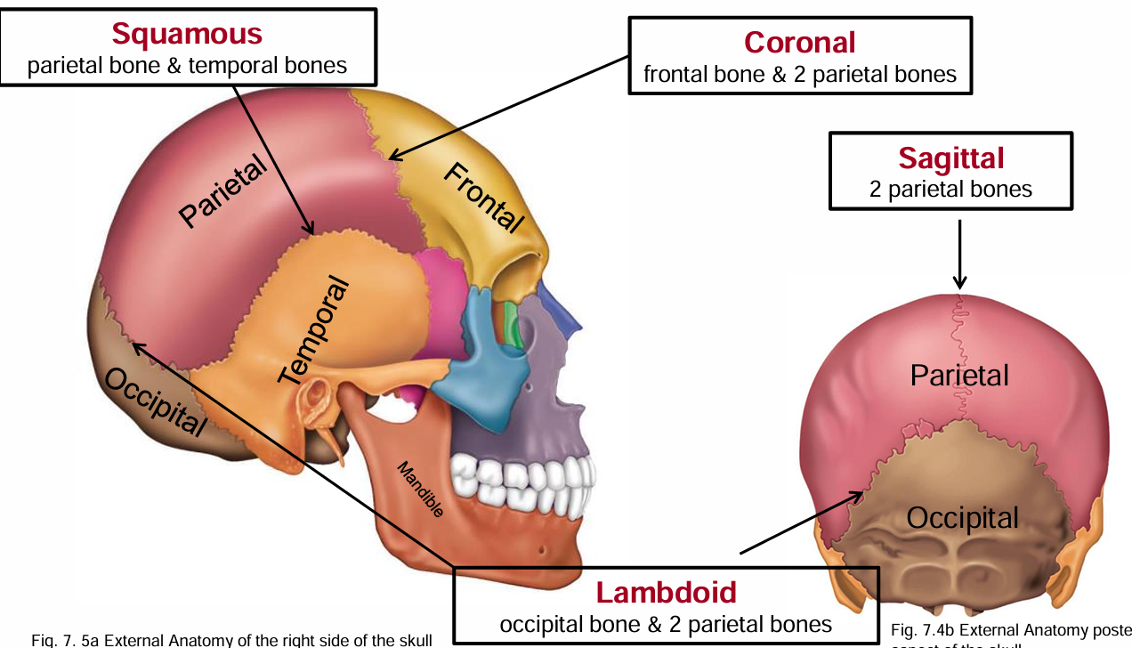
Squamous suture: parietal and temporal
Coronal: frontal and parietal
Saggital: parietal bones
Lambdoid: occipital and parietal
Sutural bones: tiny irregular obmes, additional ossifcaiton centres that appeared rapidly during fetal devel.
Facial Bones Overview
Number of Bones
14 total bones, including:
Mandible
Maxillary bones (2)
Zygomatic bones (2)
Nasal bones (2)
Lacrimal bones (2)
Palatine bones (2)
Vomer
Inferior nasal conchae (2)
Functions
Support facial structure, openings for air and food, cavities for sensory organs, and secure teeth.
Mandible Features
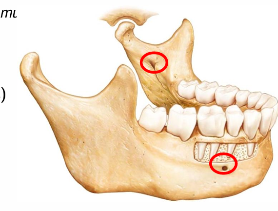
Largest and strongest facial bone.
Lower jaw, only bone that moves in cranium, allowing for the opening and closing of the mouth during speech and chewing.
Noteworthy Parts:
body: chin,
mandibular angle, where it curves up
mandibular notch, top of jaw
coronoid process, insertion point for temporalis muscle
Mandibular condyle, the rounded end of the mandible that articulates with the temporal bone at the temporomandibular joint (TMJ).
Alveolar margin contain tooth sockets
Mandibular foramina, nerves to teeth startsnear cheek
Mental foramina,openings in the mandible that allow the mental nerve and blood vessels to pass through, providing sensation to the lower lip and chin.
Maxillary Bones
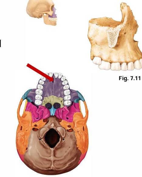
Characteristics
Fused medially, housing sinuses that may become infected.
Keystone facial bone
Palatine processes of the maxilla contribute to the formation of the hard palate
Frontal process: upward projection connect to frontal bone
Zygomatic process: side projection, connect to zygomatic bone
Incisive fossa
Infra orbital foramen: below orbit hole
Zygomatic Bones
Form cheekbones and articulate with various other bones.
Connected through zygomatic processes:
Project upwards connect to frontal bone
Project laterally connect to temporal
Project medially and connect to nasal
“The zygomatic process of the temporal bone, connects to the temporal process of the zygomatic bone.”
and “the zygomatic process of the frontal bone connects with the frontal process of the zygomatic bone.”
Nasal Bone

Small bones forming the bridge of the nose.
articulate wit frontal, maxillary, and ethmoid bones
Additional Facial Bones
Palatine Bones
Form parts of the hard palate and contribute to the nasal cavity and orbit.
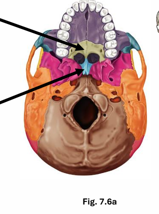
Vomer
Thin bone forming a part of the nasal septum.
Lacrimal Bones
Involved in forming the orbit and housing the lacrimal sac.
where tears collect
Inferior Nasal Conchae
Contribute to the structure of the nasal cavity.
Hyoid Bone
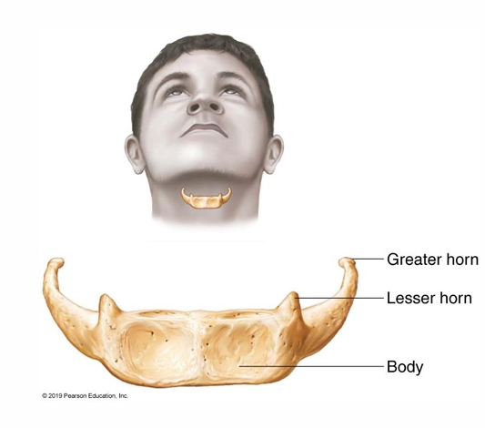
Characteristics
Located in the neck, supports tongue and is unique as it does not articulate with any other bone.
Not bone of skull
Support tongue, attachment for swallow muscles and speech
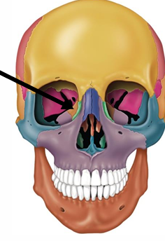
Orbits and Nasal Cavity
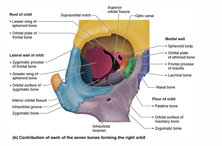
Orbital Structure
Formed by contributions from seven different facial bones.
Frontal
Zygomatic
Sphenoid
Lacrimal
Maxilla
Palatine
Ethmoid
Nasal Cavity Anatomy
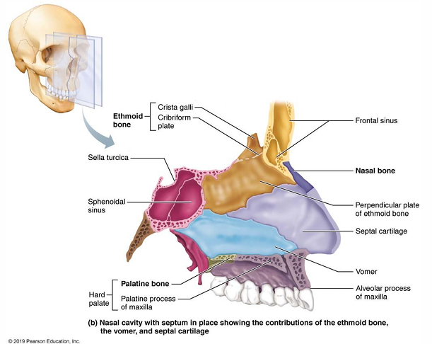
Formed by components from different bones and contributes to airflow and respiratory efficiency.
“Broken noses”
cartilage broken
“Deviated septums”
doesnt run along the midline
Paranasal Sinuses
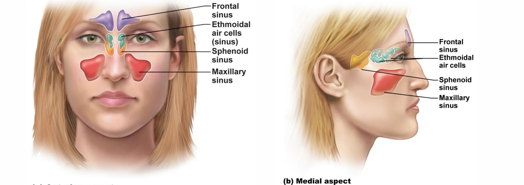
Air-filled spaces lined with mucous membranes. maximizing skull lightness and enhancing voice resonance.
Major paranasal Sinus containing bones
Frontal, Maxillary, Sphenoid, Ethmoid.
Vertebral Column Overview
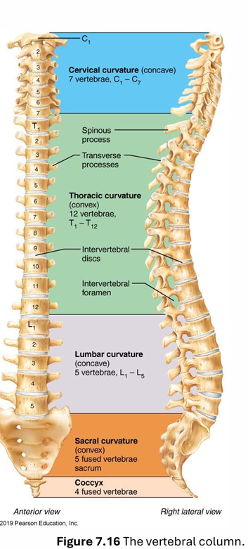
Functions
Acts as a weight-bearing structure, anchors muscles, and protects the spinal cord.
Composition
Comprises 33 vertebrae with 24 free and 2 fused structures (sacrum and coccyx).
Major Regions
brekfes lunch dinne.
breakfas at 7, lunch at 12, din at 5
cerv, thora, lum
Cervical: 7 vertebrae.
Thoracic: 12 vertebrae.
Lumbar: 5 vertebrae.
Sacrum: 1 fused bone.
Coccyx: fused from 3-4 bones.
Curvatures of the Vertebral Column
primary, born with
secondary, after birth
Main Curvatures
Cervical and Lumbar: Concave.
secondary
come from first: lift head, then walking
Thoracic and Sacral: Convex.
primary
Abnormal Curvatures
Can arise from congenital issues or lifestyle factors.
Scoliosis
abnormal lateral rotation, usually in thoracic area
lungs and heart affected by reposition of thoracic cage.
Kyphosis
hunch back
rounding of upper back
Lordosis
accentuated lumbar curvature- Swayback posture, which can lead to discomfort and pain in the lower back.
could be disease, or pregnant, or fat stomach
Supporting Elements of the Vertebral Column
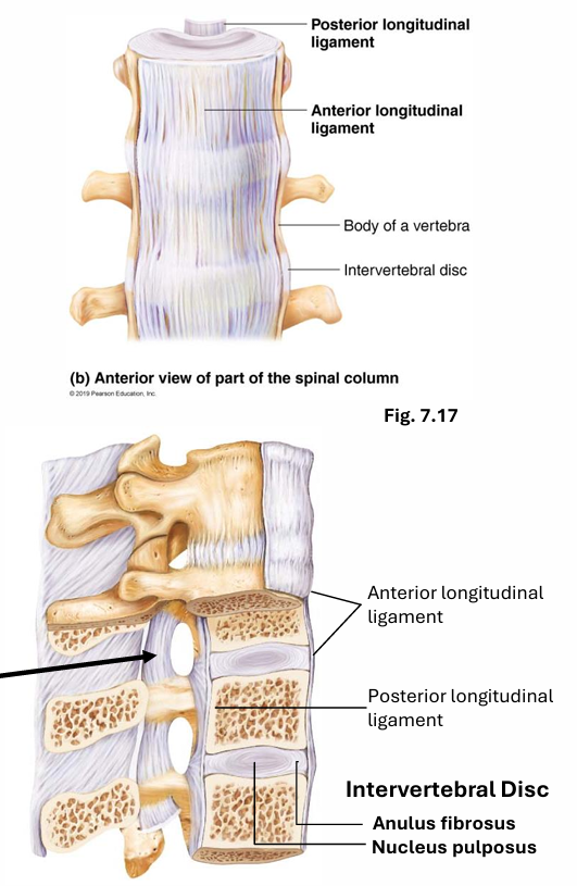
Ligaments
Play a critical role in maintaining posture and stability.
Protect against over flection and overextension
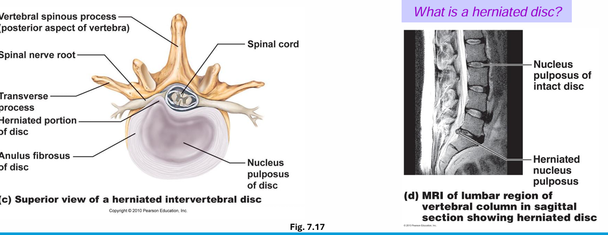
Intervertebral Discs
Act as shock absorbers and cushioning elements.
Structure: Nucleus pulposus (inner) and annulus fibrosus (outer).
General Structure of Vertebrae
Vertebra Components
Weight-Bearing Body: Anterior aspect.
Vertebral Arch: Made up of pedicles and laminae.
Vertebral canal and foramina: Allow passage for nerves and vessels.
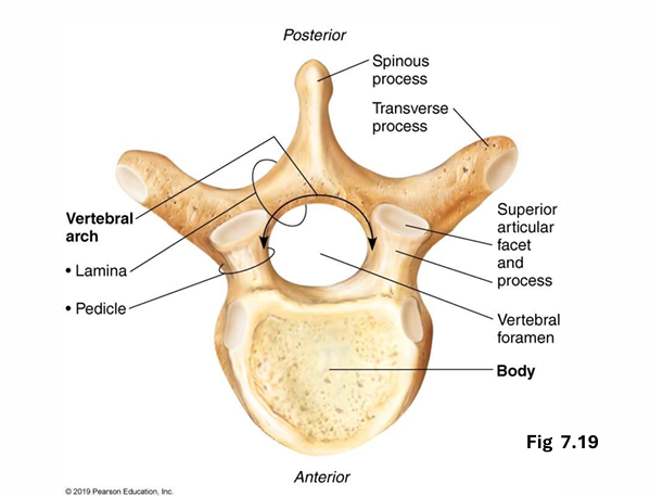
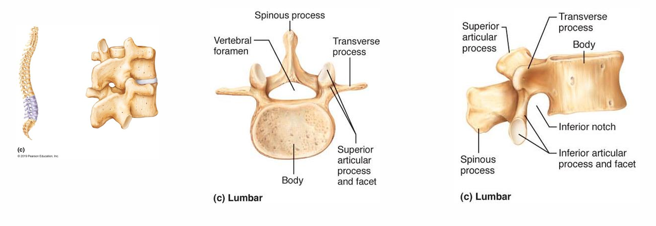
Spinous process
project posteriorly; muscle attachment
2 transverse processes
Paired superior processes
Inferior articular proceses
Superior and inferior articular processes link vertebrae above & below; smooth, collagen coated facets for articulation
Individual Vertebrae Characteristics
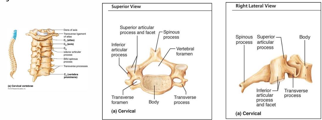
Cervical Vertebrae (C1-C7)
Unique structures such as transverse foramina and split spinous processes for muscle attachment.
C3 to C7 traditional
Cervical vertebrae have hole in transverse processs, which allow for the passage of vertebral arteries and veins, playing a crucial role in supplying blood to the brain.
Atlas and Axis
C1 (Atlas): No body, allowing nodding motion say yes.
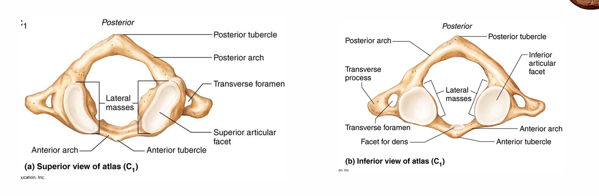
neural arches
no spinous process
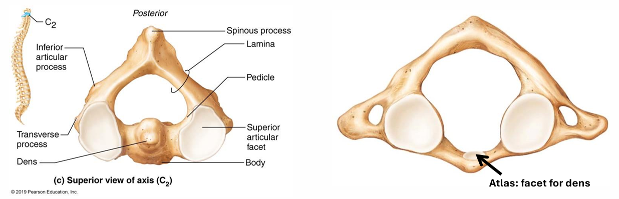
C2 (Axis): Contains dens for rotational movement.
allow head movement say no
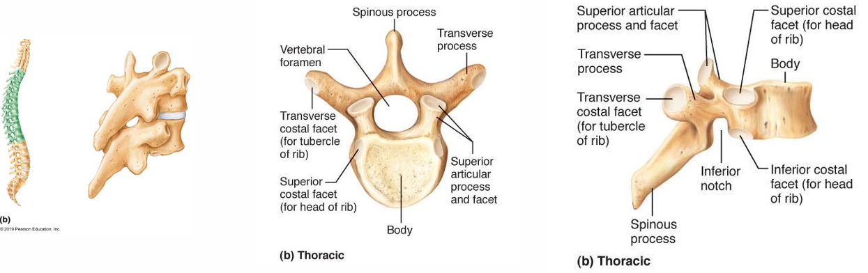
Thoracic Vertebrae
T1 to T12
These vertebrae support the rib cage and protect the thoracic organs, while also allowing for limited movement in the upper back.
Articulate with ribs and show heart-shaped bodies and longer spinous processes.
Notches on body and transverse process
Lumbar Vertebrae
Robust structure for load-bearing; unique facet orientations prevent rotation.
L1 to L5
bodies are kidney shaped, increase in size from top to bottom.
pedicles and laminae shorter and thicker than other vertebrae
Spinous processes are flat hatchet shaped
vertevral foramen is triangular
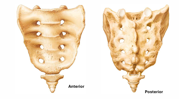
Sacrum and Coccyx
Formation from fused vertebrae articulating with pelvis and coccyx; features include sacral foramina and crests.
Sacrum
Spinal chord stops at about L1, below that the cauda equina begins, consisting of nerve roots that continue down the vertebral canal.
exit through the anterior sacral foramine
Anterior:
sacral promontory
transverse lines
anterior sacral foramina
Posterior:
median sacral crest
posterior sacral foramine
sacral canal
sacral hiatus
Coccygeal vertebrae = tailbone 3 or 4 fused coccygeal vertevrae. attachent for some pelvic ligaments
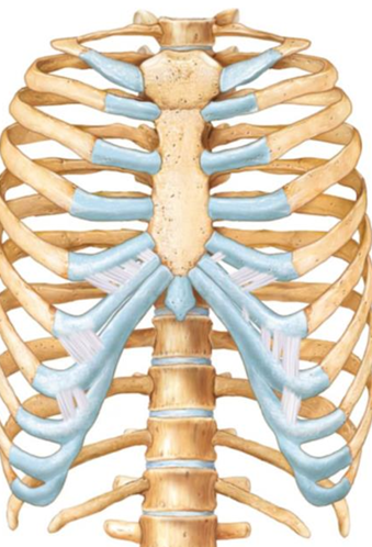
Thoracic Cage Structure
Overview
Composed of thoracic vertebrae, ribs, costal cartilage, and sternum.
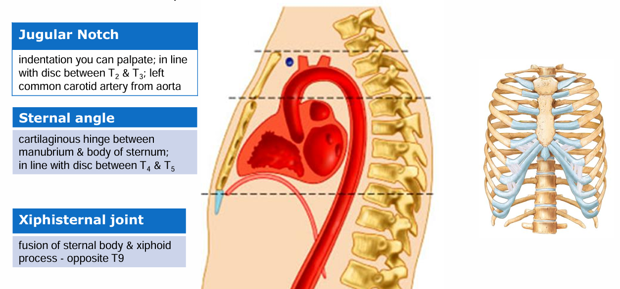
Sternum Anatomy
Three fused sections:
Manubrium
articulate with clavicles and ribs 1 and 2
body
notches forarticulation with ribs 2 to 7
xiphoid process.
attach some abdominal muscles
Ribs Overview
True Ribs: Direct sternum attachment.
Vertebrosternal
ribs 1-7 attach direcyly by individual costal cartilages
False Ribs: Indirect or no attachment.
Vertebralchondral
ribs 8-10 attach to sternum indirectly via costal cartilages of rib above and rib7
ribs 11 and 12 are not attached anterioirly to sternum (floating ribs)
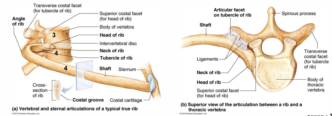
Typical rib is a bowed, flat bone.
Shaft:
main part of rib
costal groove: a shallow indentation along the inner surface of the rib that houses the intercostal vessels and nerves.
Head:
2 facets. one articulates with the demi fact on the body of the same-numbered thoracic vertebra. other with that on body of superior vertebra.
therefore rib 4, would connect to superior vert. 3 and demi facet of 4
Tubercle: connects with transverse process of the corresponding thoracic vertebra.