OSPE feedback session
**NO MARK FOR HALF ANSWERS ANYMORE
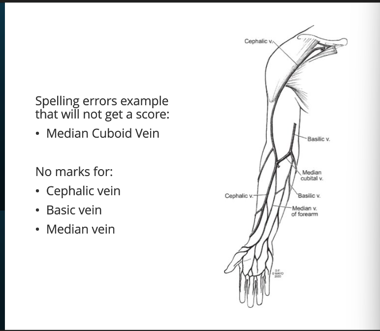
question arrow was pointing at median cubital vein
basilic vein medial side
cephalic vein lateral side? from the thumb up
these two are connected by median cubital vein
median cubital vein connects cephalic and basilic cubital fossa
study superficial veins of upper limb and lower limb
T4 vertebral level, known due to bifurcation of trachea (two black circles, left and right bronchus)
descending thoracic aorta on left side of vertebral body
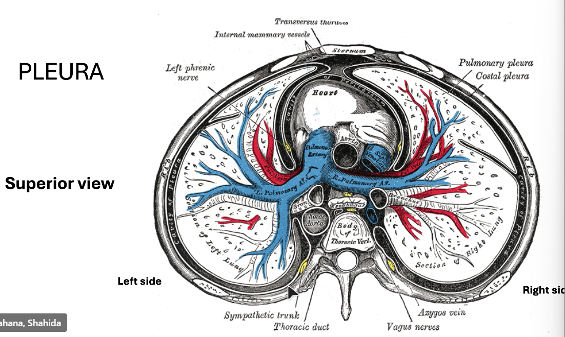
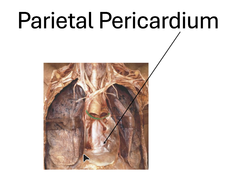
shiny lining/layer is parietal pericardium
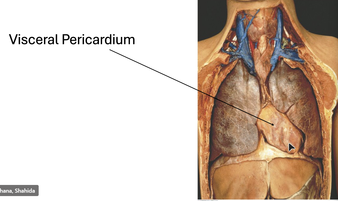
visceral pericardium is the innermost layer of the pericardium, whereas parietal pericardium is the outer layer. Visceral and parietal pericardium are two layers of the serous pericardium, a thin, fibrous membrane
outer layer made of strong inelastic connective tissue (fibrous pericardium), and an inner layer made of serous membrane (serous pericardium)
pleural effusion is accumulation of fluid, if specified in question blood then haemothorax
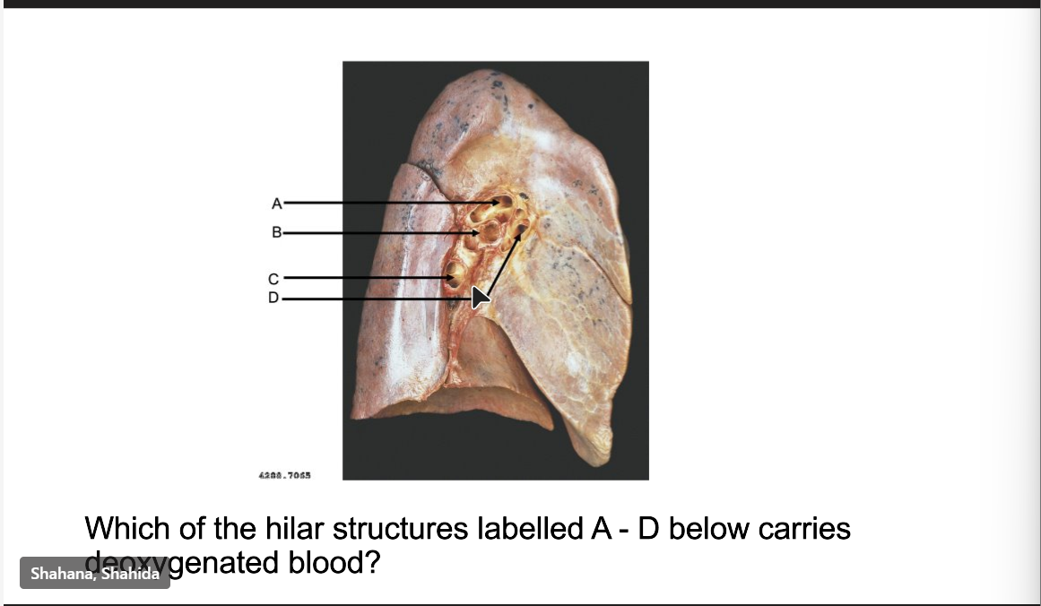
C-inferior pulmonary vein
D-posterior pulmonary vein
make a big list from structures originating from ectoderm, mesoderm, endoderm
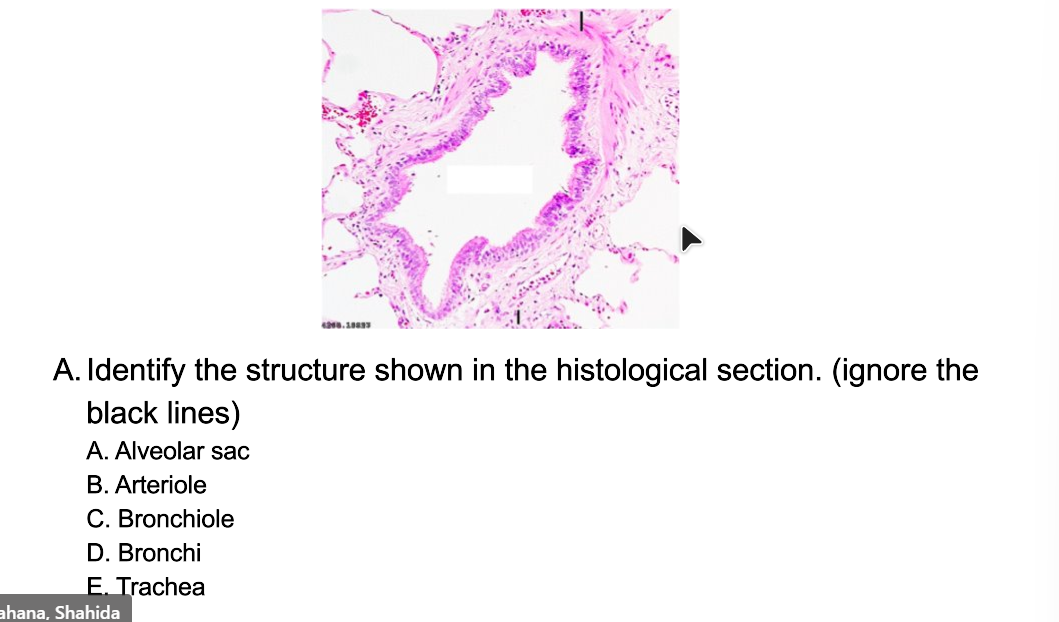
bronchiole because no cartilage or would have cartilage if bronchi and if trachea would have been c shaped cartilage
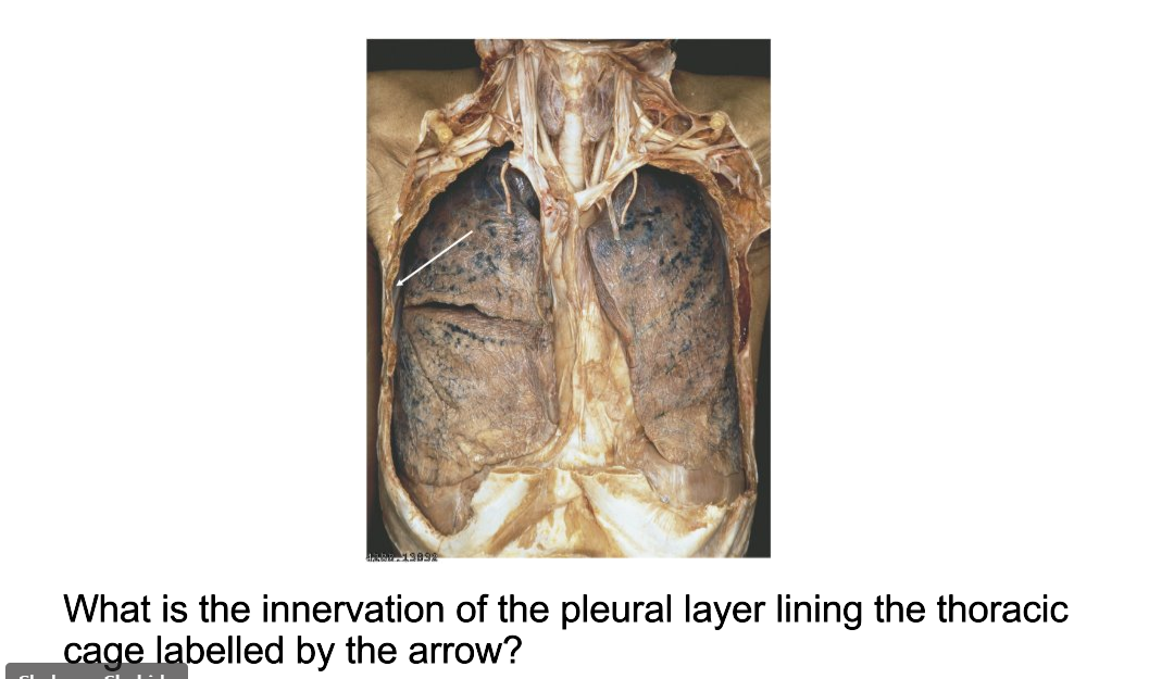
intercostal nerves T1-T11
or somatic sensory
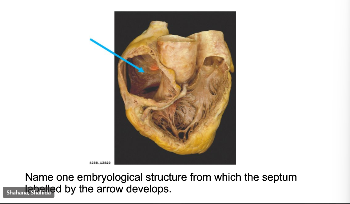
secundum of septum primum
when they mention a word like septum or artery the answer is probably what they mention
