Sen Exam 1
Introductory Class: Course is focused on the readings
Multiple Choice Quizzes to help you study
Office Hours are before class
Tests can include diagrams, short answers and long answers aside from the usual multiple choice.
Practice Tests are posted to help assist with the studying process
Overview:
Evolution has solved a difficult problem. (we haven’t solved it yet)
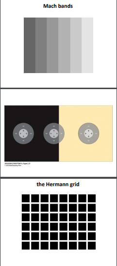
checkerboard-shadow illusion (same grey but it seems lighter, same colour properties)
Anderson’s Lightness Illusion
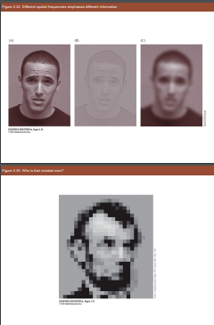
Face Recognition
Object Recognition
Light (3) God said, “Let there be light,” and there was light. 4 God saw that the light was good, and he separated the light from the darkness
a. Light is made of photons
b. Every photon has a wavelength
c. nm is a nanometre (1 billionth)
Wavelength Diagram is shown
Shorter wavelengths are blue and longer are red
Spectrum of Sunlight
Some light blue is scattered, a lot of blue light gets scattered and mostly reddished orange light remains
Bee spectral sensitivity (Human and Bee)
Imagining a UV World
Types of ways light can be reacted
Reflection, reaction and absorption
 Light bends depending on our perceptions of what we are using thicker and thinner lens.
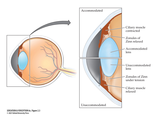
Accomdation (focusing)

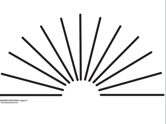 Astigmatism
Reading notes:
To clean up your notes, you can organize the information into a more structured format. Here's a suggestion:
Cranial Nerves
Olfactory (I) nerves: The first pair of cranial nerves. They conduct impulses from the olfactory epithelia in the nose to the olfactory bulb.
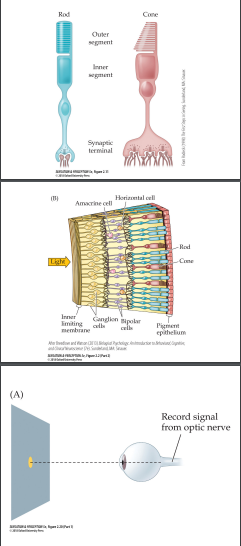
Optic (II) nerves: The second pair of cranial nerves. They carry visual information from the retina to the thalamus and other parts of the brain.
Oculomotor (III) nerves: The third pair of cranial nerves. They innervate most of the extrinsic muscles of the eye, as well as the muscles controlling the upper eyelid, ciliary muscle, and pupil.
Trochlear (IV) nerves: The fourth pair of cranial nerves. They innervate the superior oblique muscles of the eyeballs.
Abducens (VI) nerves: The sixth pair of cranial nerves. They innervate the lateral rectus muscle of the eyeballs.
Vestibulocochlear (VIII) nerves: The eighth pair of cranial nerves. They connect the inner ear with the brain, transmitting impulses related to hearing and spatial orientation.
Additionally, you may want to remove the unrelated terms "polysensory" and "vitalism" from your notes.
To clean up your notes, you can organize the information into a more structured format. Here's an example:
Magnetic Resonance Imaging (MRI)
An imaging technology that uses the responses of atoms to strong magnetic fields to form images of structures like the brain.
Can be adapted to measure activity in the brain.
Functional Magnetic Resonance Imaging (fMRI)
A variant of MRI that measures localized patterns of activity in the brain.
Activated neurons provoke increased blood flow, which can be quantified by measuring changes in the response of oxygenated and deoxygenated blood to strong magnetic fields.
Blood Oxygen Level-Dependent (BOLD) Signal
The ratio of oxygenated to deoxygenated hemoglobin that allows the localization of brain activity.
Feel free to customize and expand upon this structure based on your needs.
Method of Limits: A psychophysical method where the intensity of a stimulus or the difference between two stimuli is gradually increased or decreased until the participant perceives a change.
Method of Adjustment: A variation of the method of limits where the participant controls the change in the stimulus.
Magnitude Estimation: A psychophysical method where participants assign values to stimuli based on their perceived magnitudes.
Steven's Power Law: A principle stating that the magnitude of subjective sensation is proportional to the stimulus magnitude raised to an exponent.
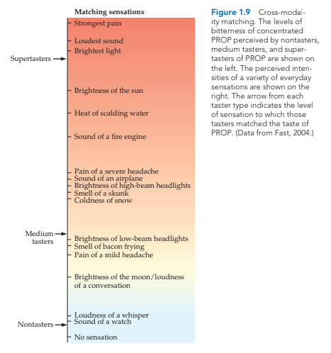
Cross-Modality Matching: The ability to match the intensities of sensations from different sensory modalities, providing insight into sensory differences.
Supertaster: An individual who experiences the most intense taste sensations, often perceiving stimuli as more intense compared to others.
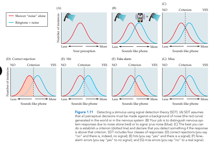
Signal Detection Theory: A theory that quantifies an observer's response to a signal presented in the presence of noise, measured by sensitivity (d') and criterion.
Criterion: An internal threshold set by the observer in signal detection theory, determining their response based on whether the internal response is above or below the criterion.
Sensitivity: A value in signal detection theory that defines an observer's ability to differentiate between the presence and absence of a stimulus or between different stimuli.
Receiver Operating Characteristic (ROC) Curve: A graphical plot of hit rate against false-alarm rate, used to assess an observer's ability to distinguish signal from noise.
Sine Wave: A simple oscillation that repeats across space or time, often used to describe waveforms in hearing or patterns in vision.
Wavelength: The distance required for one full cycle of oscillation for a sine wave.
Period: The time required for a full wavelength of an acoustic sine wave to pass by a point in space.
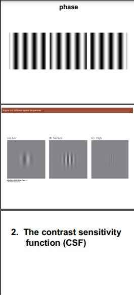
Phase: A fraction of the cycle of a sine wave described in degrees or radians, used to describe fractions of a period that relate to time.
Fourier Analysis: A mathematical procedure that separates a signal into component sine waves at different frequencies.
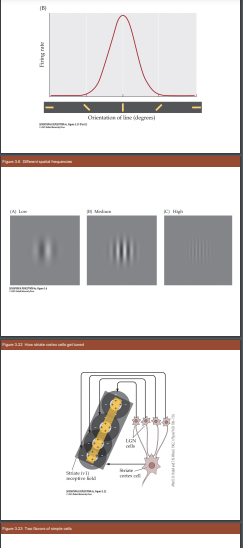
Spatial Frequency: The number of cycles of a grating per unit of visual angle, often specified in cycles per degree.
Cycles per Degree: The number of pairs of light and dark bars (cycles of grating) per degree of visual angle.
Doctrine of Specific Nerve Energies: A doctrine stating that the nature of a sensation depends on which sensory fibers are stimulated, rather than how they are stimulated.
**Cranial Nerves
rons that are most involved in a task.


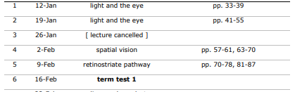
 pp 57-61, 63-70
Summary:
The chapter discusses the journey of light from distant stars to our perception. It explains how light is absorbed by photoreceptors in the retina and converted into neural signals.
Light can be absorbed, scattered, reflected, transmitted, or refracted on its way to becoming a visual sensation.
Vision begins in the retina, where light is absorbed by rods or cones. The retina acts as a minicomputer, converting light energy into neural energy.
The retinal periphery has high sensitivity to light but poor acuity due to a high degree of convergence.
The fovea has low convergence, resulting in high acuity but poor sensitivity to light.
The fovea has one-to-one pathways between cones and ganglion cells, explaining why images are seen most clearly in this area.
The visual system regulates light entering the eyeball, uses different photoreceptors, and discards unnecessary photons to deal with variations in light intensity.
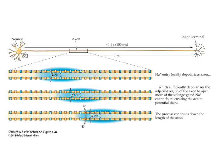
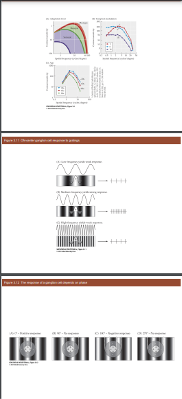
Retinal ganglion cells with center-surround receptive fields transmit information to the brain via the optic nerves and are sensitive to contrast changes.
Age-related macular degeneration (AMD) affects the macula and causes gradual loss of central vision.
Retinitis pigmentosa (RP) is a hereditary disease that leads to the death of photoreceptors, primarily affecting peripheral vision and low-light conditions.
There are ongoing developments to restore sight in individuals with retinal diseases.
Please note that this is a summary of the text content provided.
 January 26:
 Practice problem:
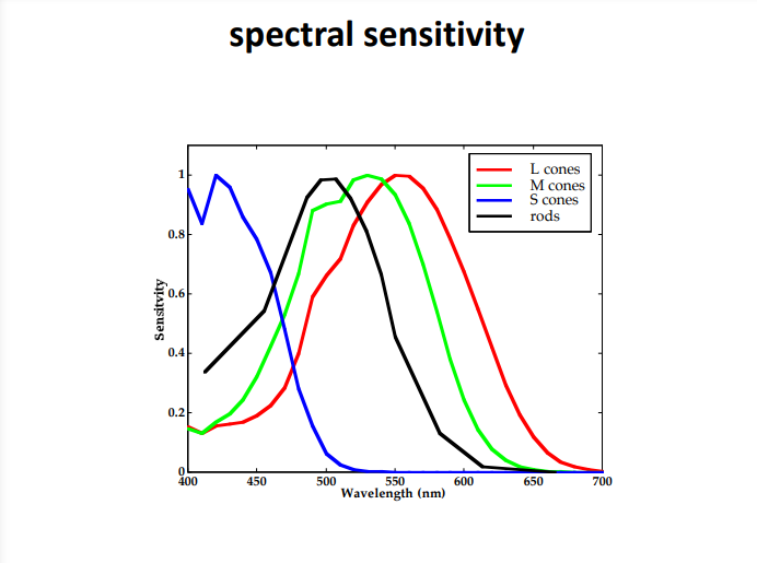
1. What range of photon wavelengths is the human eye sensitive to? 400 – 700 nm
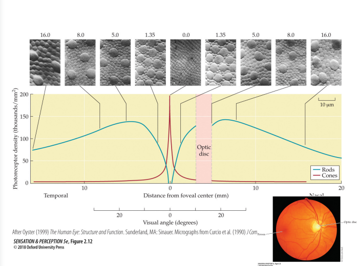
 2. Sketch a plot showing the density of the rods and the three types of cones in the human retina, as a function of eccentricity. Include separate lines on the plot for each of the three types of cones. Label the axes. See Figure 2.12. The L and M cone lines are like the cone line on the plot, although the L line is higher as the L cones are more dense than the M cones. The S cone line is also like the line on the plot, except that it has lower density overall and drops to zero at the fovea'
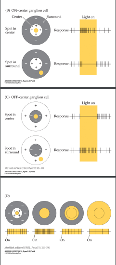
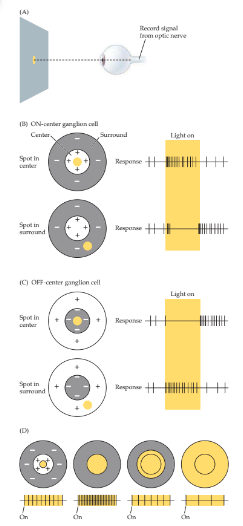 3. Sketch the receptive field of a ganglion cell. See Figure 2.20. Either an ON-centre or OFF-centre receptive field is fine.
4. Is the crystalline lens thickest or thinnest when you are looking at a faraway object? thinnest
5. What is astigmatism? Astigmatism is a visual defect caused by unequal curving of the cornea or lens in different directions, i.e., their surfaces are not spherical. Astigmatism causes the focusing power of the eye to be different at different orientations, so not all orientations can be in focus at once.
6. What is emmetropia? the condition where the strength of the lens is properly matched to the length of the eye
7. What is hyperopia? farsightedness. the condition where the lens is too weak for the length of the eye. 2
8. What is accommodation? the process in which the eye adjusts the thickness of the lens, to focus on objects at various distances
9. Name a kind of neuron that does not spike. photoreceptors, i.e., rods and cones
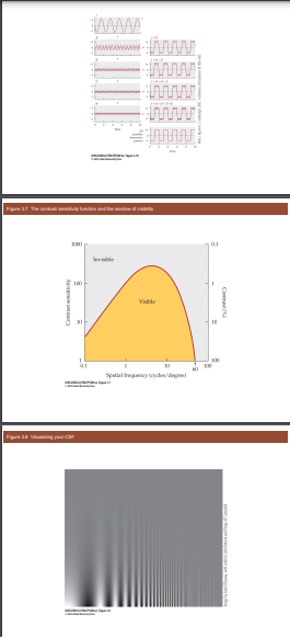
10. Sketch a contrast sensitivity function. Be sure to label the axes. See Figure 3.7
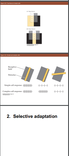
 11. What is the main difference between a simple cell and a complex cell? A simple cell responds to oriented lines at a particular location in its receptive field, whereas a complex cell responds to oriented lines at any location in its receptive field.
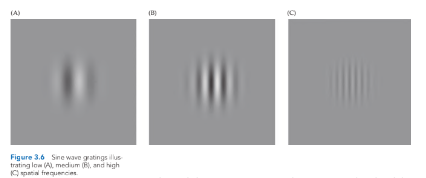 12. Sketch the receptive field of a simple cell. See Figure 3.6.
13. At what point in the visual pathway do cells first exhibit orientation tuning? Centresurround receptive fields? Binocular responses? Spiking responses? Motion tuning?
Spiking responses first appear in the collector cells (i.e., horizontal, bipolar, and amacrine cells). Centre-surround receptive fields first occur in the ganglion cells. (Actually, they first appear in collector cells, but we didn’t discuss that.) Orientation tuning, motion tuning, and binocular responses first appear in V1 cells. (We haven't discussed motion tuning in detail, so ignore that part of the question.)
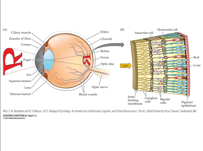
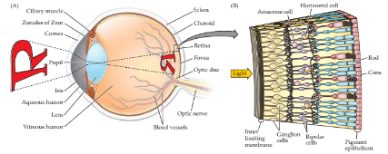
1. Sketch a diagram of the eye. Include and label the following parts: optic disc, crystalline lens, photoreceptors, ganglion cells, aqueous humour, vitreous humour, sclera, fovea, cornea, ciliary muscles, iris, choroid, pupil, zonules of Zinn, optic nerve. See Figure 2.2A for the gross anatomy of the eye, and Figure 2.2B for the layers of the retina.
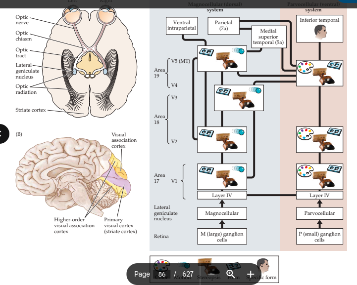
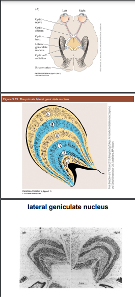
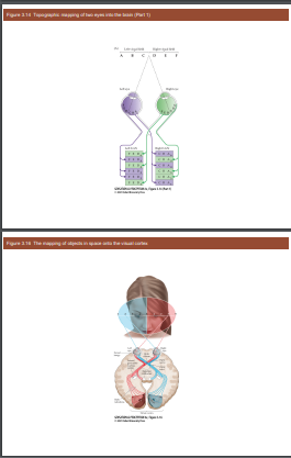
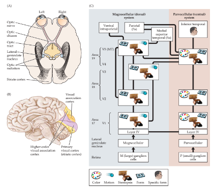
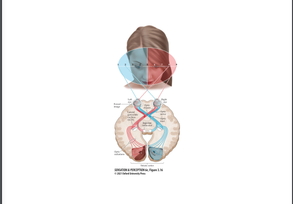
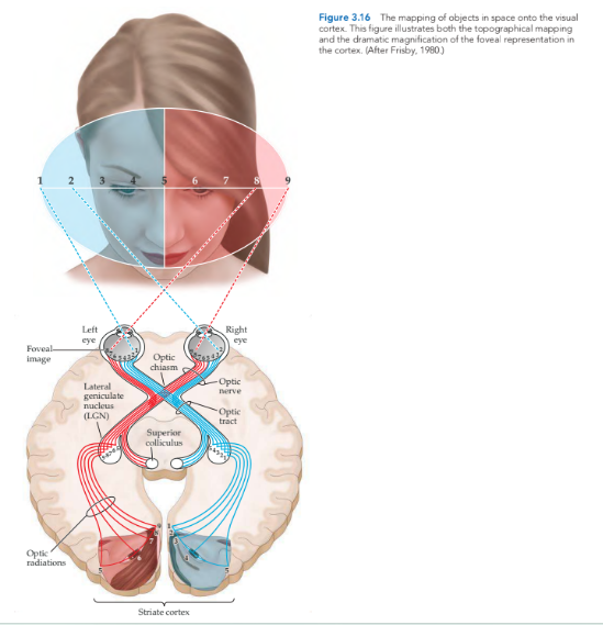
 3. Sketch a diagram of the visual pathway from the retina to striate cortex. Include and label the following parts: optic radiation, retina, complex cells, striate cortex, optic tract, LGN, optic chiasm, ganglion cells, simple cells, optic nerve. Indicate which axons from each eye cross over to the opposite side of the brain, and which stay on the same side. Sketch or briefly describe the receptive field of each type of neuron in the diagram.
See Figure 3.16. Indicate that the ganglion cells are in the retina, and simple and complex cells are in the striate cortex. Ganglion cells and LGN neurons have centresurround receptive fields. Simple cells have striped, phase-sensitive receptive fields. Complex cells have striped, phase-insensitive receptive fields.
Organized Notes:
Explain why contrast sensitivity is lower at high spatial frequencies.
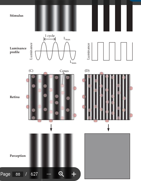
Cones in the fovea cannot accurately represent a sine wave grating with a spatial frequency higher than around 60 cycles/degree.
How do the spatial frequencies in an image change as the image moves farther away from you?
As an image moves farther away, the spatial frequencies that make it up move into a region where human visual sensitivity decreases.
Mark the layers that receive signals from the nasal half of the left retina in the monkey's lateral geniculate nuclei.
Left LGN: Layers 1, 4, and 6
Right LGN: Layers 1, 4, and 6
Mark the layers that receive signals from the retinal ganglion cells of the left eye that respond to color in the monkey's lateral geniculate nuclei.
Left LGN: Layers 3 and 5
Right LGN: Layers 4 and 6
Mark the layers that receive signals that pass through the right optic nerve in the monkey's lateral geniculate nuclei.
Left LGN: Layers 1, 4, and 6
Right LGN: Layers 2, 3, and 5
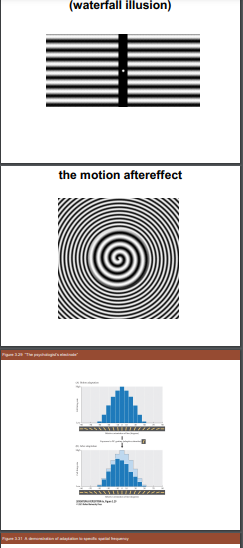
Relative activation of channels when looking at a vertical sine wave of spatial frequency 5 cycles/degree.
4 cycle/degree channel activation = 5
5 cycle/degree channel activation = 10
6 cycle/degree channel activation = 5
Relative activation of channels after adapting to a vertical sine wave of spatial frequency 4 cycles/degree and looking back at a sine wave of 5 cycles/degree.
4 cycle/degree channel activation = 2
5 cycle/degree channel activation = 10
6 cycle/degree channel activation = 5
The sine wave will appear to have a higher spatial frequency than 5 cycles/degree.
Relative activation of channels after adapting to a complex pattern containing vertical sine waves at all spatial frequencies and looking back at a sine wave of 5 cycles/degree.
4 cycle/degree channel activation = 3
5 cycle/degree channel activation = 6
6 cycle/degree channel activation = 3
The original sine wave may appear dimmer, but its perceived spatial frequency will not be affected
Vision Blurry Underwater and Swimming Goggles
Human eye has two lenses: cornea and crystalline lens
Cornea provides most of the optical power
Crystalline lens is adjustable and important for focusing
Lenses rely on refraction of light when it travels between different transparent mediums
When opening eyes underwater, water is both outside and inside the cornea, causing loss of focusing power and blurry vision
Wearing swimming goggles preserves the air-water boundary, allowing the cornea to maintain its focusing power
Positioning Light for Faint Light Perception Experiment
Rods are more sensitive to faint light than cones
Rod density peaks away from the fovea, except at the optic disc on the nasal side
Optimal position for the light stimulus is on the high-density region, about 15 degrees from the fovea
Light should be positioned on the nasal side of straight ahead, falling on the temporal side of the retina to avoid the optic disc
Pigeons' Unusual Eyes
Pigeons peck at the ground for food while watching for distant predators from above
Pigeon's lens has a single focal power, making it challenging to focus on nearby and distant objects simultaneously
Top part of the retina receives images from nearby objects on the ground, while the bottom part receives images from distant objects in the sky
By having the top half of the retina farther from the lens than the bottom half, pigeons can focus on both nearby and distant objects simultaneously
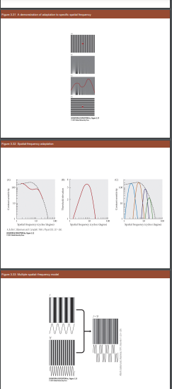
Adapting to a 5 Cycle/Degree Sine Wave Grating
(a) Observer with many spatial frequency channels:
Adapting to a 5 cycle/degree grating only fatigues channels sensitive to that grating
Contrast sensitivity function shows a dip around 5 cycles/degree, but sensitivity remains the same at other spatial frequencies
(b) Observer with one spatial frequency channel:
Adapting to a 5 cycle/degree grating fatigues the single channel responsible for perceiving all spatial frequencies
Sensitivity at all spatial frequencies decreases
Adapting to a Pattern with 2.0 and 6.0 Cycle/Degree Sine Waves
Adapting to the pattern fatigues neurons detecting 2.0 and 6.0 cycle/degree spatial frequencies
Sensitivity at these frequencies declines, resulting in dips in these spatial frequencies
 Feb9:
Feb9:

 Practice Quiz + Notes:
Light can be described as a stream of photons or - a wave
Light cannot be - dissolved
Refraction of a wave of energy means - bending or spreading out of waves as they pass from one medium to another.
When light strikes a surface and bounces off at a well-defined angle, it is being - reflected
. The transparent “window” on the outer part of the eye that allows light into the eyeball is called the - cornea
The Aqueous Humor is a - watery fluid between the cornea and iris
The vitreous humor is - a gel-like fluid between the lens and retina.
The dark, circular opening at the center of the eye, where light enters the eye, is called the - pupil
The colored part of the eye, consisting of a muscular diaphragm, is called the - iris
The structure that becomes thicker or thinner to allow images to be focused onto the back of the eye is called the - lens
The light-sensitive membrane at the back of the eye that contains rods and cones is called the - retina
The retina - contains rods and cones
People with ______ do not require an optical correction to see normally - emmetropia
Which of the following is a unit of measurement of the optic power of a lens? - Diopter
Which of the following refers to nearsightedness? - Myopia
Accommodation is the process during which the _______ of the eye changes its shape - lens
Literally meaning “old sight,” this term refers to age-related loss of accommodation, which makes it difficult to focus on near objects. - Presbyopia
In presbyopia, the lens becomes stiff with age and cannot change its shape. What is the perceptual consequence of this change? - It may become difficult to focus on objects at certain depths.
The retina is analogous to the ______ in a camera - film
1. The _____ is a light-sensitive surface at the back of the eye. E. retina
2. The _____ is a tough outer covering that protects the eye. B. sclera
3. Which of the following cell types are light-sensitive? A. cones
4. Rods are specialized for _____. C. night vision
5. Cones are specialized for _____. A. daytime vision
6. The human retina is more sensitive in the _____ than in the _____. D. fovea; optic disk
7. Light and dark adaptation are partly due to changes in _____. E. photopigment regeneration
8. The _____ of a photoreceptor contains photopigment molecules. A. outer segment
9. Which of the following terms describes a typical ganglion cell receptive field? D. centre-surround .
The optic nerve is made up of _____. B. axons
11. When a doctor looks through your pupil in to your eye, he or she sees the _____. A. fundus
12. The _____ is the region on the retina that influences a neuron's firing rate. E. receptive field
13. There are three types of cones in the human retina: _____, _____, and _____. A. long-, medium-, short-wavelength
14. Which of the following is not a type of ganglion cell? E. B type 15.
An ON-center ganglion cell: A. responds most to light in the center of its receptive field
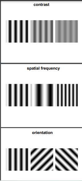
1. If a figure is much brighter than the surrounding area, we say it has high _____. A. contrast
2. We measure the smallest detail that a person can perceive. We are measuring their _____. C. acuity
3. A vision researcher measures visual angle in order to quantify the _____. E. size an object takes up on the retina
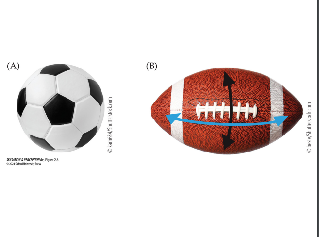
4. How can you increase the visual angle of a balloon? C. Inflate the balloon
5. What is the spatial frequency of a square wave grating? E. the number of cycles of the grating per unit of visual angle
6. What are the units we use to measure of spatial frequency? E. cycles per degree
7. When we plot sensitivity versus spatial frequency, we are plotting a _____. B. contrast sensitivity function
8. What do we call the lowest contrast level at which a person can detect a pattern? D. contrast threshold
9. Where do the axons of most reginal ganglion cells terminate? E. the lateral geniculate nuclei
10. The lateral geniculate nucleus contains which of the following types of neurons? E. magnocellular, parvocellular, and koniocellular
11. An object on your right-hand side is imaged on the _____ half of the retina, and then is represented in the LGN on the _____ side of your brain. A. left; left
12. How many lateral geniculate nuclei are there in a typical human visual system? C. two
1. All these names refer to the same structure except: C. LGN.
2. The amount of _____ corresponding to a region of the visual field is called its ‘cortical magnification’. B. cortical area
3. The phenomenon that makes peripheral object recognition difficult in the presence of clutter is known as E. visual crowding.
4. Important properties of the _____ of neurons in striate cortex were discovered by Hubel and Wiesel. E. receptive fields
5. Neurons in primary visual cortex respond more strongly to some orientations than to others. This is known as: A. orientation tuning.
6. What visual pattern would you use to make a neuron in striate cortex respond most strongly? A. An oriented bar of light
7. Many neurons in striate cortex respond more strongly to stimuli in one eye than to stimuli in the other eye. This is called: B. Ocular dominance
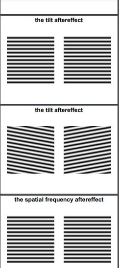
8. When a sense organ or neuron responds less strongly after sustained stimulation, this is called _____. C. adaptation.
9. To produce a tilt aftereffect, you would have an observer: A. adapt to a pattern of a given orientation.
10. The earliest part of the visual pathway that contains neurons that receive information from both eyes is the: B. striate cortex.
11. A blurry image of an object contains mostly _____ spatial frequencies. A. low
Study notes from Feb12:
Blue is the weaker colour on the wavelength spectrum from blue to red.
Middle colours include lighter blue, green, yellow than orange to the strongest being red.
The cornea is the transparent window of the eyeball
The front part of the eye consists of the Clinary muscle at the very top, the Zinn below the clinary, Cornea below that than the pupil at the very middle, than below it is the Iris, Aqueous humor (easy way to remember is by remembering the a.h is on the actual eyelens) , the Lens of the eye and the Vitreous humor on the actual eyeball.
The back of the eye (top to bottom) consists of the Sclera than the Choroid, Retina, Fovea, Optic Disc, The optic nerve and then the blood vessels.
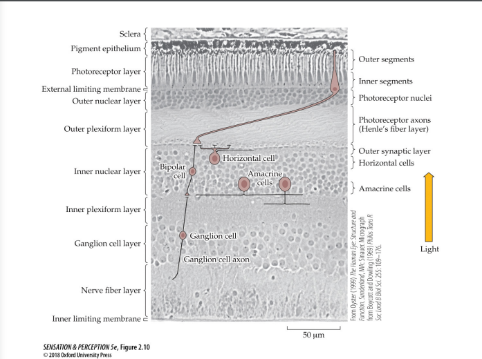
The anatomy of the structure of light-receiving aspect to the eye (Retina) is as follows: The Amacrine Cell and the Horiztonal Cell is at the top of the Retina, than to the opposing side you have the Rod (blue thing) than the Cone (black looking thing.) They act as connectors to the Rod and Cone. The wall on the light receiving side is the inner limiting membrane, the connectors of such thing is the Ganglion cells which act as connectors to the A and H cells. The Bipolar cells located near the A and H Cells and the Pigment Epithelium which is essentially the back wall of the Retina.
Aqueous humor: A clear fluid that fills the front portion of the eye, maintaining its shape and providing nutrients to the cornea and lens.
Lens: A transparent structure behind the iris that focuses light onto the retina, allowing for clear vision.
Pupil: The opening in the center of the iris that regulates the amount of light entering the eye.
Iris: The colored part of the eye that controls the size of the pupil, adjusting the amount of light that enters the eye.
Vitreous humor: A gel-like substance that fills the back portion of the eye, helping maintain its shape and transmitting light to the retina.
Retina: The innermost layer of the eye that contains light-sensitive cells (rods and cones) and converts light into electrical signals, which are then sent to the brain for visual processing.
Cataract: A cataract is a clouding of the lens in the eye, which leads to a decrease in vision.
Refractive error: Refractive error refers to a condition in which the shape of the eye prevents light from focusing correctly on the retina, resulting in blurred vision. Common refractive errors include myopia, hyperopia, and astigmatism.
Emmetropia: Emmetropia is a term used to describe normal vision, where the eye is able to focus light precisely on the retina without the need for corrective lenses.
Myopia: Myopia, also known as nearsightedness, is a refractive error where distant objects appear blurry, while close objects can be seen clearly.
Hyperopia: Hyperopia, also known as farsightedness, is a refractive error where close objects appear blurry, while distant objects may be seen more clearly.
Astigmatism: Astigmatism is a refractive error caused by an irregularly shaped cornea or lens, resulting in distorted or blurred vision at all distances.
Transduce: Transduce refers to the process of converting one form of energy into another. In the context of vision, it refers to the conversion of light energy into electrical signals by the photoreceptor cells in the retina.
Fundus: The fundus refers to the back portion of the eye, which includes the retina, optic disc, blood vessels, and other structures that can be observed during an eye examination.
Photoreceptor: Photoreceptors are specialized cells in the retina that are responsible for detecting and responding to light stimuli. They include rods, which are sensitive to low light levels and help with night vision, and cones, which are responsible for color vision and visual acuity in bright light conditions.
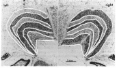
A photomicrograph of the retina is an image taken using a microscope that captures the detailed structure and cellular components of the retina. The retina is the light-sensitive tissue lining the inner surface of the eye. It contains specialized cells called photoreceptors, including rods and cones, which convert light into electrical signals that are transmitted to the brain via the optic nerve. A photomicrograph of the retina allows for the visualization and analysis of the different layers of the retina, such as the outer nuclear layer, inner nuclear layer, ganglion cell layer, and the presence of blood vessels. It provides valuable information for studying retinal anatomy, identifying abnormalities, and understanding various eye diseases.
Receptive Field: In neuroscience, a receptive field refers to the specific area of the sensory space in which a stimulus can elicit a response from a sensory neuron or a group of neurons.
Age-Related Macular Degeneration (AMD): AMD is a progressive eye condition that affects the macula, the central part of the retina. It leads to a loss of central vision, making it difficult to read, recognize faces, or perform tasks that require detailed vision.
Retinitis Pigmentosa: Retinitis pigmentosa is a genetic disorder that causes the degeneration of photoreceptor cells in the retina. It leads to progressive vision loss, starting with night blindness and peripheral vision loss, and eventually affecting central vision as well.
Chromophore: A chromophore is a molecule or part of a molecule that is responsible for its color. It absorbs certain wavelengths of light and reflects or transmits others, giving the molecule its characteristic color.
Rhodopsin: Rhodopsin is a light-sensitive protein found in the rod cells of the retina in the eye. It plays a crucial role in vision by capturing light and initiating the process of visual signal transduction.
Melanopsin: Melanopsin is a photopigment found in specialized cells called intrinsically photosensitive retinal ganglion cells (ipRGCs) in the retina. It is involved in non-image-forming functions of the eye, such as regulating circadian rhythms and pupillary reflexes.
Photoactivation: Photoactivation refers to the process by which a molecule or a biological system is activated or undergoes a change in response to light exposure.
Hyperpolarization: Hyperpolarization is a change in the electrical potential across a cell membrane, making the inside of the cell more negative relative to the outside. It occurs when the cell's membrane potential becomes more negative than its resting potential.
Graded potential: Graded potential refers to a change in the membrane potential of a cell that varies in magnitude and can be either depolarizing (making the inside of the cell more positive) or hyperpolarizing (making the inside of the cell more negative). Graded potentials are used for short-distance communication within cells and can summate to generate action potentials.
Horizontal cell: A type of neuron found in the retina of the eye that receives input from multiple photoreceptor cells and modulates the communication between them.
Lateral inhibition: A process in which the activity of one neuron inhibits the activity of neighboring neurons, enhancing the contrast and sharpness of visual signals.
Diffuse bipolar cell: A type of bipolar cell in the retina that receives input from multiple photoreceptor cells and distributes the signal to multiple ganglion cells.
On bipolar cell: A type of bipolar cell in the retina that responds to increased light intensity and transmits excitatory signals to downstream neurons.
Off bipolar cell: A type of bipolar cell in the retina that responds to decreased light intensity and transmits inhibitory signals to downstream neurons.
Ganglion cell: A type of neuron in the retina that receives visual information from bipolar cells and transmits it to the brain via the optic nerve.
P ganglion cell: A subtype of ganglion cell in the retina that is responsible for processing color and fine details in the visual scene.
M ganglion cell: A subtype of ganglion cell in the retina that is responsible for processing motion and spatial information in the visual scene.
Koniocellular cell: A type of ganglion cell in the retina that is involved in color processing and projects to different visual areas in the brain.
Optic chiasm: The optic chiasm is a structure located at the base of the brain where the optic nerves partially cross over. It is responsible for the partial decussation (crossing) of the optic nerves, allowing visual information from each eye to be transmitted to the opposite side of the brain.
Optic tract: The optic tract is a bundle of nerve fibers that carries visual information from the optic chiasm to the brain. It consists of axons from the retinal ganglion cells and continues from the optic chiasm to various visual processing centers in the brain.
Lateral geniculate nucleus: The lateral geniculate nucleus (LGN) is a relay station in the thalamus of the brain that receives visual information from the optic tract. It plays a crucial role in processing and relaying visual signals to the primary visual cortex.
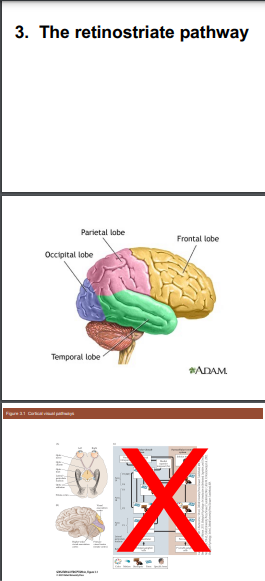
Optic radiation: The optic radiation, also known as the geniculocalcarine tract, is a collection of nerve fibers that carry visual information from the lateral geniculate nucleus to the primary visual cortex (striate cortex) in the occipital lobe of the brain. It is responsible for transmitting visual signals for further processing and interpretation.
Striate cortex: The striate cortex, also called the primary visual cortex or V1, is located in the occipital lobe at the back of the brain. It is the first cortical area that receives and processes visual information from the optic radiation. The striate cortex plays a crucial role in visual perception, including the analysis of basic visual features such as orientation, motion, and color.
The visual cortex is like a special part of our brain that helps us see things. It's like a movie theater in our head where all the pictures from our eyes go. When we look at something, our eyes send the pictures to the visual cortex, and it helps us understand what we are seeing. It's like a super smart computer that can recognize shapes, colors, and even faces! So, the visual cortex is really important for us to be able to see and understand the world around us.
Sure! Here are simplified definitions for each term:
Visual Crowding: When objects that are close together make it difficult to see and identify them individually.
Orientation Tuning: The ability of our eyes and brain to be more sensitive to certain orientations (like vertical or horizontal lines) than others.
Ocular Dominance: The tendency of one eye to be more dominant than the other in terms of visual processing.
Simple Cell: A type .
of neuron in our brain that responds to specific features of an image, like edges or lines.
Complex Cell: Another type of neuron that responds to more complex features, like movement or patterns.
End Stopping: When a neuron responds strongly to a line or edge that ends at a specific point.
Cytochrome Oxidase: An enzyme found in our brain that helps with energy production and is used to study brain organization.
Adaptation: When our eyes and brain adjust to changes in the environment, like getting used to a bright light after being in the dark.