3.3-3.4: Visual Anatomy + Perception

HUE (color): dimension of color determined by wavelength of light.
WAVELENGTH: is the distance from the peak of one wave to the peak of the next. - Notice the longer waves of red at the top, and the tighter (shorter) waves of blue and violet at the bottom.

INTENSITY (BRIGHTNESS)
INTENSITY: amount of energy in a wave determined by amplitude; related to perceived brightness. AMPLITUDE: how high each wave is.
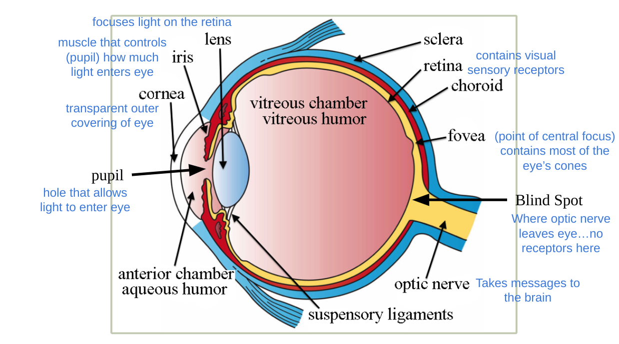
NEARSIGHTEDNESS: A condition in which nearby objects are seen more clearly than distant objects because eye is elongated in shape, so the image focuses before it hits the retina.
FARSIGHTED: A condition in which faraway objects are seen more clearly than near objects because the eye is shortened and the image focuses after it hits retina.
RETINA: The light-sensitive inner surface of the eye, containing receptor rods and cones plus layers of other neurons (bipolar and ganglion cells) that process visual information.
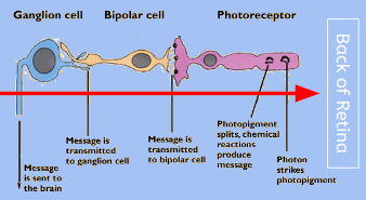
Light travels to the back of the retina, then moves forward, then to the optic nerve.

FOVEA:
Central point in the retina, within which the eyes cones cluster together..because of the cones here, there are little color vision in the farthest periphery of our vision.
PHOTORECEPTORS:
Rods: sensitive to light
Cones: sensitive to color and fine detail
- In this image, notice what the next cell that the rods connect to versus the cones… what’s different?
BIPOLAR AND GANGLION CELLS:
- Bipolar cells receive messages from photoreceptors and transmit them to ganglion cells, which then form the optic nerve
- cones each have their own bipolar cells
- multiple rods share one bipolar cell, so their is not sent as clearly
FEATURE DETECTORS:
- Nerve cells in the visual cortex that respond to specific features, like edges, angle, length and movement.
- There are even more some feature detectors that are specifically sensitive to the human face!
VISUAL INFORMATION PROCESSING:
- Processing several aspects of the stimulus simultaneously is called parallel processing. The brain divides a visual scene into subdivisions such as color, depth, form and movement, etc.
THEORIES OF COLOR VISION:
Trichromatic theory (Young-Helmholtz): Based on behavioral experiments, Helmholtz suggested that the retina contains three receptors (cones) sensitive to red, blue, and green colors.
Opponent Process Theory: Hering, proposed that we process four primary colors opposed in pairs of red-green, blue-yellow, and black-white.
COLOR DEFICIENCIES:
- Genetic disorder which prevents individuals from discriminating between certain colors due to a weakness in or lack of one of the cones.
- Most common form of color “blindness” is difficulty distinguishing between red and green
- Complete color deficiency does exist but is very rare..it would be like watching a black and white movie.