W.2.1- Vertebral column musculature
PLANES AND MOVEMENTS
Muscle Groups summary
Vertebral Column Muscles
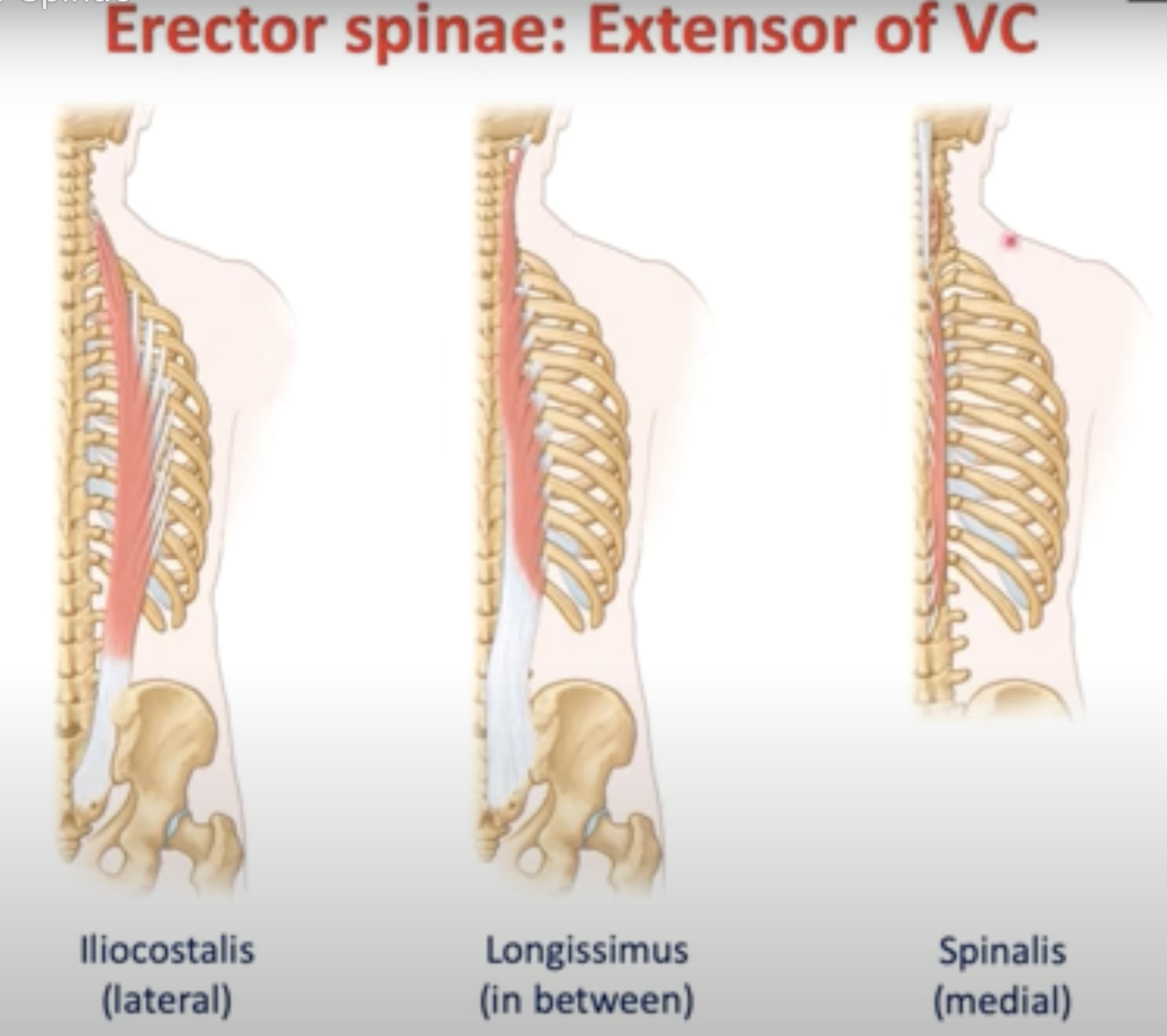
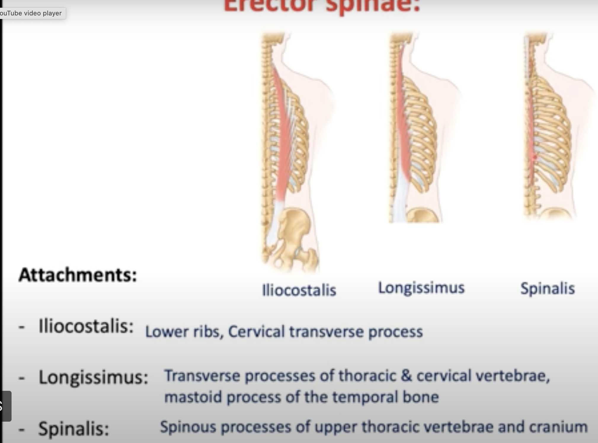
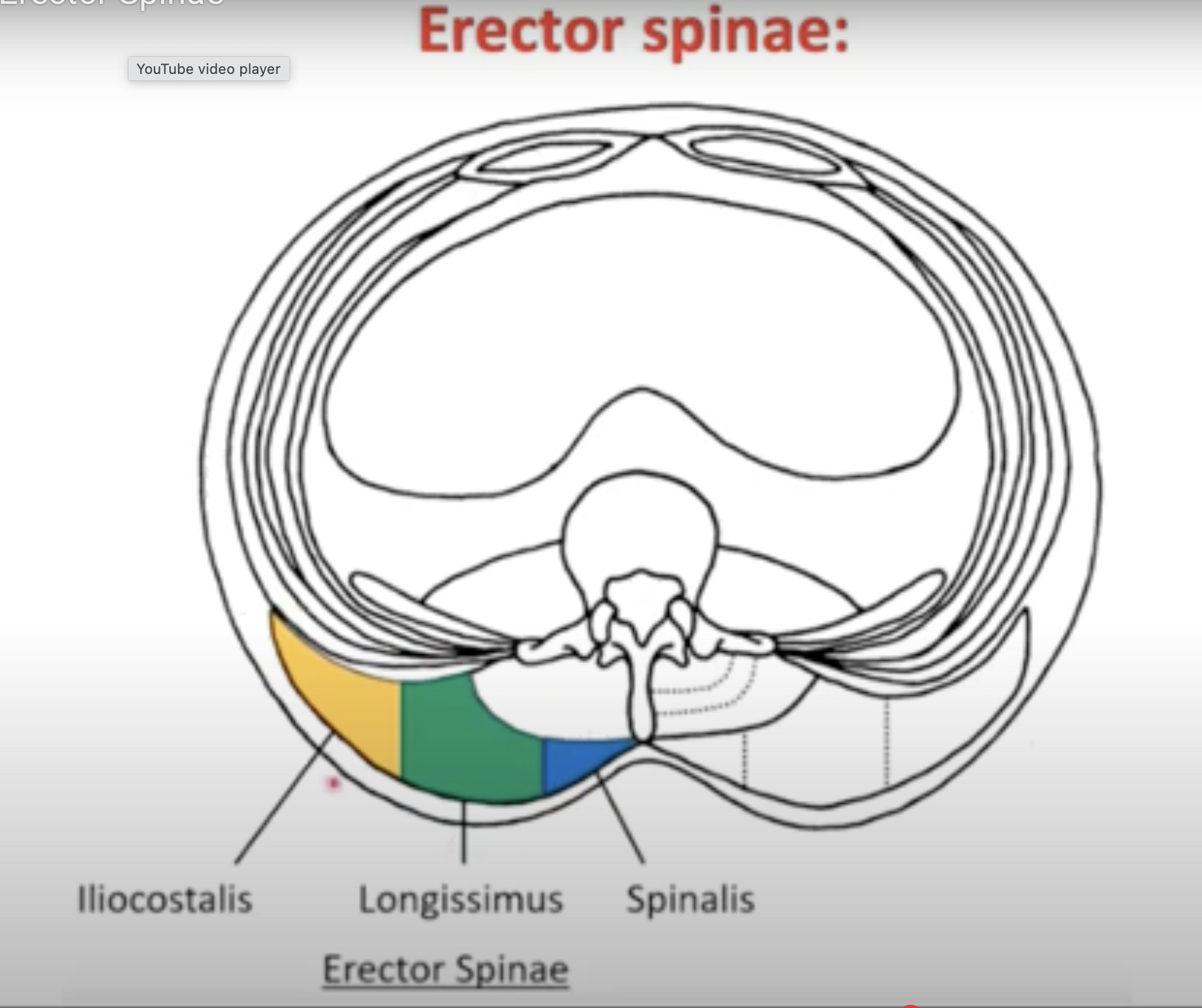
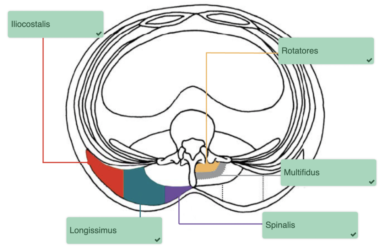
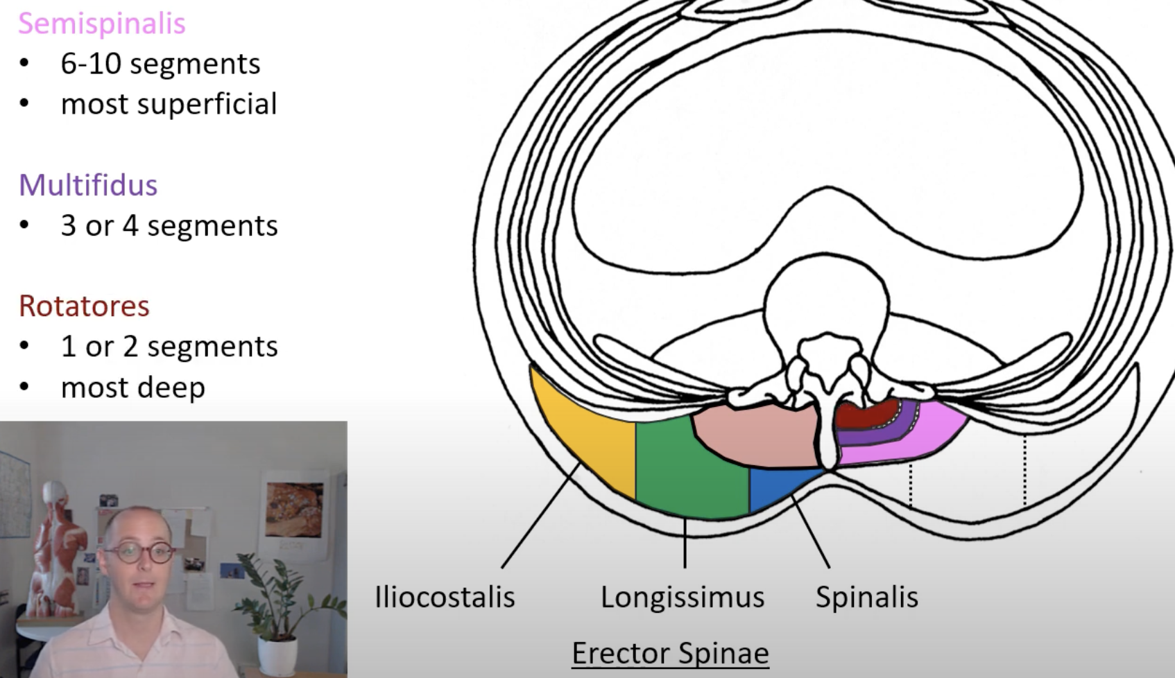
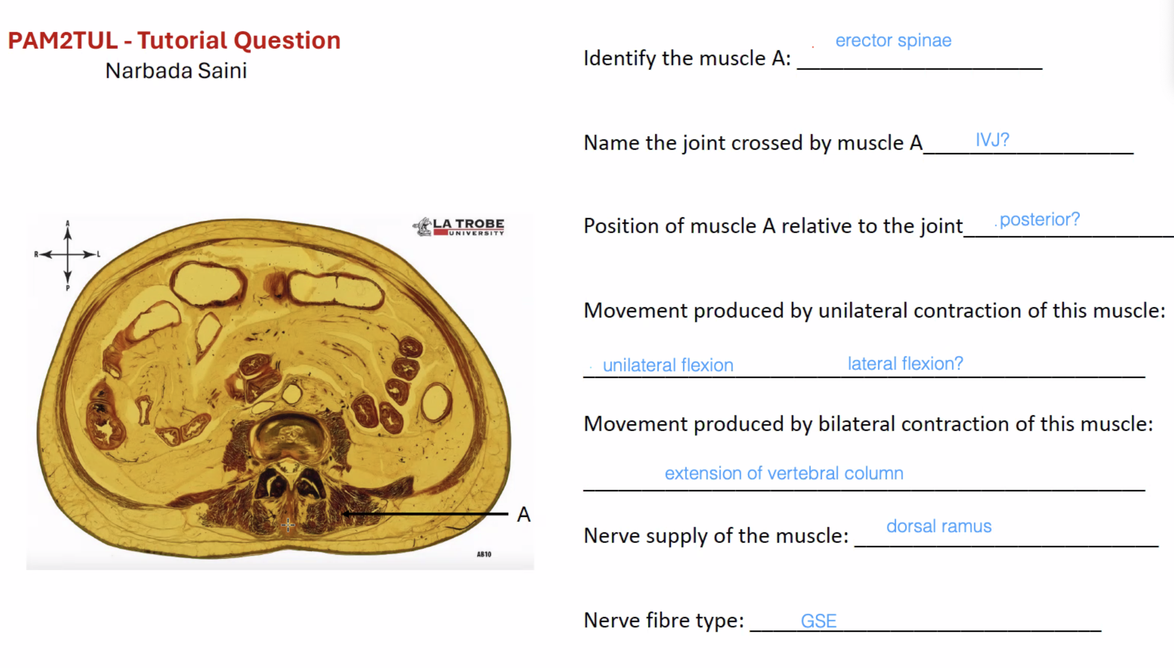
Erector Spinae: Responsible for extension and lateral flexion of the vertebral column. Includes Iliocostalis, Longissimus, and Spinalis.
Transversospinales: Includes Semispinalis, Multifidus, and Rotatores, responsible for stabilization and movements of the spine.
Anterolateral Abdominal Muscles
Rectus Abdominis: Anterior orientation, aids in flexion and increasing intra-abdominal pressure.
External Oblique: Anterolateral, most superficial layer, assists in lumbar flexion.
Internal Oblique: Anterolateral, middle layer, also contributes to abdominal pressure.
Transversus Abdominis: Deepest layer, primarily increases intra-abdominal pressure.
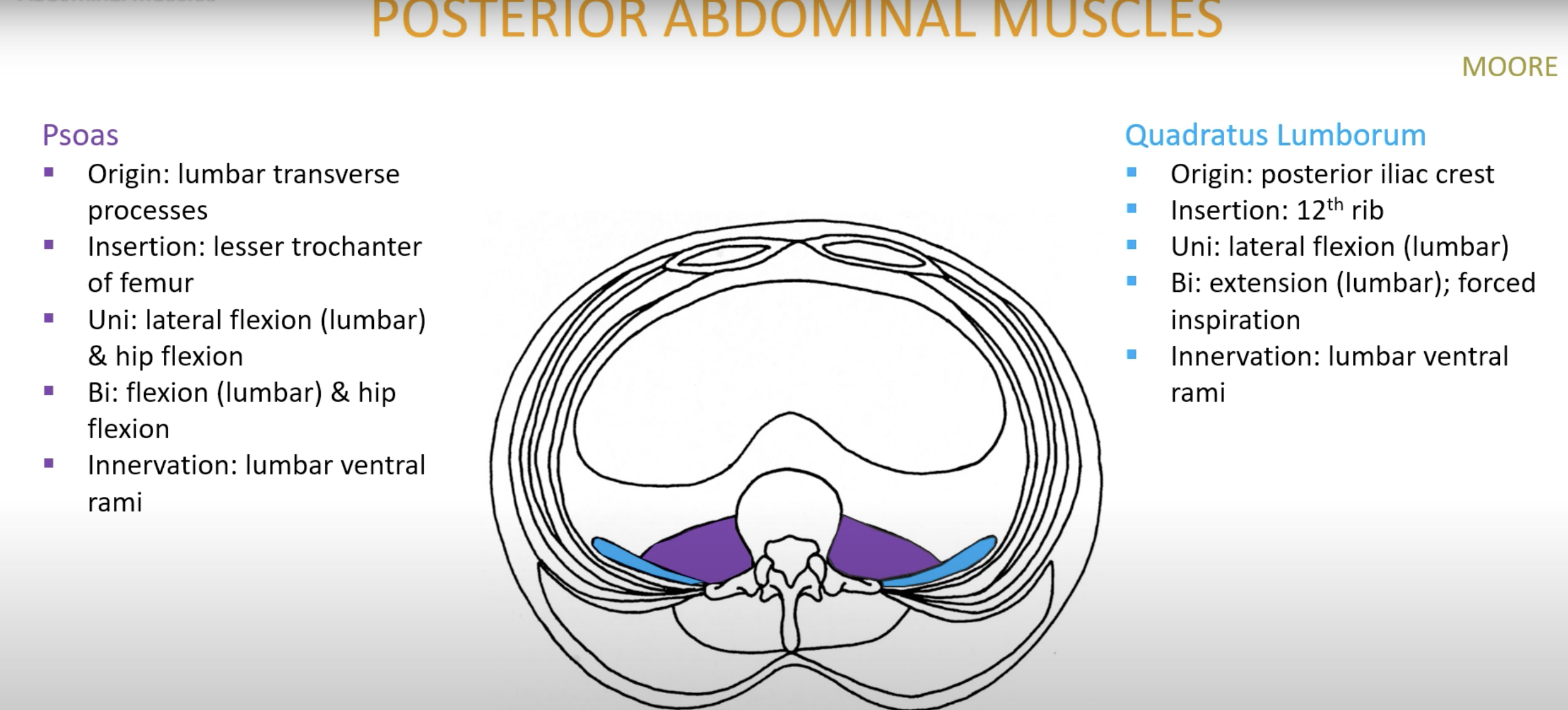
Posterior Abdominal Muscles
Psoas: Originates from lumbar transverse processes, involved in hip flexion and lateral flexion of the lumbar spine.
Quadratus Lumborum: Aids in lateral flexion and stabilization of the pelvis.
Cervical Muscles
Anterior Cervical Muscles: Include Sternocleidomastoid, Scalenes, Longus Capitis, and Longus Colli, involved in neck flexion, rotation, and stabilization.
Posterior Cervical Muscles: Include Trapezius and Splenius muscles, responsible for neck extension and lateral flexion.
Sub-occipital Muscles
Include Rectus Capitis Posterior Major/Minor, Obliquus Capitis Inferior/Superior, involved in movements at the atlanto-occipital and atlantoaxial joints.
Muscle Groups Lecture:
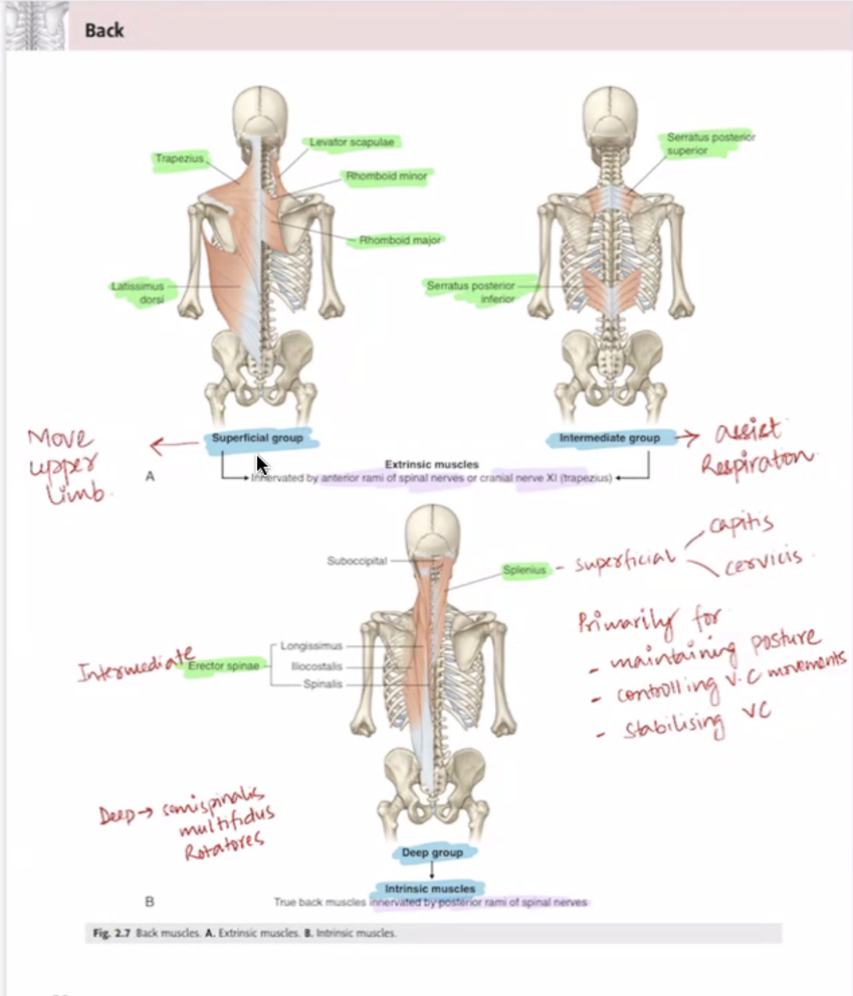
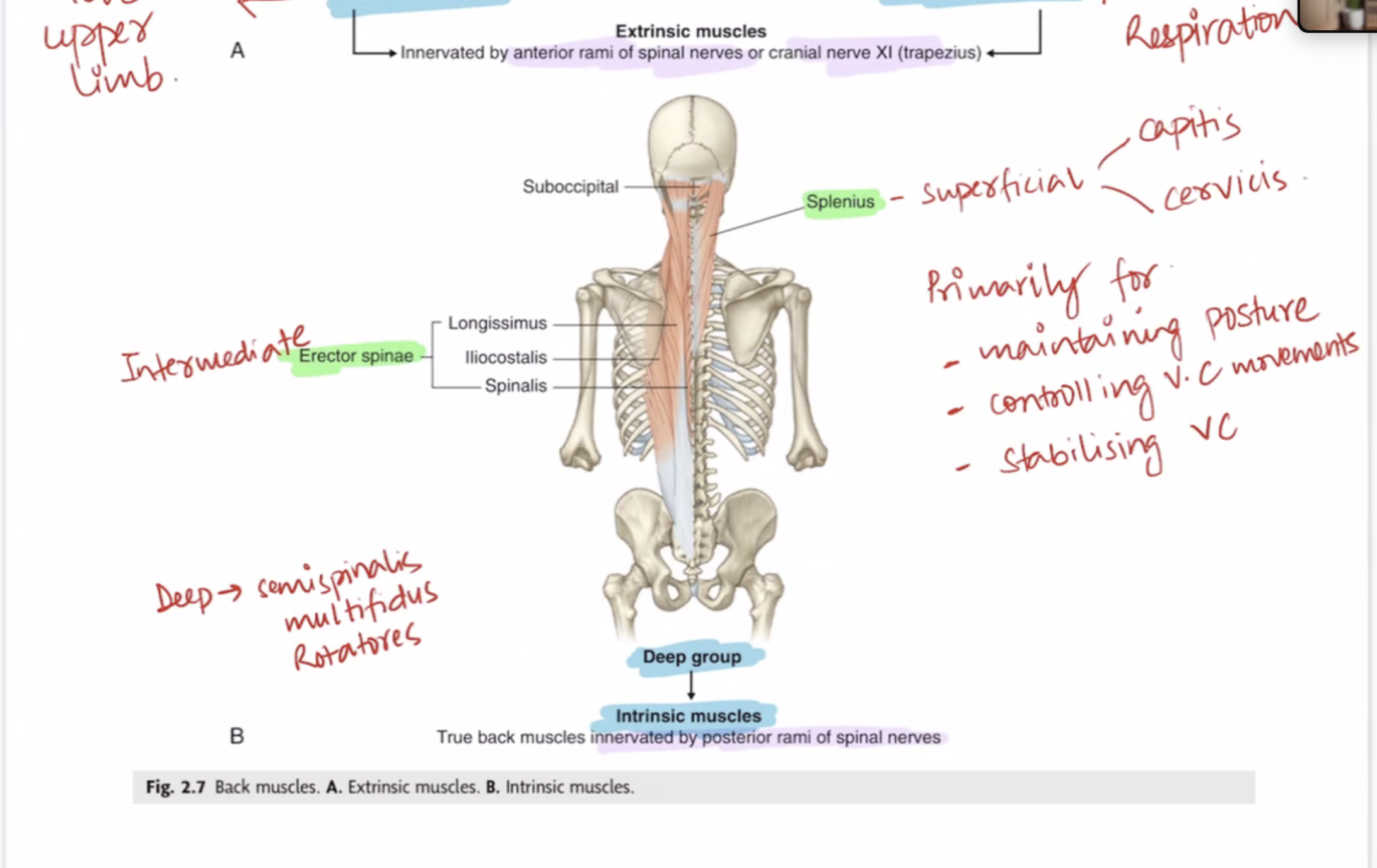
- There are two major group of muscles in the back.
(i) extrinsic muscles
- superficial and intermediate
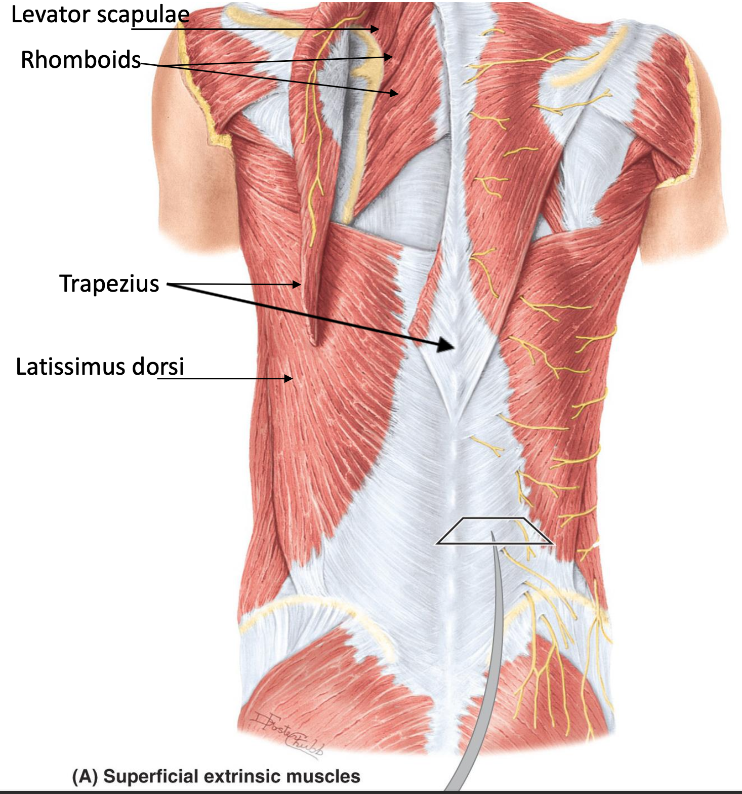
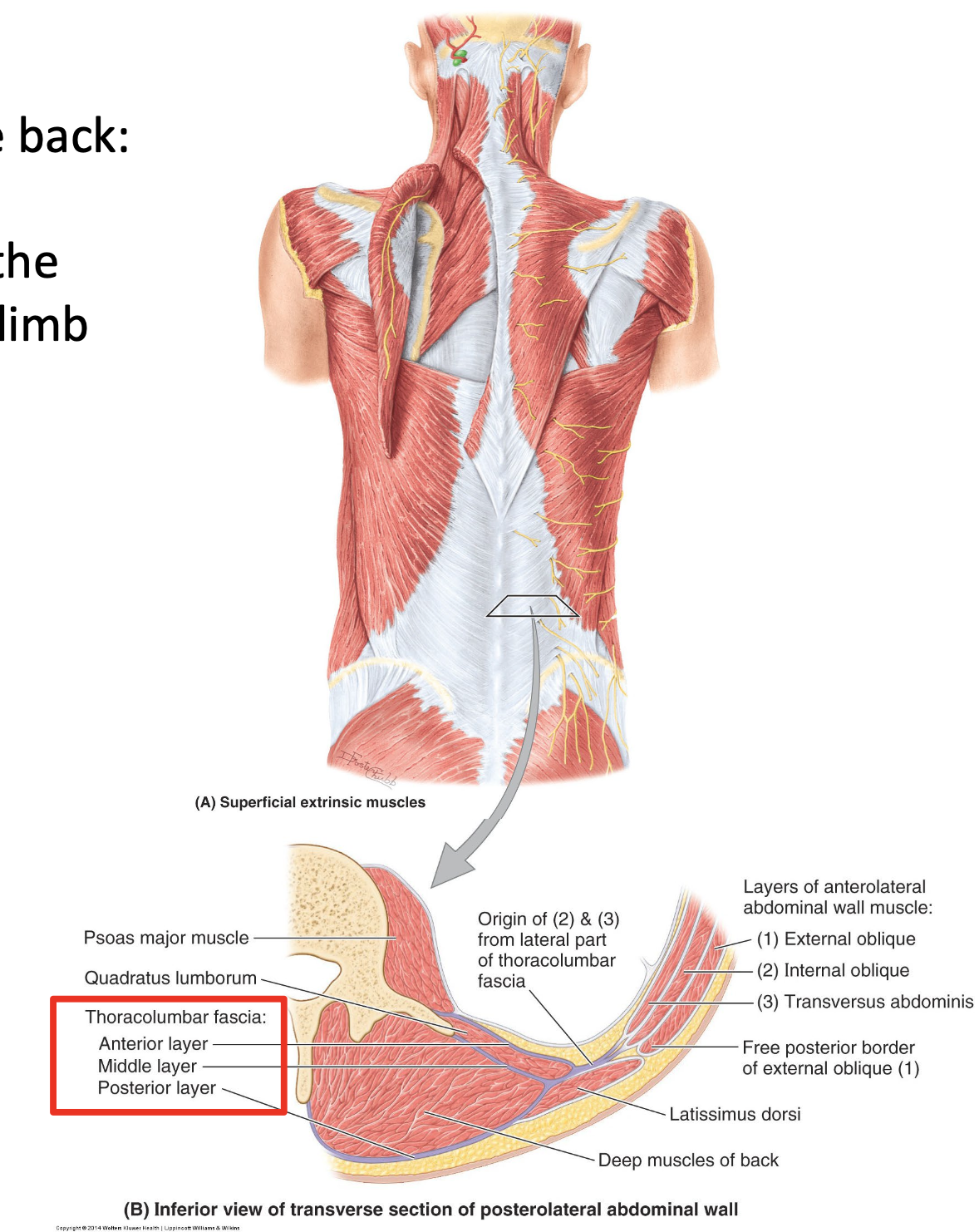
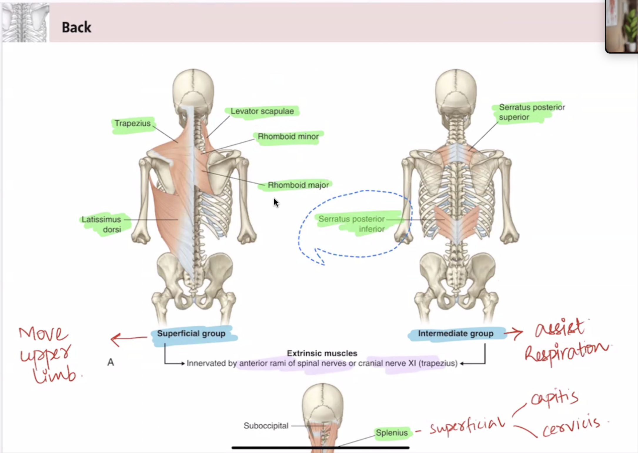
Superficial extrinsic muscles
* although located in the back region, for most part of these muscles receive their innervation from anterior rami of cervical nerves and act on the upper limb.
Extrinsic muscles produce and control limb and respiratory movements.
(ii) intrinsic muscles
Intrinsic muscles include muscles that specifically act on the vertebral column producing its
movements and maintain posture.
Intermediate extrinsic muscles
* are thin muscles that are mostly designated as
superficial respiratory muscles.
E.g. serratus posterior superior and serratus
posterior inferior.
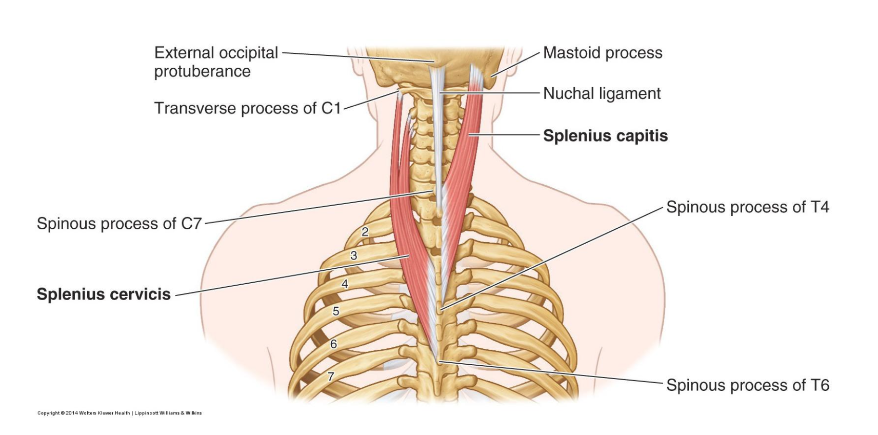
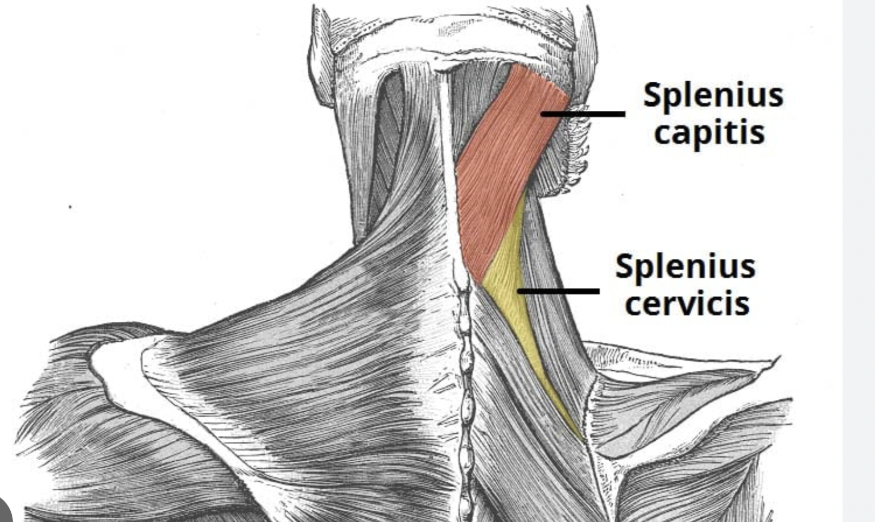
The splenius cervicis and splenius capitis are two muscles located in the posterior neck region, both part of the splenius muscle group. Here are their key differences:
Location:
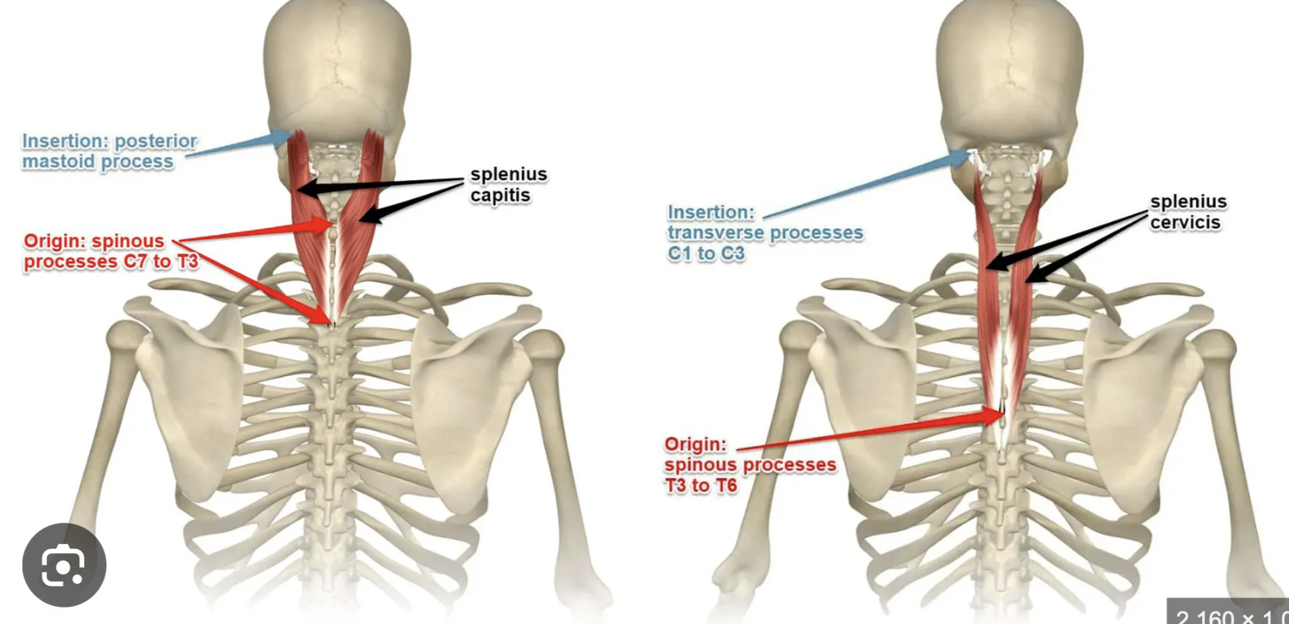
Splenius Cervicis: This muscle is situated lower and is responsible for connecting the cervical vertebrae to the upper thoracic vertebrae. It originates from the spinous processes of T3-T6 and inserts into the transverse processes of C1-C3.
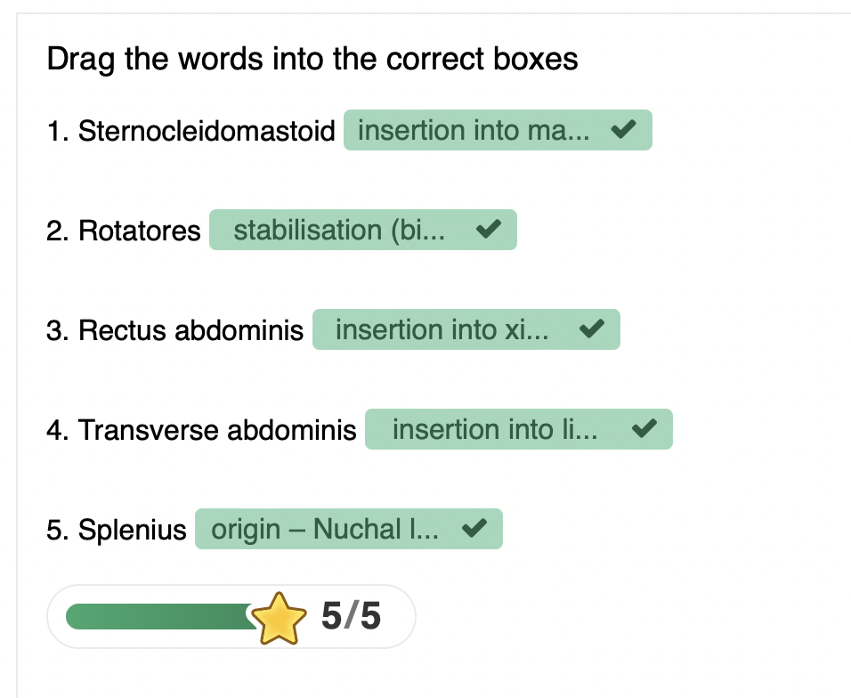
Splenius Capitis: This muscle is located higher and primarily connects the skull to the cervical region. It originates from the lower half of the nuchal ligament and the spinous processes of C7-T3 (or T4) and inserts into the mastoid process of the temporal bone and the lateral part of the superior nuchal line of the occipital bone.
Function:
Splenius Cervicis: Primarily responsible for neck extension and rotation to the same side (ipsilateral rotation) when contracted unilaterally.
Splenius Capitis: Functions similarly in neck extension and also aids in the rotation of the head toward the same side when contracted unilaterally.
In summary, while both muscles serve similar functions in head and neck movements, they differ in their location, origin, insertion, and specific action contributions.
VERTEBRAL COLUMN MOVEMENTS & MUSCLE LOCATIONS
Sagittal Plane
**Movements::Flexion and extension, as well as movements in the anterior-posterior direction.
**Muscle positions:** Muscles that act in this plane primarily include those that facilitate flexion and extension, such as the rectus abdominis, erector spinae, and hip flexors.Coronal Plane
Movements: Abduction and adduction, as well as movements in the lateral direction.
Muscle positions: Muscles that act in this plane primarily include those that facilitate abduction and adduction, such as the latissimus dorsi and the deltoid.Horizontal / Transverse Plane
**Movements:** Rotation and twisting actions, involving movements parallel to the ground.
**Muscle positions:** Muscles that act in this plane primarily include those that facilitate rotational movements, such as the obliques (external and internal), multifidus, and transversospinales.
PRINCIPLES OF MUSCLE ACTIONS
M4: Muscle names reflect structure/function (shape, size, orientation, location, attachments, action)
M5: Muscles must attach to two structures for movement
Note that origin typically refers to inferior attachment
M6: Must cross a joint to act at that joint
M7: Muscle position relative to joint affects movement direction and plane
INNERVATION OF MUSCLES OF THE VERTEBRAL COLUMN
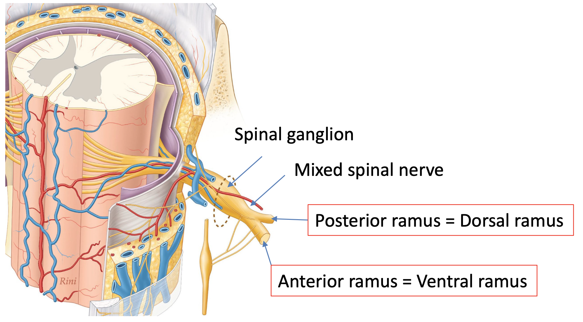
NP8: All spinal nerves divide into posterior and anterior primary rami:
posterior primary rami supply the structures of the back; anterior
primary rami supply all other somatic structures including the limbs
All spinal nerves split into posterior and anterior primary rami
Posterior Primary Rami: supply back structures
Anterior Primary Rami: supply other somatic structures including limbs
Spinal ganglion connections:
Posterior ramus = Dorsal ramus
Anterior ramus = Ventral ramus
TERMINOLOGY FOR VERTEBRAL COLUMN MUSCLE ACTIONS
Bilateral vs Unilateral Contractions
Bilateral: movements parallel to the sagittal section
Causes synergistic actions that cancel out other plane movements
Unilateral: movements in transverse and/or coronal planes
Ipsilateral vs Contralateral Movements
Ipsilateral: same side body movement (e.g., left erector spinae = left flexion)
Contralateral: opposite side movement (e.g., left external oblique = right rotation)
Bilateral Contractions (both sides working together):
When you nod your head up and down, like when you say "yes." Both sides of your neck muscles are working to help you move your head straight up and down.
Unilateral Contractions (one side working alone):
When you tilt your head to the left or right, like when you try to listen to a sound on one side. Only the muscles on that one side of your neck are working.
Ipsilateral Movements (same side working together):
If you tilt your head to your right and raise your right shoulder at the same time. The muscles on the right side of your neck and right shoulder are working together.
Contralateral Movements (opposite sides working together):
When you turn your head to the left while looking down with your right shoulder. Here, the muscles on the left side of your neck and the right side of your shoulder are working together to make that movement happen.
VERTEBRAL COLUMN MUSCLES
The sternocleidomastoid muscle has bones related to its origin and insertion; the sternum, the clavicle and the mastoid process. Can you identify the action and the position of the muscle? Use the arrows or click the icons to move through the pages. You can revisit the previous videos to help you answer this question .The primary action of the sternocleidomastoid muscle is to facilitate neck flexion and rotation, allowing the head to turn towards the opposite side when one side contracts. Additionally, when both sides of the sternocleidomastoid contract simultaneously, they assist in flexing the neck forward, contributing to overall posture and stability.
Sternocleidomastoid
Attachments:
Origin: Sternum and Clavicle
Insertion: Mastoid process
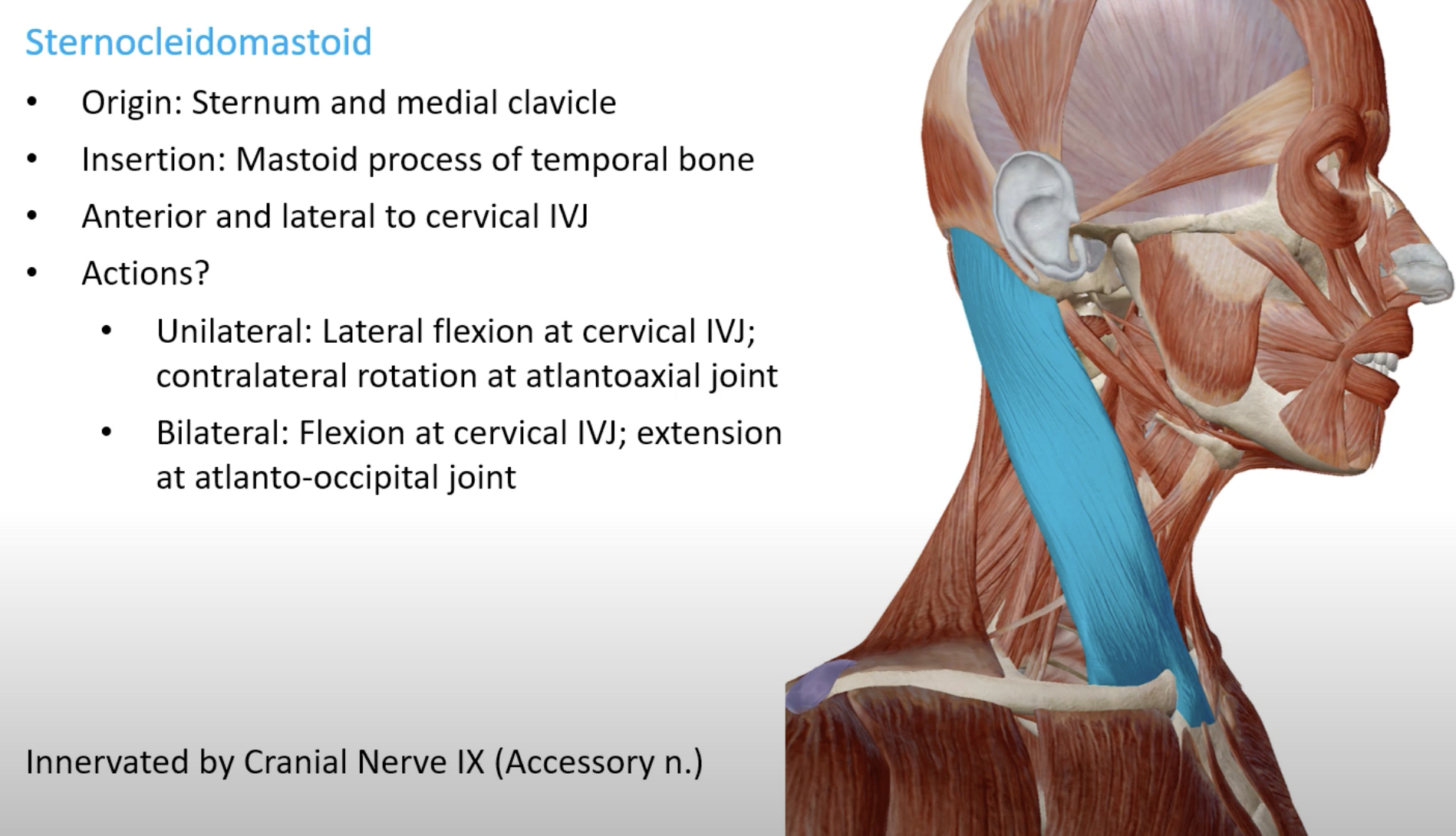
The sternocleidomastoid muscle crosses the cervical intervertebral joints and the atlanto-occipital joint.
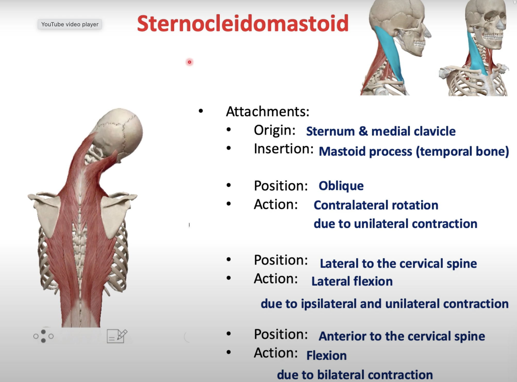
The sternocleidomastoid muscle is positioned anterior and lateral to the cervical intervertebral joints. It crosses the atlanto-occipital joint, allowing for its actions in neck flexion and rotation.
The primary movement produced by unilateral contraction of the sternocleidomastoid muscle is rotation of the head towards the opposite side. Additionally, it assists in lateral flexion of the neck towards the same side.
The primary movement produced by bilateral contraction of the sternocleidomastoid muscle is neck flexion. When both sides contract simultaneously, it also contributes to overall posture and stability of the cervical spine.
The sternocleidomastoid muscle is primarily supplied by the accessory nerve (cranial nerve XI) for motor innervation. Additionally, sensory innervation is provided by the cervical plexus, particularly the ventral rami of C2 and C3.
The nerve supply to the sternocleidomastoid muscle primarily involves the accessory nerve (cranial nerve XI), which carries motor nerve fibers. Additionally, sensory innervation is provided by the cervical plexus (ventral rami of C2 and C3), which consists of sensory fibers. Therefore, the nerve fiber types associated with the sternocleidomastoid muscle include both motor fibers from the accessory nerve and sensory fibers from the cervical plexus.
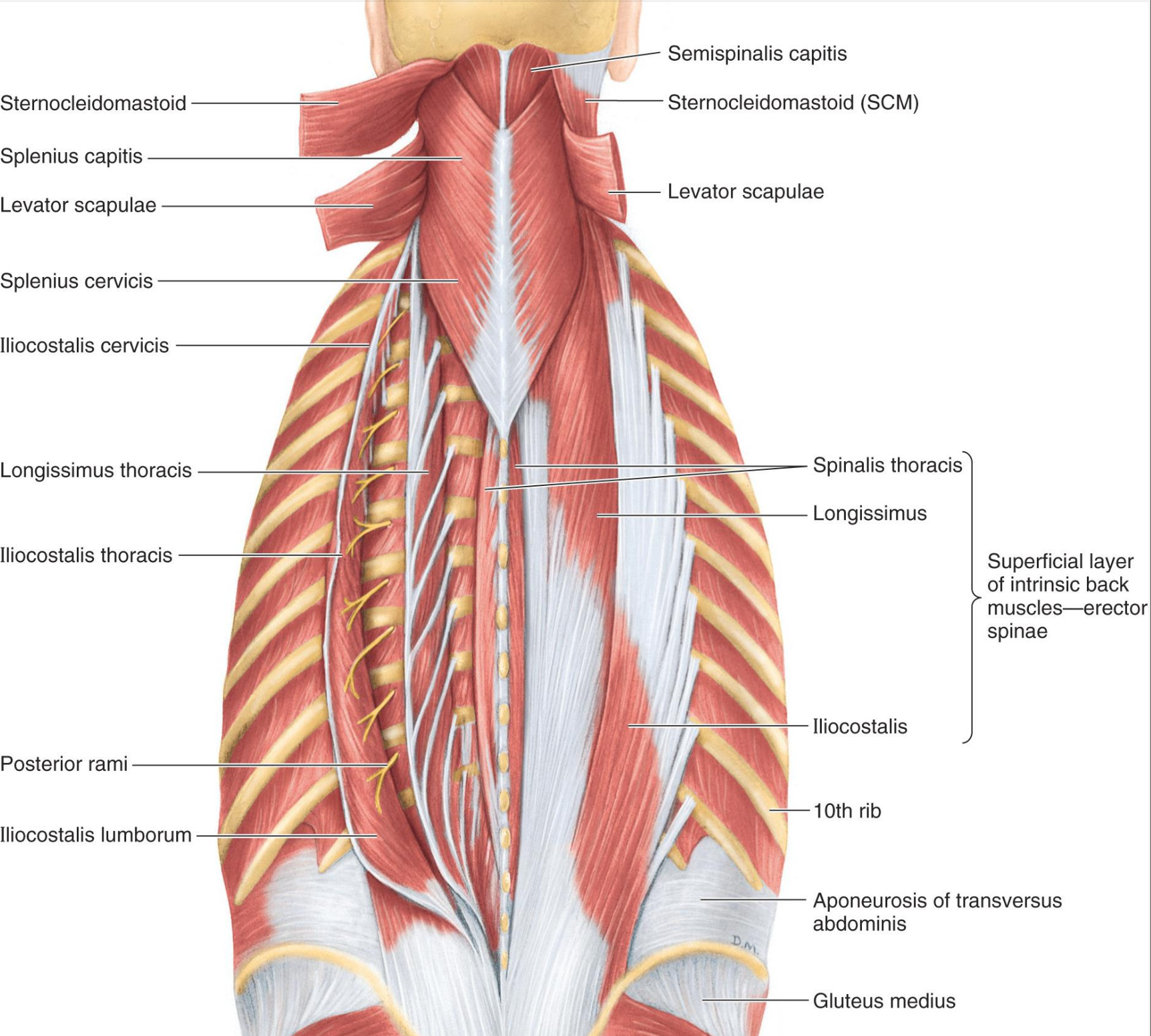
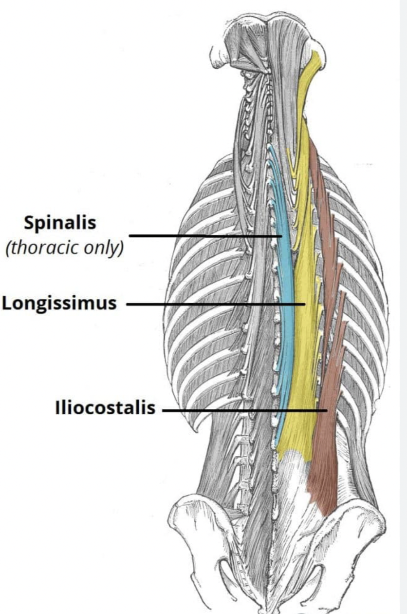
ERECTOR SPINAE
ERECTOR SPINAE STRUCTURE
Origin (common):
tendon from iliac crest,
posterior sacrum,
sacroiliac ligaments,
spinous processes of sacral and lumbar vertebrae
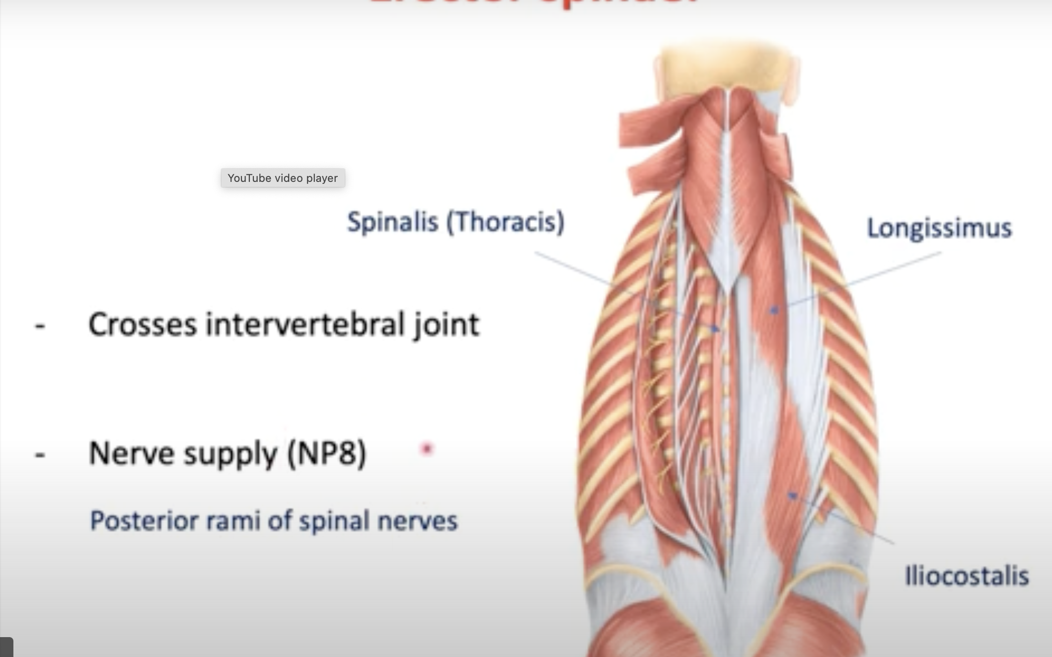
Innervation:
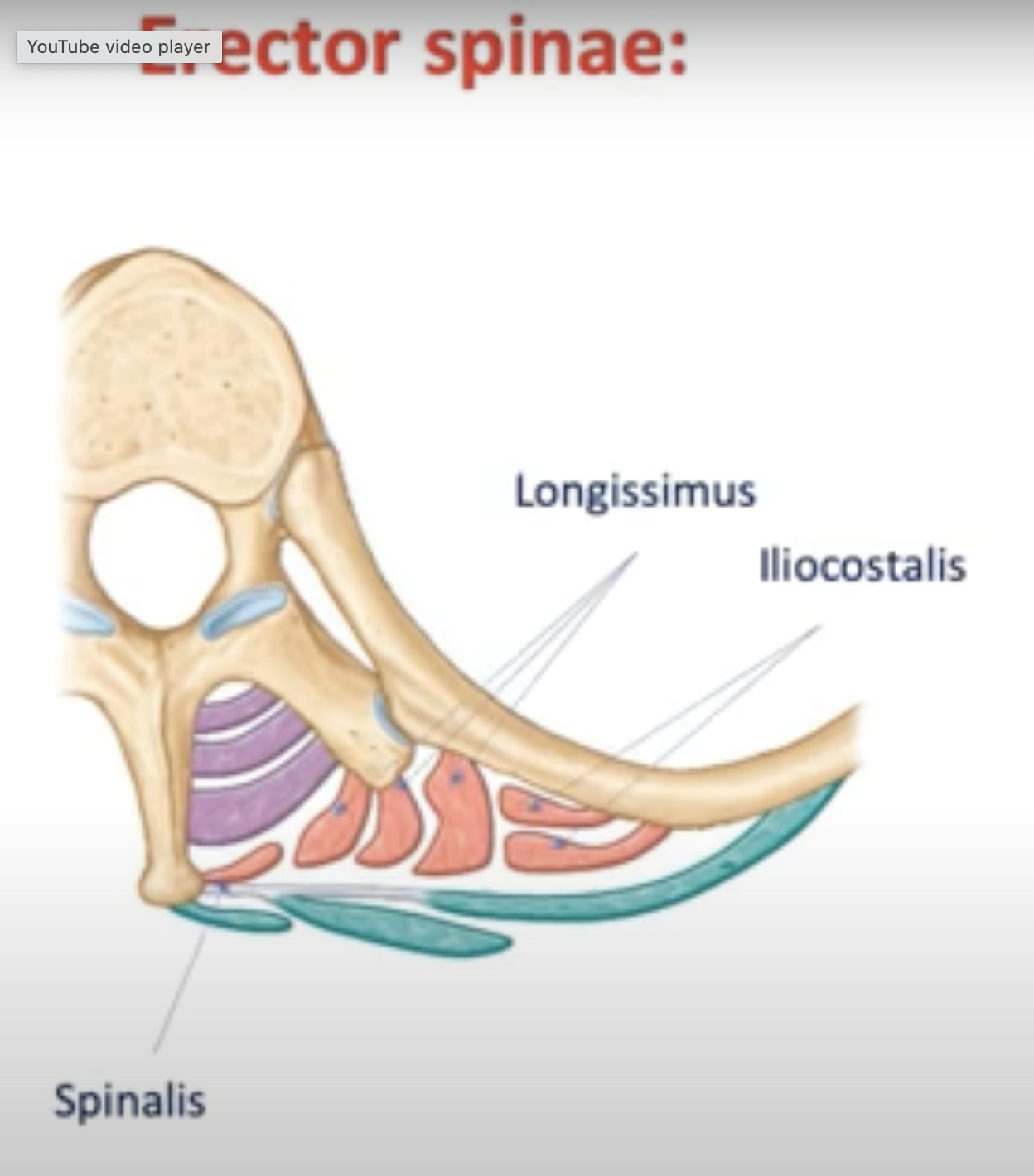
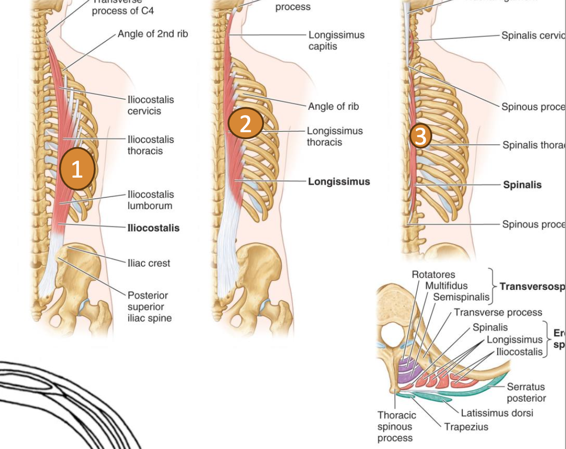
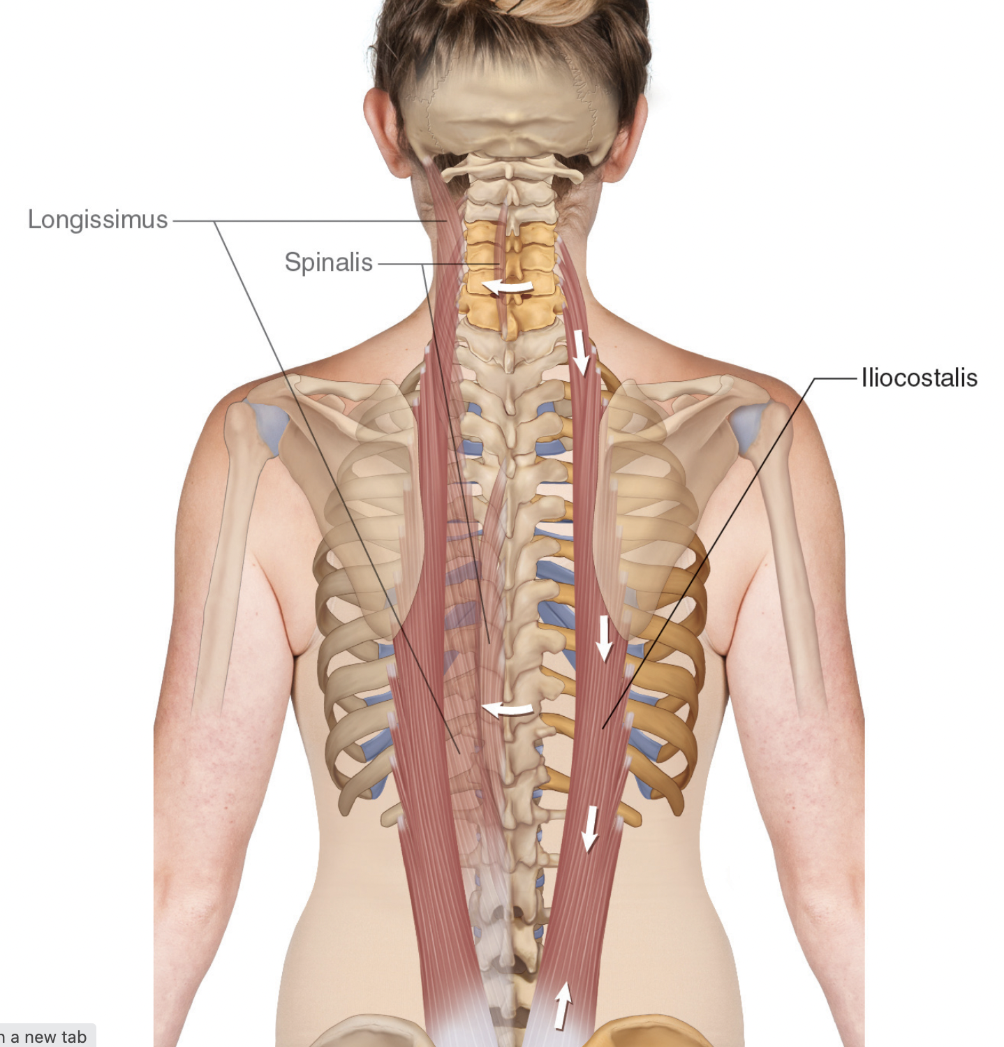
Iliocostalis ( lateral)
Longissimus ( middle)
Spinalis (Thoracis) ( medial)
Movements:
The erector spinae muscles are responsible for the extension of the vertebral column and assistance in lateral flexion.
Muscle Actions:
Unilateral Actions: Lateral flexion of the vertebral column
Bilateral Actions: Extension of the vertebral column, contributing to posture and balance.
THORACOLUMBAR FASCIA
The thoracolumbar fascia is a layer of connective tissue that covers the muscles of the back, extending from the pelvis to the cranium. It plays a crucial role by enclosing intrinsic back muscles, separating the extensor muscles of the back from those of the upper limbs, and consists of three layers in the lumbar region: anterior, middle, and posterior.
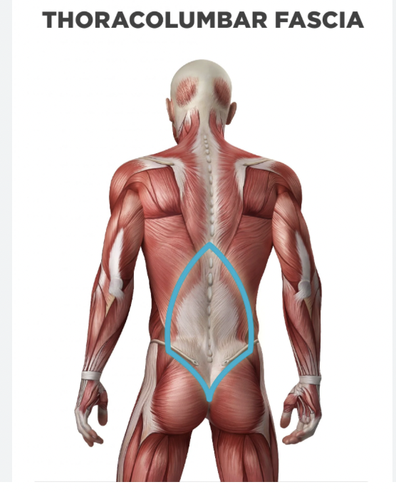
Encloses intrinsic back muscles, extends from pelvis to cranium
Separates extensor muscle of the back and upper limb muscles
Composed of three layers in lumbar region: Anterior, Middle, Posterior
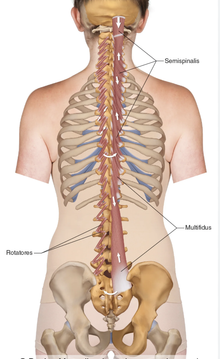
TRANSVERSOSPINALES
Components:
Semispinalis ( longest- most superficial ( outer layer))
Multifidus
Rotatores
Innervated by segmental dorsal rami
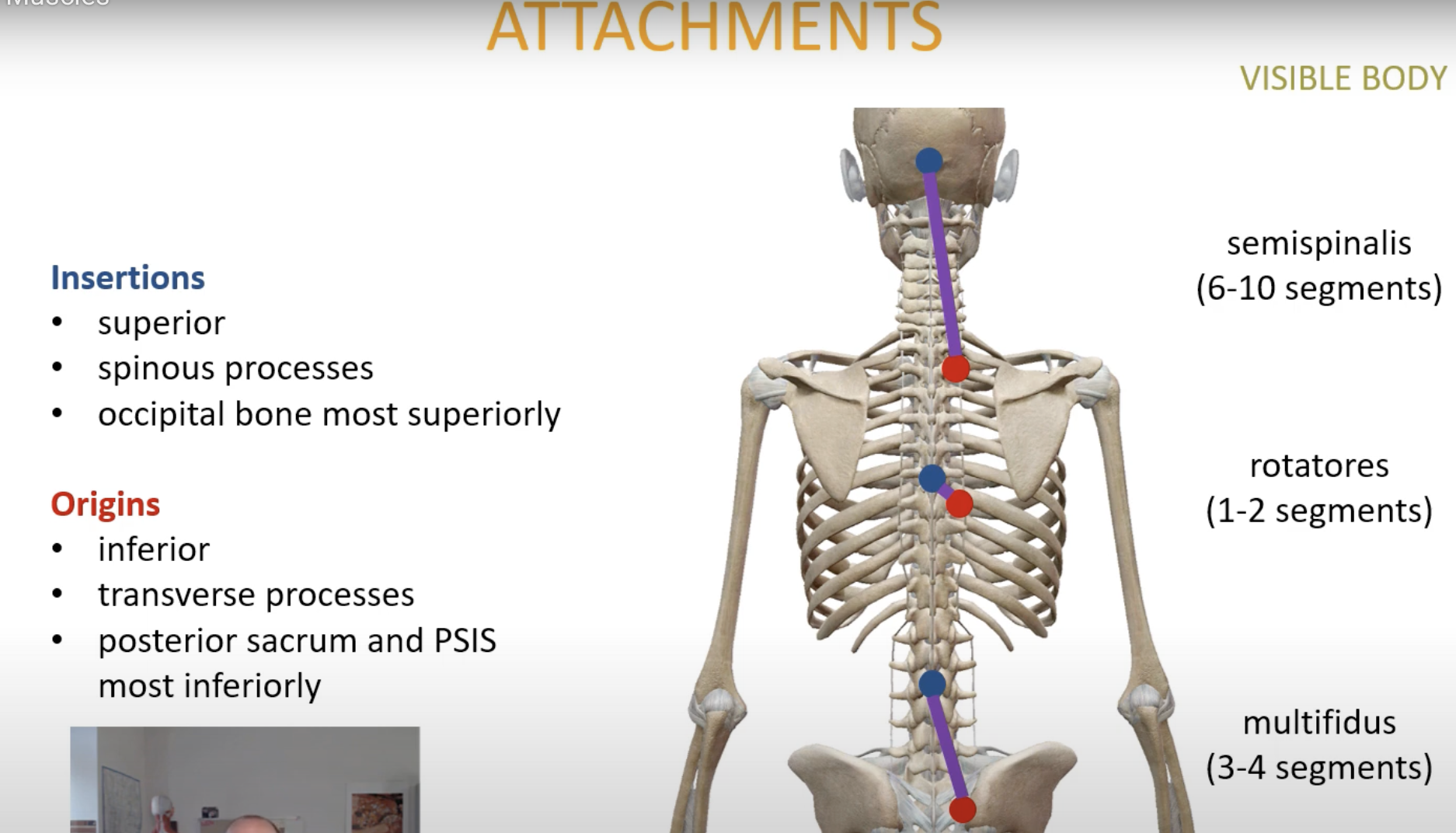
ATTACHMENTS
Insertions: superior
Origins: inferior
Muscle Distribution:
Semispinalis (6-10 segments)
Multifidus (3-4 segments)
Rotatores (1-2 segments)
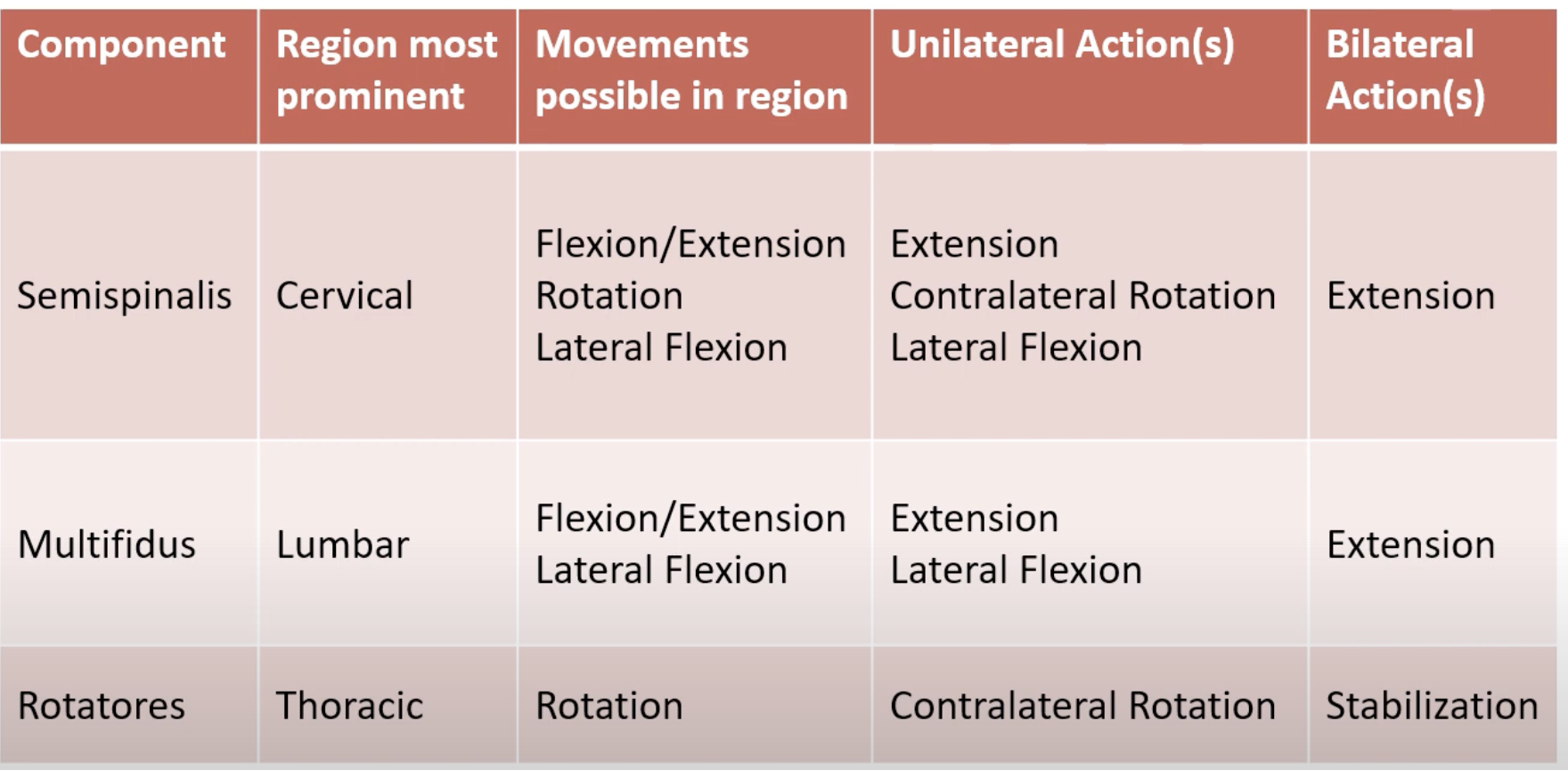
ACTIONS OF TRANSVERSOSPINALES
Unilateral Action(s): Varies by muscle
Bilateral Action(s): Flexion, extension, rotation stabilization
he transversospinales muscles and erector spinae muscles have distinct roles in spinal movement and stabilization:
Transversospinales Muscles
Components: Semispinalis, Multifidus, Rotatores.
Location: Deep layer of back muscles, situated between the spinous and transverse processes of the vertebrae.
Function: Primarily responsible for stabilization of the spine and contributing to rotation and extension movements. They typically span 1-2 vertebrae for rotatores, 3-4 for multifidus, and 6-10 for semispinalis.
Innervation: Innervated by segmental dorsal rami.
Erector Spinae Muscles
Components: Iliocostalis (lateral), Longissimus (middle), Spinalis (medial).
Location: More superficial than transversospinales, running along the length of the vertebral column.
Function: Responsible for extending the vertebral column and assisting with lateral flexion. They play a crucial role in maintaining posture and balance of the spine during movement.
Innervation: Also innervated by segmental dorsal rami.
In summary, the transversospinales provide stabilization and fine-tuned movements, while the erector spinae are primarily involved in larger movements and posture support.
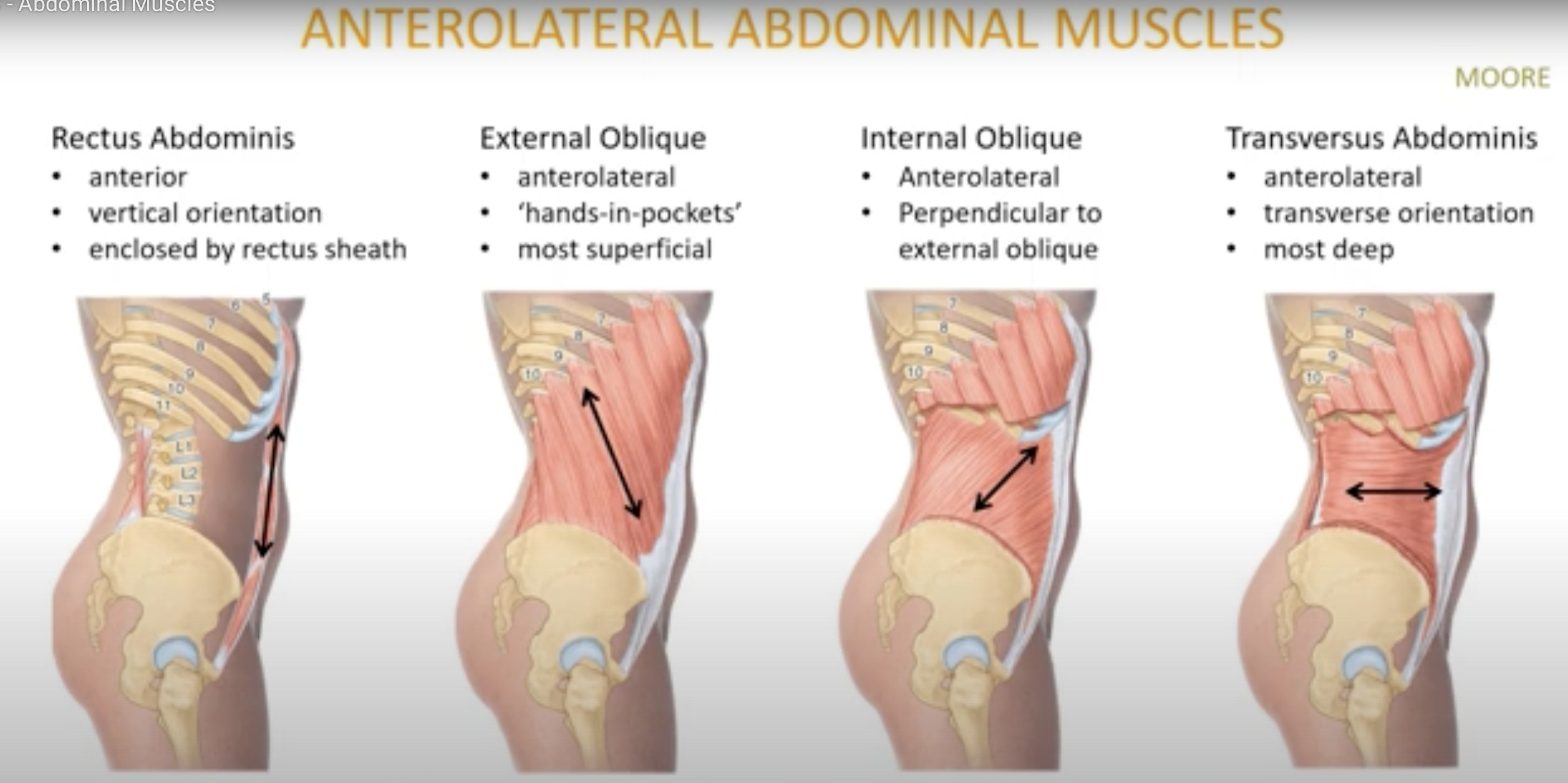
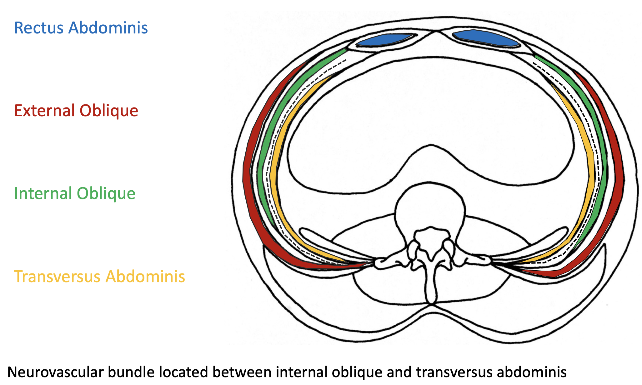
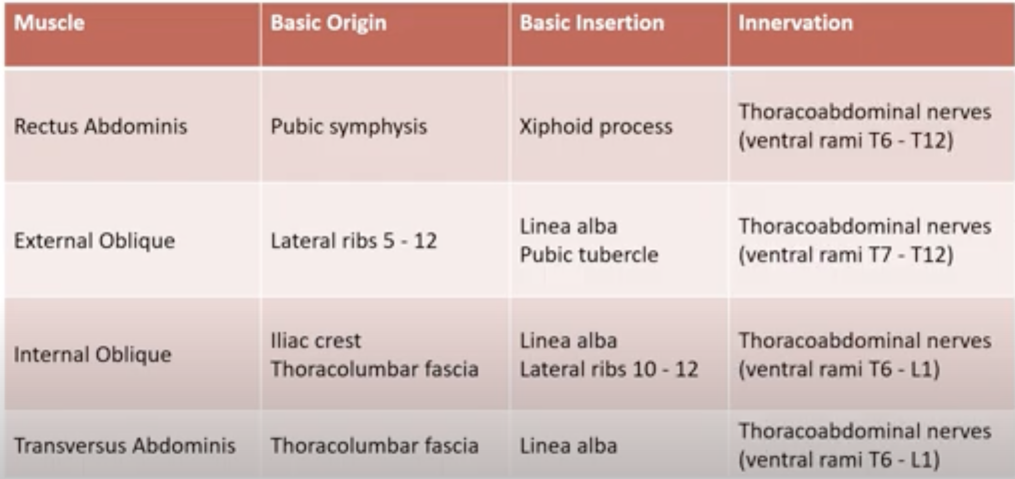
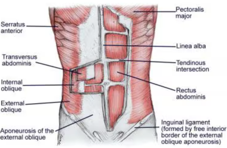
ANTEROLATERAL ABDOMINAL MUSCLES
Rectus Abdominis:
Orientation: Anterior
External Oblique:
Orientation: Anterolateral, most superficial
Internal Oblique:
Orientation: Anterolateral, middle layer
Transversus Abdominis:
Orientation: Anterolateral, deepest layer
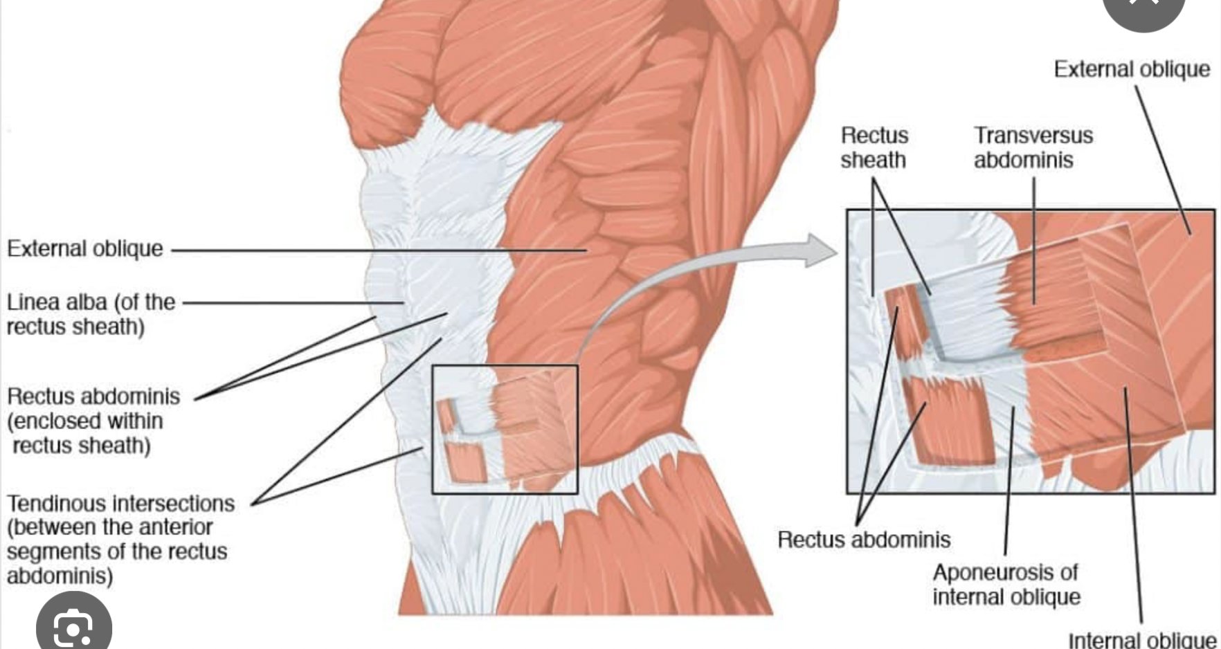
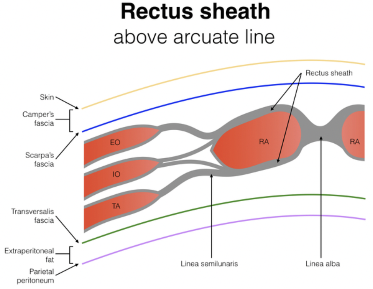
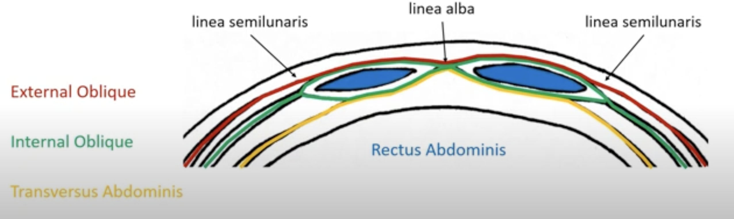
ANTEROLATERAL MUSCLE ATTACHMENTS
Rectus Sheath
Aponeuroses: Aponeuroses are flattened tendon-like structures that serve as connective tissues. They attach muscles to bones or to other muscles. They function similarly to tendons but are broader and provide a greater area of attachment for muscles, often covering a muscle group or fascia, contributing to structural support for muscle actions.
ANTEROLATERAL MUSCLE ACTIONS
Support and stabilize the trunk during movement
Aid in respiration by compressing the abdominal cavity
Assist in lumbar spine flexion and rotation
Contribute to pelvic stability and posture by controlling the position of the pelvis and maintaining alignment during various activities.
Rectus Abdominis:
Unilateral: Flexion (lumbar IVJ)
Bilateral: Increase intra-abdominal pressure
External Oblique:
Unilateral: Flexion (lumbar IVJ)
Internal Oblique: Increase intra-abdominal pressure
Transversus Abdominis: Increase intra-abdominal pressure

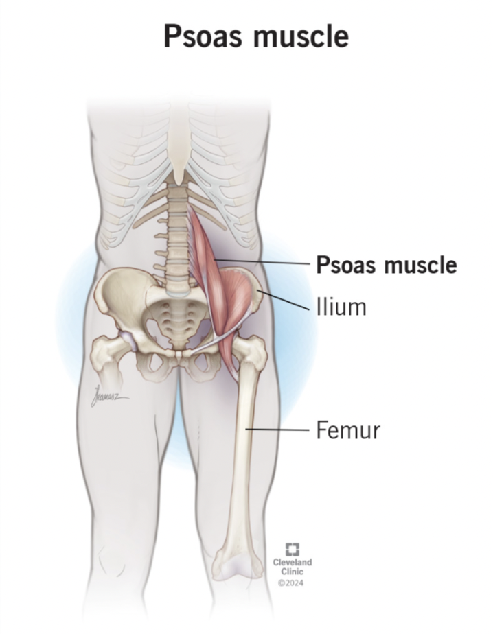
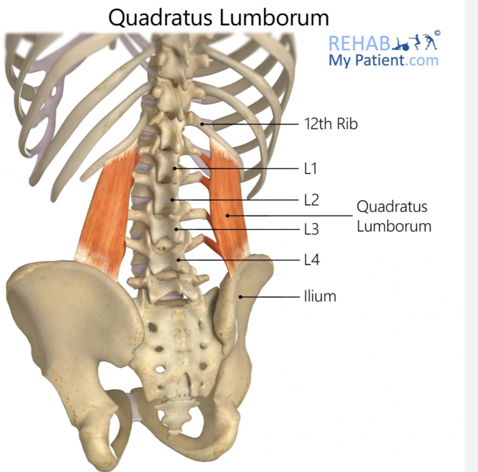
POSTERIOR ABDOMINAL MUSCLES
POSTERIOR ABDOMINAL MUSCLES
Psoas
Origin: Lumbar transverse processes
Insertion: Lesser trochanter of femur
Unilateral actions:
- Hip flexion: Assists in lifting the thigh towards the abdomen.
- Lateral flexion of the lumbar spine: Contributes to bending the torso to the side.
Bilateral actions:
-Extension of the lumbar spine: Aids in straightening the torso back to an upright position.
- hip Flexion of the thoracic spine: Facilitates bending forward at the waist.
Innervation: Lumbar ventral rami
Quadratus Lumborum
Origin: Posterior iliac crest
Insertion: 12th rib
Unilateral actions:
Lateral flexion of the vertebral column to the same side
Elevation of the pelvis on the same side
Bilateral actions:
Extension of the vertebral column
Flexion of the vertebral column
Stabilization of the pelvis during movement.
Innervation: Lumbar ventral rami
This section focuses on the posterior abdominal muscles, which play vital roles in various movements and stability of the lumbar region.

/
CERVICAL MUSCLES
Groups:
Trunk to Neck
Neck to Head
Trunk to Head
The groups you mentioned act on different sets of joints:
Trunk to Neck: This group primarily acts on the cervical vertebrae and the cervico-thoracic joints. These muscles assist in movements like flexion, extension, and rotation of the neck.
Neck to Head: This group acts on the atlanto-occipital joint (AO joint) and the atlantoaxial joint (AA joint). They facilitate movements such as head nodding (flexion/extension) and rotation of the head.
Trunk to Head: This group acts on the cervical spine, particularly engaging with the upper cervical joints, including the AO and AA joints, allowing complex movements that involve both neck and head positioning.
Relationships to joints vary these groups act on.
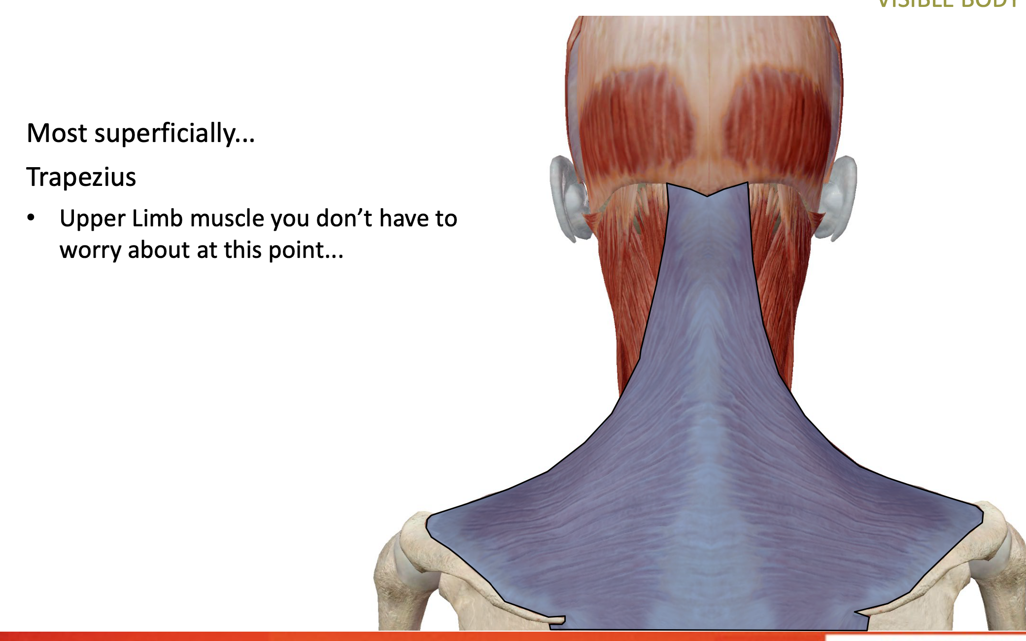
POSTERIOR CERVICAL MUSCLES
Trapezius: most superficial msucle
from shoulder blade to head
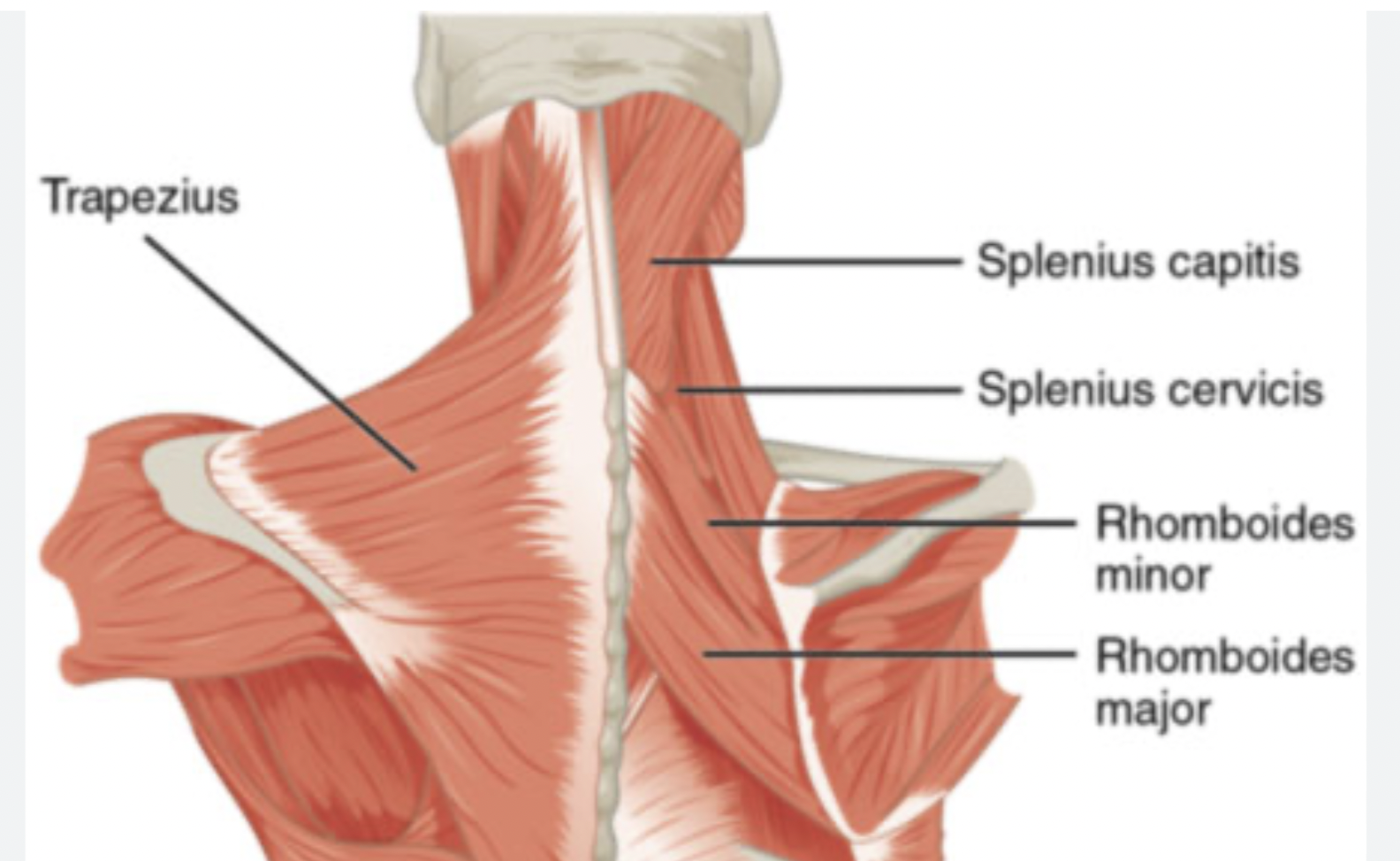
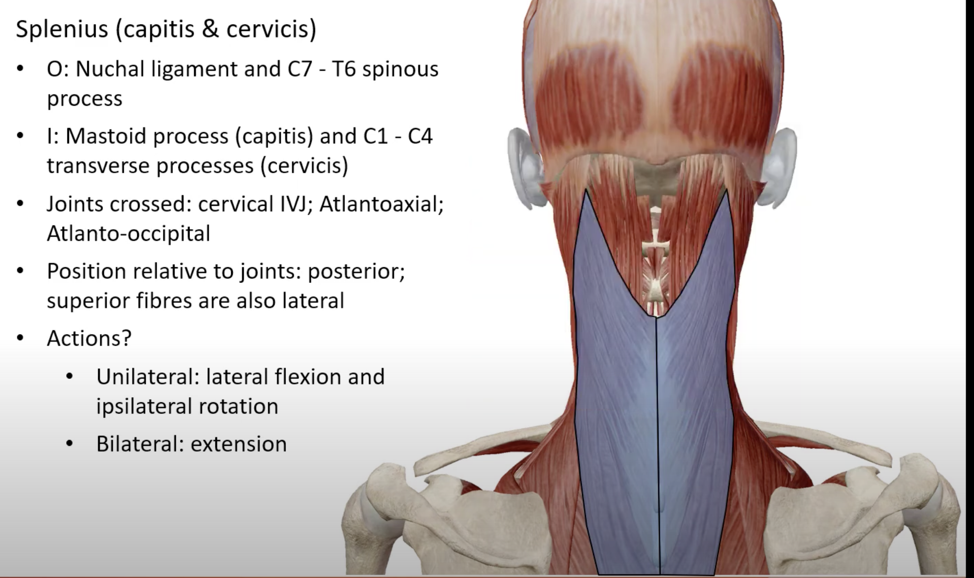
Splenius (capitis & cervicis):
O: Nuchal ligament, C7 - T6 spinous process
I: Mastoid process, C1 - C4 transverse processes
Unilateral: Lateral flexion and ipsilateral rotation
Bilateral: Extensio
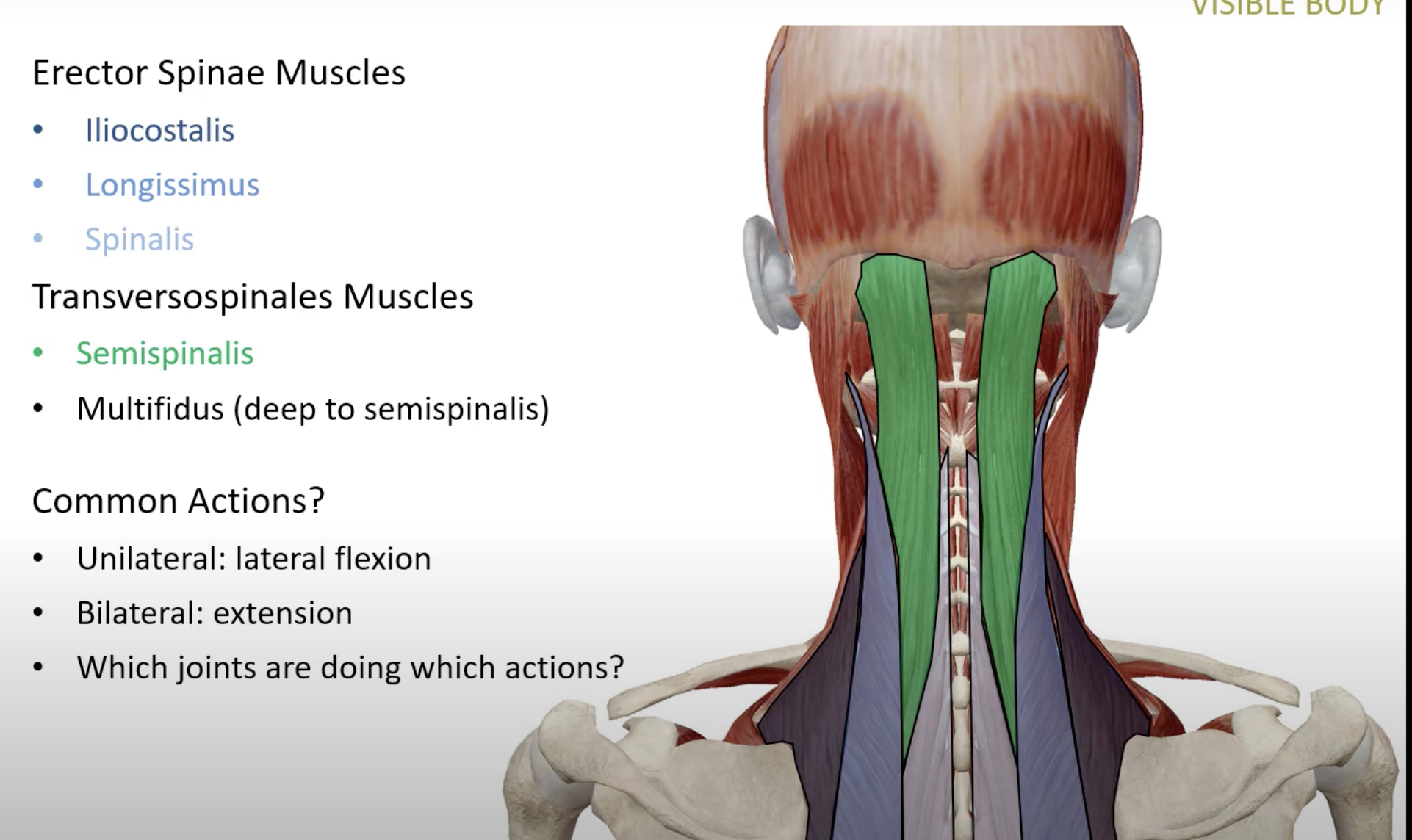
COMMON ACTIONS
Erector Spinae Muscles: Iliocostalis, Longissimus, Spinalis
Transversospinales Muscles: Semispinalis, Multifidus (deep to semispinalis)
Actions vary: Unilateral and bilateral across respective joints.
SUB-OCCIPITAL MUSCLES
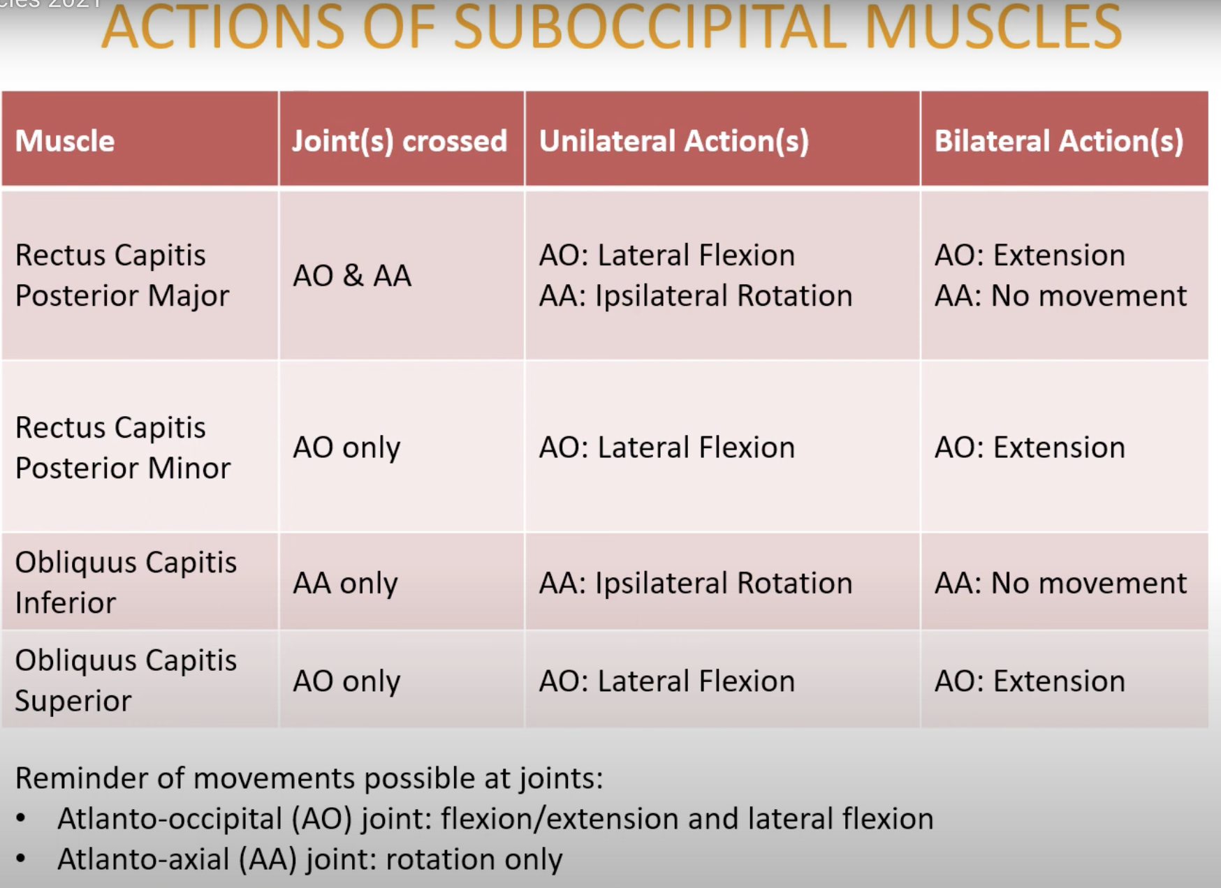
Muscles: Rectus Capitis Posterior Major/Minor, Obliquus Capitis Inferior/Superior
Actions measured across AO and AA joints
Unilateral and bilateral actions vary per muscle
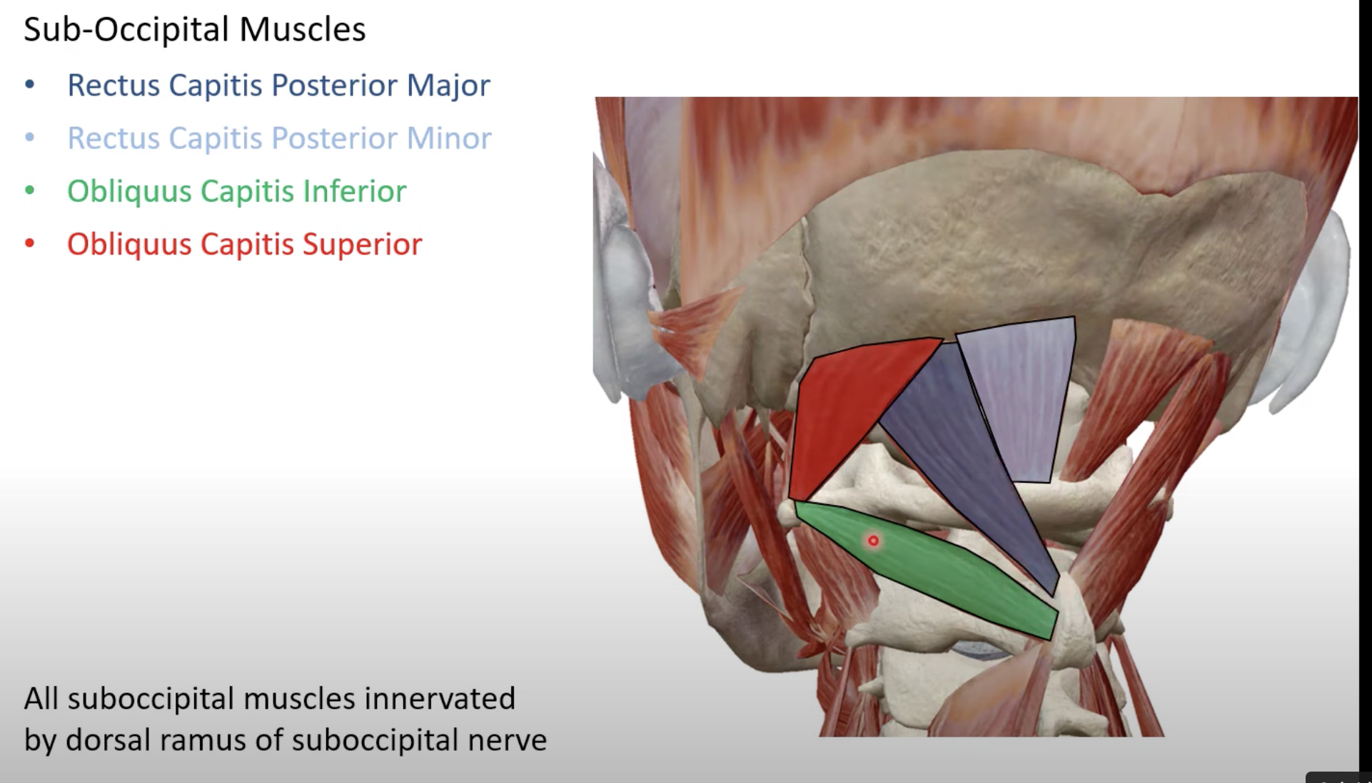
ANTERIOR CERVICAL MUSCLES
Sternocleidomastoid:
Origin: Sternum and medial clavicle
Insertion: Mastoid process
Anterior/lateral to cervical IVJ
Unilateral and bilateral actions analyze per muscle
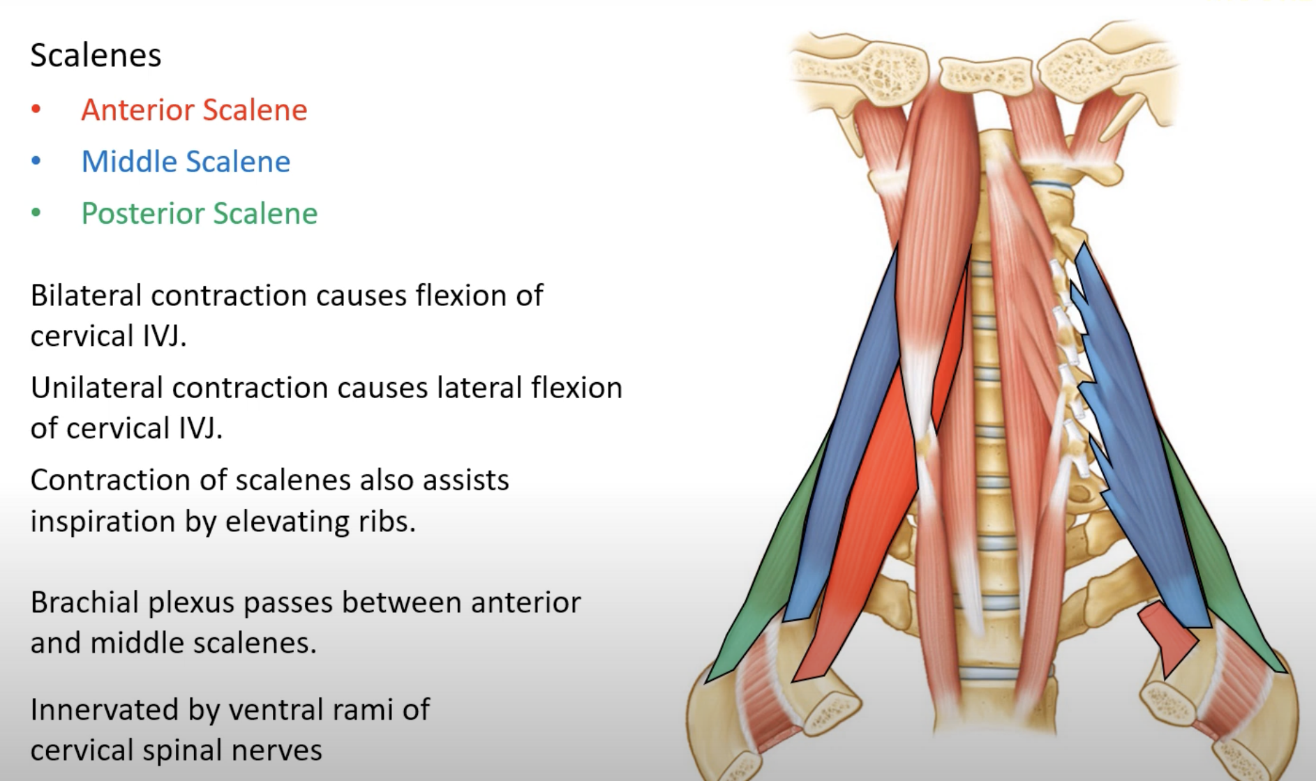
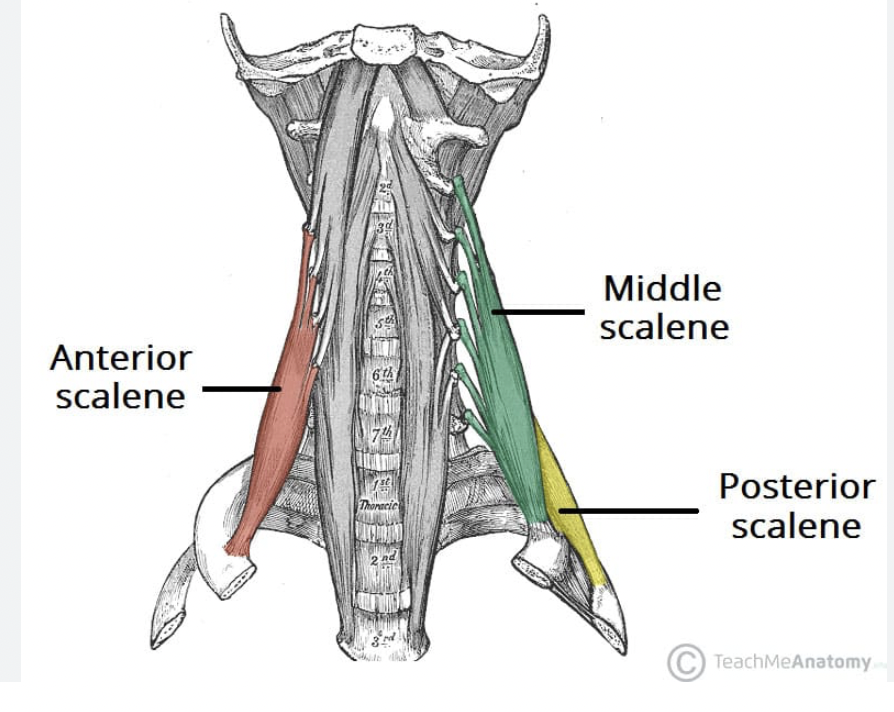
Scalenes: Anterior, Middle, Posterior
Anatomy of brachial plexus connection between anterior and middle scalenes
Actions characterized by contraction types
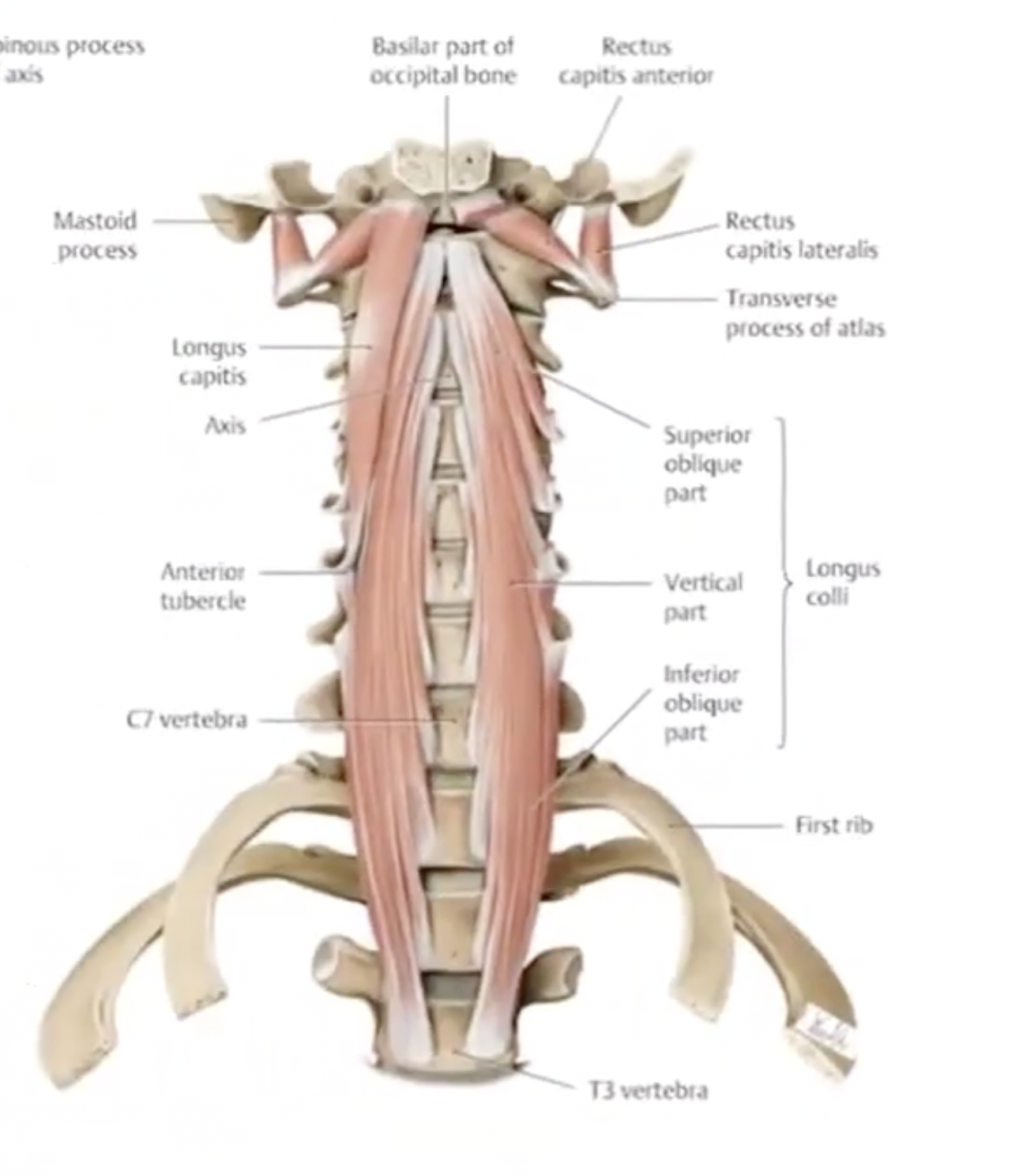
Longus Capitis and Longus Colli:
Actions and joints crossed specified for muscular functionality
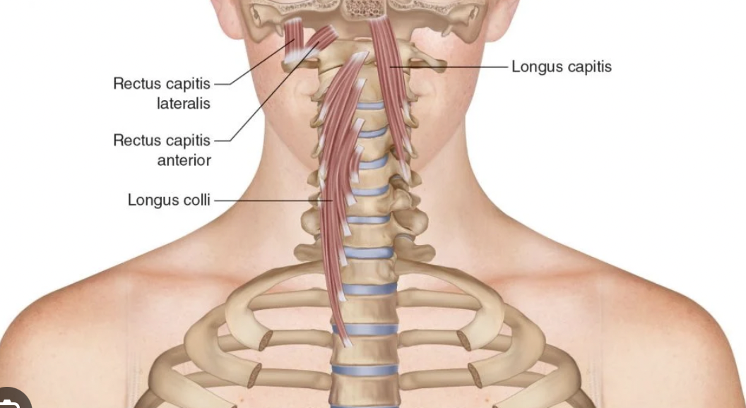
Longus Capitis
Function: The Longus Capitis muscle assists in flexing the head and neck forward and aids in stabilizing the cervical spine.
Location: It is located in the anterior part of the neck, extending from the cervical vertebrae to the base of the skull.
Joints Crossed: It crosses the cervical intervertebral joints and the atlanto-occipital joint.
Actions: Bilateral contraction results in flexion of the neck, while unilateral contraction allows for head tilting and rotation towards the same side.
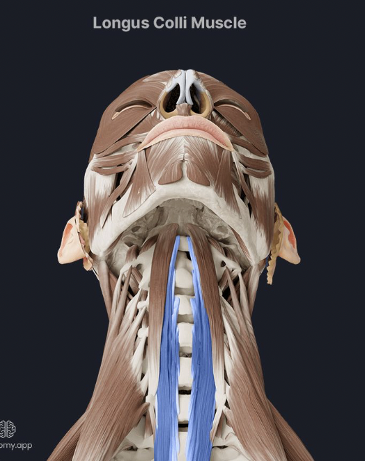
Longus Colli
Function: The Longus Colli muscle functions to flex and stabilize the cervical spine.
Location: It is situated in the anterior neck, running from the upper cervical vertebrae to the lower cervical vertebrae and upper thoracic vertebrae.
Joints Crossed: It crosses multiple cervical intervertebral joints.
Actions: Bilaterally, it facilitates neck flexion, while unilaterally, it allows for rotation and lateral flexion of the neck.
Histological Classification of Atlanto-Occipital Joint
Type: Synovial Joint
Specifics: It is characterized as a condylar joint, which allows for flexion and extension movements (nodding) and slight lateral flexion.
Functional Classification of Atlanto-Occipital Joint
Function: This joint primarily facilitates head movements. It allows for the nodding motion of the head (such as when saying "yes") and a slight amount of lateral bending. The joint contributes significantly to the overall range of motion of the cervical spine.
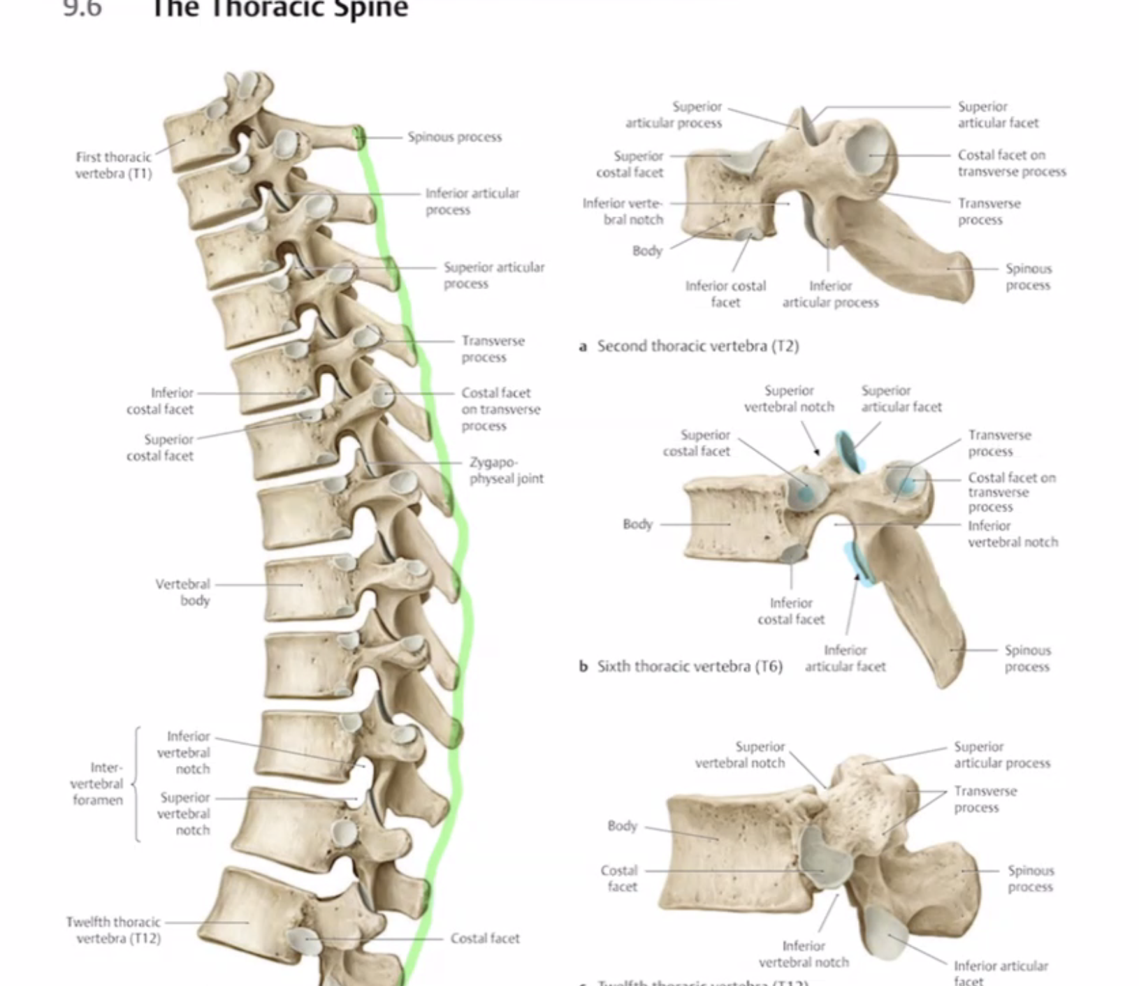
Histological Classification of Thoracic Joints
Type: Synovial Joint
Specifics: Thoracic joints, particularly the costovertebral and costotransverse joints, are classified as synovial joints which allow for various movements, primarily gliding actions.
Functional Classification of Thoracic Joints
Function: These joints play a crucial role in facilitating the movement of the thoracic cage during respiration and trunk mobility. They allow for limited movement needed during breathing, as well as aiding in rotations and lateral flexions of the thorax.
Histological Classifications:
Fibrous Joints: These joints are held together by dense connective tissue and allow for minimal to no movement, such as sutures in the skull.
Cartilaginous Joints: These joints are connected by cartilage, allowing slight movement, as seen in the intervertebral discs and pubic symphysis.
Primary Cartilaginous Joints:
Connected by hyaline cartilage.
Allow slight movement.
Serve as temporary connections during growth, such as the epiphyseal plate in growing bones.
Secondary Cartilaginous Joints:
Connected by fibrocartilage.
Provide stability and support.
Found in areas like the intervertebral discs and the pubic symphysis.
Allow for limited movement.
Synovial Joints: These are the most movable joints, characterized by a fluid-filled joint cavity and include types like hinge, ball-and-socket, and pivot joints.
Fibrous Joints: These joints are held together by dense connective tissue and allow for minimal to no movement, such as the sutures in the skull and the syndesmoses between the radius and ulna.
Vertebral Examples: Sutures of the skull
Rib Examples: Costochondral joints
Muscle Examples: Connective tissue at the muscle's origin and insertion points.
Cartilaginous Joints: These joints are connected by cartilage, allowing slight movement, as seen in the intervertebral discs and the pubic symphysis.
Vertebral Examples: Intervertebral discs
Rib Examples: Costosternal joints where ribs attach to the sternum via costal cartilage
Muscle Examples: Cartilage between ribs and sternum.
Synovial Joints: These are the most movable joints, characterized by a fluid-filled joint cavity and include types like hinge, ball-and-socket, and pivot joints.
Vertebral Examples: Zygapophyseal (facet) joints between vertebrae
Rib Examples: Costovertebral joints where ribs attach to the thoracic vertebrae
Muscle Examples: Hinge joints of the elbow (between the humerus and ulna).