Topic 17- Action Potentials
Outcomes:
Compare and contrast neuron structures
Sequence the process of generating a membrane potential
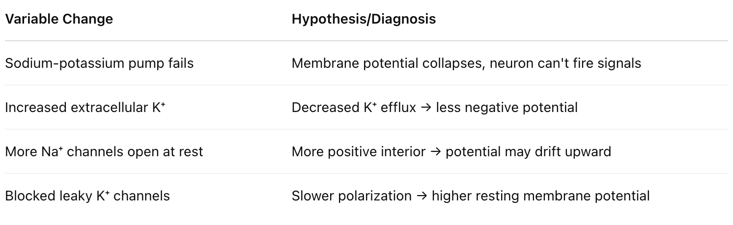
Hypothesize and diagnose the impact of variability on the generation of a membrane potential
Compare and contrast hyperpolarization, depolarization, and threshold
Sequence action potentials
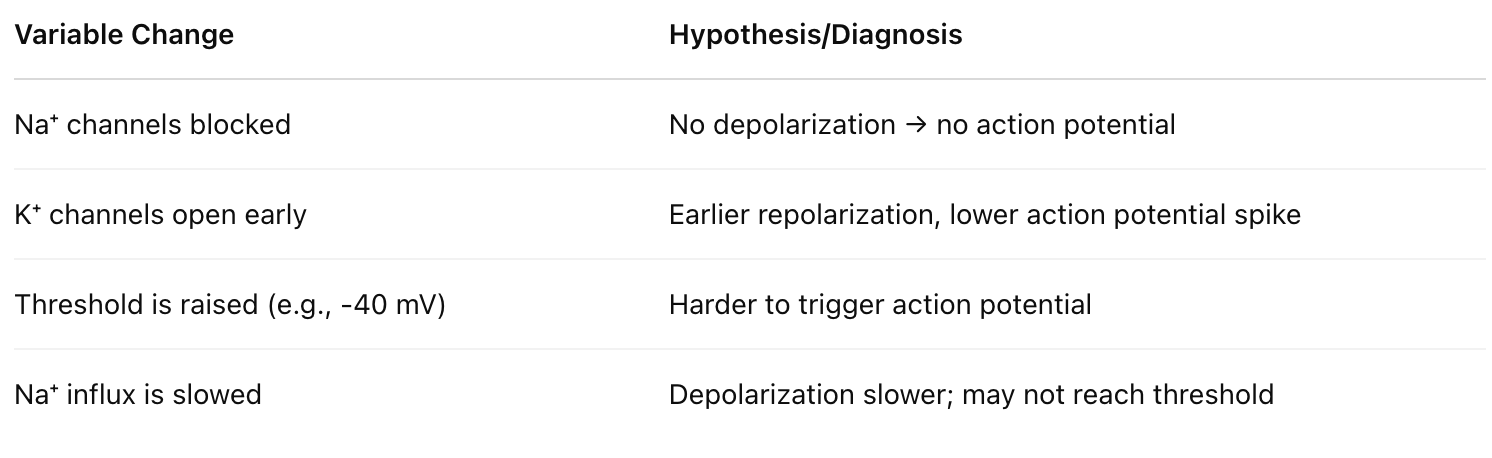
Hypothesize and diagnose the impact of variability on action potentials
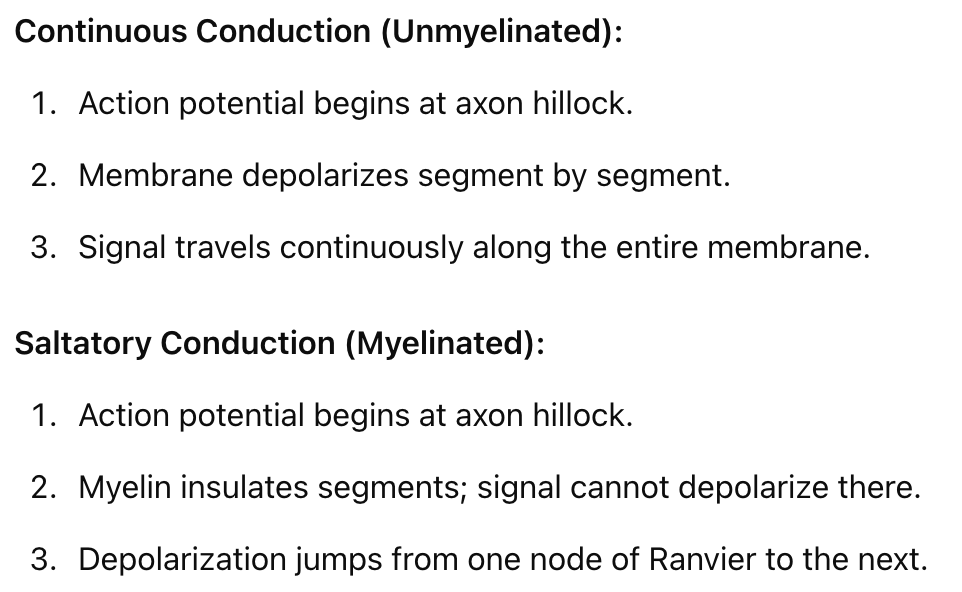
Sequence the processes of continuous and saltatory conduction
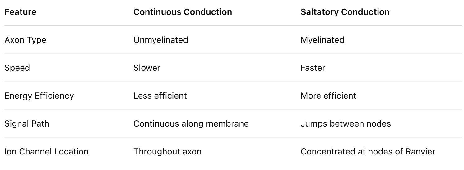
Compare and contrast continuous and saltatory conduction
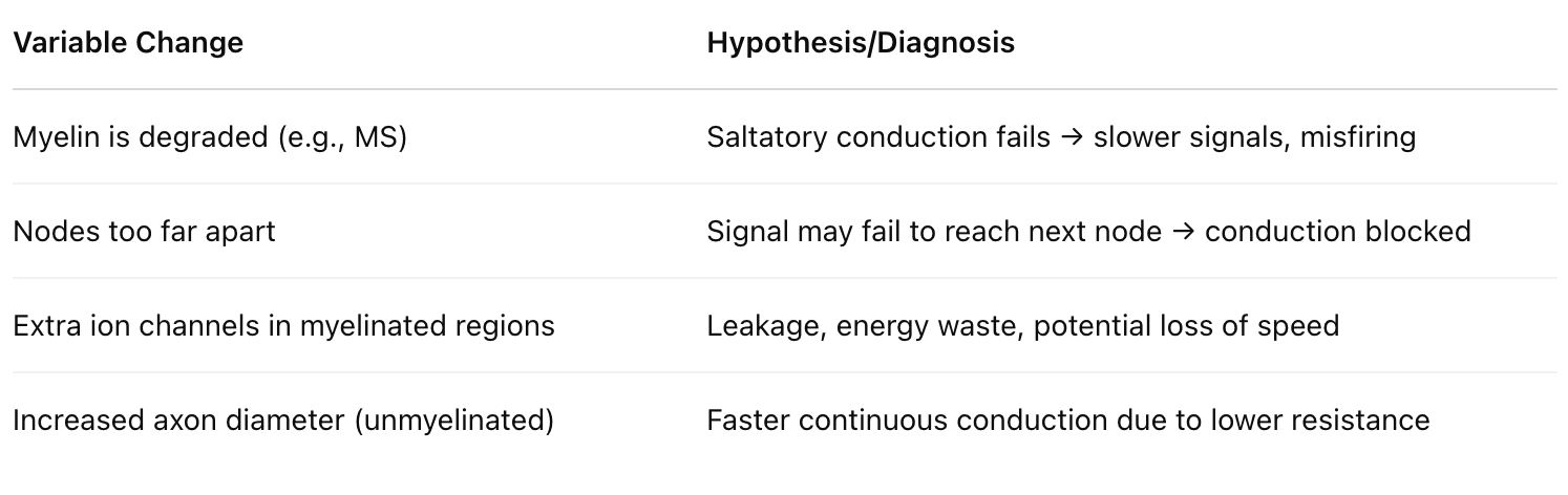
Hypothesize and diagnose the impact of variability on continuous and saltatory conduction
I. Neurons
A. Structure and Function
Neuron = nerve cell.
Specialized to conduct messages as electrical signals in one direction only.
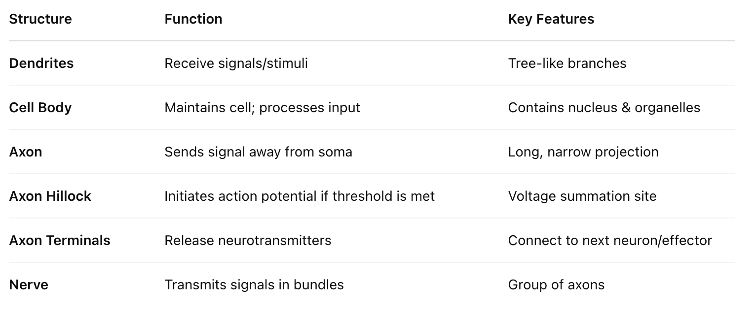
Cell body (soma): Contains the nucleus and organelles.
Dendrites: Receive incoming signals/stimuli; branched like little trees.
Axon: Long projection that transmits the signal away from the cell body to an effector (muscle/gland) or another neuron.
Axon hillock: Located at the base of the axon; this is where the signal is initiated if the threshold is reached.
Synaptic terminals (axon terminals): End of axon branches that release neurotransmitters to the next cell.
Nerve: A bundle of axons from many neurons.
Signals move like a wave—each segment of the membrane depolarizes and repolarizes, creating the illusion of movement, like lights on a marquee.
II. Membrane Potential
A. Introduction
All animal cells have a selectively permeable membrane.
Ions are charged and cannot diffuse freely through the membrane.
They move either through:
Passive transport: via ion channels (facilitated diffusion, no ATP).
Active transport: using energy (ATP), e.g., sodium-potassium pump.
Ion channels allow ions to move down their concentration gradient until dynamic equilibrium is reached.
Gated channels stay closed until triggered:
Voltage-gated channels open in response to a change in membrane voltage.
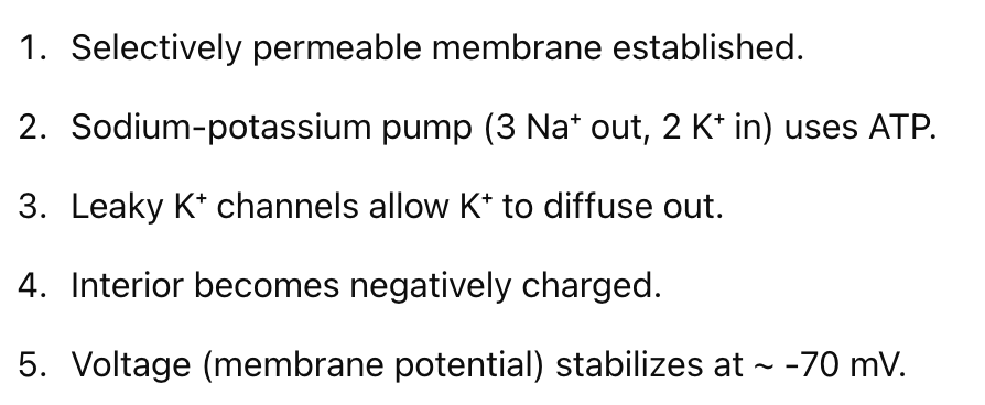
Sodium-Potassium Pump (Na⁺/K⁺ Pump):
Works against the concentration gradient.
Requires ATP.
Pumps 3 Na⁺ out and 2 K⁺ in, maintaining a net positive charge outside the cell.
Always active to keep neurons ready for signal transmission.
B. Voltage
Ion movement causes differences in charge inside vs. outside the cell → creates membrane potential.
Measured in millivolts (mV).
Excitable cells (neurons and muscle cells) can rapidly change membrane potential by opening and closing ion channels.
Voltage is key to triggering signals—neurons communicate by changing voltage.
C. Resting Potential
The resting potential is the charge across the membrane of a neuron at rest, typically -60 to -80 mV.
Maintained by:
Closed gated ion channels (except slightly leaky K⁺ channels).
Sodium-potassium pump, which maintains ion gradients.
No signal is being sent, but the neuron has potential energy.
To send a signal: voltage must change enough to reach the threshold.
D. Changes to Membrane Potential
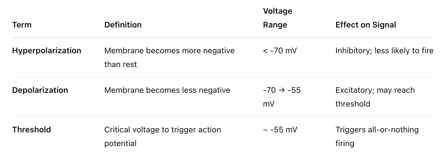
Hyperpolarization:
Membrane becomes more negative than resting potential (e.g., -70 to -90 mV).
Makes the neuron less likely to fire a signal (inhibitory).
Depolarization:
Membrane becomes less negative (e.g., -70 to -56 mV).
Brings the membrane closer to threshold.
May be insufficient on its own—subthreshold.
E. Threshold Potential
A strong enough depolarization can hit the threshold of about -55 mV.
When threshold is reached:
Voltage-gated sodium channels open → Na⁺ rushes in.
This triggers the action potential.
III. Action Potential
A. Change in Membrane Potential
Action potential: A rapid, massive depolarization once the threshold is reached.
Sodium rushes in → voltage spikes to about +35 mV.
All-or-nothing response: if the threshold is hit, the action potential always occurs.
B. Voltage-Gated Ion Channels
Two types involved:
Voltage-gated Na⁺ channels:
Open at threshold (-55 mV), close rapidly.
Allow Na⁺ to flood in, depolarizing the cell.
Voltage-gated K⁺ channels:
Open more slowly, after Na⁺ channels.
Allow K⁺ to exit, repolarizing the membrane back to resting potential.
Facilitated diffusion: Ion channels let ions move passively—no ATP needed.
The Sodium-Potassium Pump resets the gradient afterward using ATP.
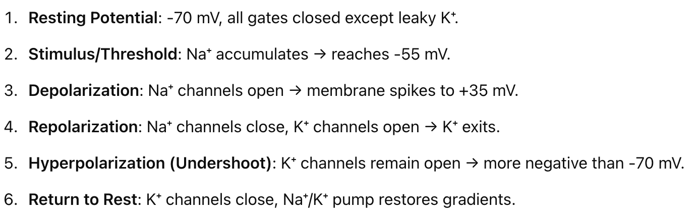
C. Events of an Action Potential

1. Neuron at Rest (Resting Potential)
No signal is being transmitted.
Voltage-gated Na⁺ channels are closed.
K⁺ channels are mostly closed, with some leaky K⁺ channels.
The membrane potential is at -70 mV.
2. Stimulus (Signal)
Na⁺ ions accumulate at the axon hillock.
The membrane depolarizes as potential approaches -55 mV (threshold potential).
If threshold (-55 mV) is not reached → the neuron remains at rest.
If threshold is reached → action potential is triggered (all-or-nothing response).
3. Rising Phase (Depolarization)
Voltage-gated Na⁺ channels open, causing a rapid influx of Na⁺.
Membrane potential spikes to around +35 mV.
K⁺ channels begin to open around +30 mV, preparing for repolarization.
4. Falling Phase (Repolarization)
Na⁺ channels close and become temporarily inactivated (refractory state).
K⁺ channels are fully open, and K⁺ exits the cell.
Membrane potential begins to return to negative.
5. Refractory Period
Period when the neuron cannot fire another action potential.
Ensures unidirectional flow of the signal along the axon.
6. Undershoot (Hyperpolarization)
Some K⁺ channels remain open, causing excessive K⁺ to leave.
Membrane potential becomes more negative than -70 mV.
Eventually, K⁺ channels close, and the neuron returns to resting potential.
Neuron is less likely to fire during this hyperpolarized state.
IV. Conduction of an Action Potential
A. Introduction
Action potential starts at the axon hillock.
The refractory period prevents backflow of the signal.
Results in a wave of depolarization moving along the axon.
B. Continuous Propagation
Occurs in unmyelinated axons (gray matter).
Signal moves smoothly and continuously down the axon.
C. Saltatory Propagation
Occurs in myelinated axons.
Myelin (produced by glial cells) insulates the axon.
Nodes of Ranvier (unmyelinated gaps) are rich in voltage-gated Na⁺/K⁺ channels.
Depolarization occurs only at the nodes, causing the signal to "jump" node to node.
Faster and more energy-efficient than continuous conduction.
(Analogy: Like dribbling and passing a basketball rather than rolling it.)