Cerebellum - The Little Brain
No direct connection to lower motor neurons (exerts influence trough motor systems of cortex and brainstem)
Deficits in coordination always occur ipsilateral to the lesion
Structure of the Cerebellum
Functional Regions
Region | Functions | Motor Pathway |
Lateral Hemisphere | Motor planning for extremities | Lateral Corticospinal Tract |
Intermediate Hemispheres | Coordination of distal limb | Lateral Corticospinal Tract and Rubrospinal Tract |
Vermis and Flocculonodular Lobe (Medial Hemisphere) | Coordination of proximal limb and trunk | Anterior Corticospinal Tract, Reticulospinal Tract, Vestibulospinal Tract, Tectospinal Tract |
Balance and Eye Movement (vestibulo-ocular reflex) | Medial longitudinal fasciculus |
Lateral Hemispheres: The largest most lateral portion of the cerebellum, responsible for higher-order motor and cognitive processes.
Function: Involved in motor planning by anticipating and preparing complex voluntary movements and supports cognitive tasks.
Connections: Sends output to the dentate nucleus, relays information to the motor and premotor cortices via the thalamus.
Intermediate Hemispheres: Lies lateral to the vermis and is primarily involved in distal limb coordination.
Function: Refines fine motor movements of the arms and legs, ensuring smooth and accurate performance of tasks like grasping and typing.
Connections: Sends output to the interposed nuclei, which influence the lateral motor systems, enabling precise motor control.
Vermis: Narrow worm-like structure located along the midline of the cerebellum responsible for proximal muscle coordination.
Functions: Controls proximal muscles (those closer to the trunk) and axial movements required for maintaining posture or performing gross motor tasks and integrates sensory input to regulate balance and locomotion.
Connections: Projects to the fastigial nucleus, which sends outputs to the medial motor systems (e.g., vestibulospinal and reticulospinal tracts) responsible for trunk stability.
Flocculonodular Lobe: Found in the inferior region of the cerebellum, anterior to the posterolateral fissure, essential for maintaining balance and equilibrium.
Components:
Flocculus: Two small, rounded structures on either side of the cerebellum.
Nodulus: A central worm-like structure at the midline, forming the most caudal part of the vermis.
Function: Integration of input from the vestibular system to ensure stability during movement. Also coordinates eye movements, particularly trough the vestibulo-ocular reflex.
Connections: Receives direct input from the vestibular nuclei and sends output back to these nuclei, forming a feedback loop crucial for balance.
Cerebellar Peduncles
Peduncle | Primary Role | Connection | Main Pathways | Main Function |
Superior (SCP) | Output | Cerebellum ↔ Midbrain | Efferent: Thalamus, Red Nucleus | Sends processed motor signals to midbrain and cortex |
Middle (MCP) | Input | Cerebellum ↔ Pons | Afferent: Pontocerebellar Fibres | Brings sensory and motor instruction from cortex |
Inferior (ICP) | Input | Cerebellum ↔ Medulla | Afferent: Spinocerebellar tracts Efferent: Vestibular Nuclei | Integrates Sensory input from the spinal cord and vestibular system and sends output to stabilise posture and balance |
Cerebellar Peduncles: Three paired structures of white matter that connect the cerebellum to the brainstem and facilitate communication between the cerebellum, spinal cord, and cerebral cortex. They serve as a highway for the input and output of neural signals, enabling the cerebellum to coordinate movement, balance and motor learning.
Superior Cerebellar Peduncle: Primarily an output structure that transmits information from the cerebellum to other parts of the brain, particularly the motor areas. It facilitates motor control and coordination by sending processed cerebellar signals to the motor cortex via the thalamus.
Efferent Pathways: Originate in the deep cerebellar nuclei - especially the dentate and interposed nuclei - and project to the:
Ventral Lateral Nucleus (VLN): Coordinates voluntary motor activity with the cerebral cortex.
Red Nucleus: Influences motor control via the rubrospinal tract.
Afferent Pathways: Includes input from the ventral spinocerebellar tract which carries feedback on spinal motor activity. (Minor component)
Middle Cerebellar Peduncle: Largest of the three peduncles that serves as a major input structure, transmitting information from the cerebral cortex to the cerebellum. It acts as the main conduit for cortical information to reach the cerebellum, critical for movement and planning adjustment.
Afferent Pathways: Arise from the pontine nuclei in the pons and carry signals originating in the cerebral cortex, including motor commands and sensory input, to the cerebellar hemispheres.
Inferior Cerebellar Peduncle: Primarily an input structure that connects the cerebellum to the medulla and spinal cord. Also carries some output signals. It transmits proprioceptive information playing a key role in coordinating posture, balance, and motor control by integrating sensory input.
Afferent Pathways:
Dorsal Spinocerebellar Tract: Carries proprioceptive information from the lower limbs.
Cuneocerebellar Tract: Carries proprioceptive input from the upper limbs.
Vestibulocerebellar Fibres: Transmit balance information from the vestibular system.
Olivocerebellar Fibres: Arise from the inferior olive and provide climbing fibre input for motor learning.
Extras
Uncinate Fasciculus: Bundle of efferent fibres that originates from the fastigial nucleus and project to the vestibular nuclei in the brainstem. This pathway is part of the cerebellar influence on balance and posture. It enables the fastigial nucleus to regulate postural control by influencing the lateral vestibulospinal tract, which adjusts extensor tone in the trunk and limbs to maintain balance.
Juxtarestiform Body: Travels next to the inferior cerebellar peduncle and contains both afferent and efferent fibres connecting the cerebellum to the vestibular nulcei, thus providing a bidirectional pathway for the integration of vestibular and cerebellar signals. It plays a critical role in the vestibulo-ocular reflex (VOR) by allowing the cerebellum to fine-tune gaze stabilization during head movements and maintains equilibrium by modulating the medial vestibulospinal tract, which controls head and neck posture.
Deep Cerebellar Nuclei
Nucleus | Region | Primary Function | Output Targets |
Dentate | Lateral Hemispheres | Motor Planning and Fine Motor Control | Thalamus and Red Nucleus |
Interposed | Intermediate Zone | Distal Limb Coordination | Thalamus and Red Nucleus |
Fastigial | Vermis and Flocculonodular Lobe | Balance, Posture and Gaze Stabilisation | Vestibular Nuclei, Reticular Formation |
Deep Cerebellar Nuclei: Primary output structures of the cerebellum that receive processed information from the cerebellar cortex and relay it to other parts of the brain to influence motor control, coordination and other functions. These nuclei serve as essential relay stations, integrating input from the cerebellar cortex and external sources to refine and execute motor commands.
Dentate Nucleus: Largest and most lateral of the deep cerebellar nuclei, primarily associated with the lateral hemispheres of the cerebellum.
Function: Responsible for motor planning and coordination of fine voluntary movements. Also contributes to higher-order functions like motor learning and cognition.
Inputs: Receives input from the lateral cerebellar hemispheres.
Outputs: Sends outputs to the:
Ventral Lateral Nucleus (VLN): Relays to the motor and premotor cortex to influence voluntary movement.
Red Nucleus: Minor projection influencing motor pathways.
Interposed Nuclei: Consists of two nuclei: the emboliform and globose nuclei. These nulcei are associated with the intermediate zone of the cerebellum.
Function: Responsible for coordinating distal limb movements during voluntary actions and correct errors in ongoing movement via real-time feedback.
Input: Receives inhibitory signals from the intermediate hemispheres of the cerebellar cortex.
Outputs: Projects to the:
Ventral Lateral Nucleus: Influences the motor cortex for precise limb movement.
Red Nucleus: Plays a role in controlling limb movement via the rubrospinal tract.
Fastigial Nucleus: Most medial of the deep nuclei associated with the vermis and flocculonodar lobe.
Function: Regulates posture, balance and gaze stabilisation and controls the axial muscles for stability during movement.
Inputs: Receives inhibitory input from the vermis and flocculonodular lobe and excitatory inputs from the vestibular and proprioceptive pathways.
Outputs: Sends projections to the:
Vestibular Nuclei: Influences the vestibulospinal tract for balance.
Reticular Formation: Regulates postural control.
Medial Longitudinal Fasciculus (MLF): Coordinates head and eye movements.
Main Cerebellar Output Pathways
Region | Deep Nuclei | Cerebellar Peduncle | Main Output |
Lateral Hemispheres | Dentate Nucleus | Superior Cerebellar Peduncle | Ventrolateral Nucleus (VL) and Parvocellular Red Nucleus |
Intermediate Hemisphere | Interposed Nuclei | Superior Cerebellar Peduncle | Ventrolateral Nucleus (VL) and Magnocellular Red Nucleus |
Vermis | Fastigial Nuclei | Superior Cerebellar Peduncle | Ventrolateral Nucleus (VL) and Tectum |
Uncinate Fasciculus and Juxtarestiform Body | Reticular Formation and Vestibular Nuclei | ||
Inferior Vermis and Flocculonodular Lobe | Vestibular Nuclei | Juxtarestiform Body | Medial Longitudinal Fasciculus |
Dentate Nucleus Output Pathway: Largest and most lateral deep central nuclei whose output pathway is primarily involved in motor planning and coordination of voluntary movements.
Origin: Lateral Hemisphere of the cerebellum processed through the dentate nucleus.
Via: Superior cerebellar peduncle (SCP).
To:
Ventral Lateral Nucleus: Major relay station for cerebellar output to the primary and premotor cortex. Ensures precise timing and execution of voluntary movements.
Red Nucleus: Modulates the rubrospinal tract for fine motor control of upper limb movements.
Inerposed Nuclei Output Pathway: Consistent of the the emboliform and globose nuclei which primarily influence distal limb coordination during voluntary movements.
Origin: Intermediate zone of the cerebellar cortex, processed through the interposed nuceli.
Via: Superior cerebellar peduncle (SCP).
To:
Ventral Lateral Nucleus: Sends output to the motor cortex, influencing corticospinal tract activity for precise limb movement.
Red Nucleus (Magnocellular Portion): Modulates the rubrospinal tract, which influences distal limb motor control.
Fastigial Nucleus Output Pathway: Medial deep cerebellar nucleus and is involved in postural control, balance, and gaze stabilization.
Origin: Vermis of the cerebellum, processed through the fastigial nucleus.
Via: Inferior cerebellar peduncle (ICP)
Superior Colliculus: Involved in visual reflexes and eye movement coordination.
Venteral Lateral Nucleus: Modulates motor output for balance, posture, and coordination by integrating sensory feedback with cortical motor planning
Via: Uncinate fasciculus and juxtarestiform body.
Vestibular Nuclei: Influence the vestibulospinal tract to regulate extensor tone and balance
Reticular Formation: Modulates postural reflexes via the reticulospinal tract
Vestibular Nuclei: Nuclei in the brain stem that receive direct output from the flocculonodular lobe bypassing the deep cerebellar nuclei.
Function: Regulates balance and posture in response to vestibular (inner ear) inputs and contributes to the vestibulo-ocular reflex (VOR), which stabilizes gaze during head movements.
Origin: Flocculonodular lobe and inferior vermis.
Via: Juxtarestiform body.
Projections:
MLF: Coordinates eye movements by projecting to cranial nerves controlling the extraocular muscles.
Main Cerebellar Input Pathways
Input Pathway | Main Origin | Cells Projecting | Cerebellar Peduncle |
Pontocerebellar Fibres | Cortex | Pontine Nuclei | Midde Cerebellar Peduncle |
Spinocerebellar Pathways | |||
Dorsal | Leg Proprioceptors | Clark’s Column | Inferior Cerebellar Peduncle |
Cuneocerebellar | Arm Proprioceptors | External Cuneate Nucleus | Inferor Cerebellar Peduncle |
Ventral | Leg Interneurons | Spinal Cord Neurons | Superior Cerebellar Peduncle |
Rostral | Arm Interneurons | Spinal Cord Neurons | Superior and Inferior Cerebellar Peduncle |
Climbing Fibres | Red Nucleus, Cortex, Brainstem, Spinal Cord | Inferior Olivary Nucleus | Inferior Cerebellar Peduncle |
Vestibular Inputs | Vestibular System | Vestinular Ganglia and Nuclei | Juxtarestiform body |
Cerebral Cortex (Pontocerebellar Pathway)
Pontocerebellar Pathway: Transmits motor commands from the cerebral cortex to the cerebellum via the pons, enabling the cerebellum to adjust and refine voluntary movements.
Origin: Motor, premotor, and sensory cortices.
Relay Point: Signals synapse at the pontine nuclei in the pons.
Projection: Pontocerebellar fibers cross the midline and enter the cerebellum through the middle cerebellar peduncle.
Termination: Targets the lateral hemispheres (cerebrocerebellum).
Spinal Cord (Spinocerebellar Pathways)
Spinocerebellar Pathway: Carries proprioceptive information from the muscles, joints, and skin to the cerebellum, allowing it to monitor the position and movement of the body in real time.
Dorsal Spinocerebellar Tract: Relays proprioceptive information from the lower limbs and trunk. It originates in the Clarke’s column of the spinal cord, travels through the inferior cerebellar peduncle and projects to the vermis and intermediate zone of the cerebellum.
Ventral Spinocerebellar Tract: Transmits feedback from the interneurons spinal motor circuits about executed movements of the legs. It originates in the ventral horn of the spinal cord, ascends and enters the cerebellum via the superior cerebellar peduncle.
Cuneocerebellar Tract: Carries proprioceptive information from the upper limbs and neck. It originates in the external cuneate nucleus of the medulla and enters the cerebellum via the inferior cerebellar nucleus.
Rostral Spinocerebellar Tract: Transmits motor feedback from the interneurons of the upper limbs. Originates in the cervical spinal cord and enters via both the inferior and superior cerebellar peduncles.
Vestibular System
Vestibulocerebellar Pathway: Provides the cerebellum with information about head position, balance, and spatial orientation from the vestibular system enabling coordination of balance and posture and regulation of the vestibulo-ocular reflex to stabilise gaze during head movement.
Origin: Vestibular ganglia and vestibular nuclei in the brainstem.
Projection: Fibers travel through the juxtarestiform body to the flocculonodular lobe and vermis.
Outputs: Projects back to the vestibular nuclei for modulation of the vestibulospinal tracts.
Inferior Olive
Oluvocerebellar Pathway: Transmits information about motor errors and learning signals from the inferior olivary nucleus to the cerebellum. It is essential for motor learning and adaptation and provides error signals to modify motor output.
Origin: Inferior olivary nucleus in the medulla.
Projection: Climbing fibers enter the cerebellum via the inferior cerebellar peduncle.
Termination: Synapse on Purkinje cells in the cerebellar cortex.
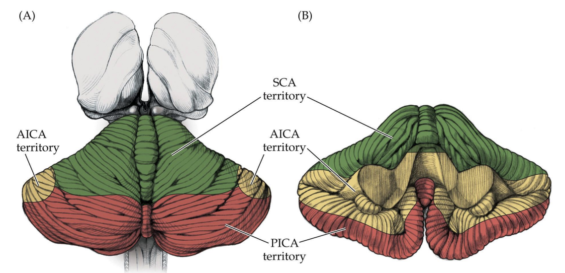
Vascular Supply
Superior Cerebellar Artery (SCA): Most superior branch supplying the upper regions of the cerebellum and nearby brainstem structures.
Origin: Arises from the basilar artery near its bifurcation into the posterior cerebral arteries.
Supplies: Superior half of the cerebellar hemispheres, vermis (upper part), deep cerebellar nuclei, particularly the dentate nucleus and portions of the midbrain (e.g., superior colliculus).
Anterior Inferior Cerebellar Artery (AICA): supplies the middle regions of the cerebellum and some structures in the brainstem. (Inferior front (anterior) part)
Origin: Arises from the basilar artery, inferior to the SCA.
Supplied Regions: Anterior inferior part of the cerebellum, flocculus, middle cerebellar peduncle, portions of the pons and inner ear structures.
Posterior Inferior Cerebellar Artery (PICA): Supplies the inferior regions of the cerebellum and a portion of the brainstem (Inferior back (inferior) part)
Origin: Arises from the vertebral artery, just before it merges with the contralateral vertebral artery to form the basilar artery.
Supplied Regions: Inferior part of the cerebellar hemispheres, inferior vermis, flocculonodular lobe, portions of the medulla, including the nucleus ambiguus and vestibular nuclei.
Cerebellar Lesions
Ataxia is ipsilateral to the side of a cerebellar lesion.
Midline lesions of the cerebellar vermis or flocculonodular lobes mainly cause unsteady gait (truncal ataxia) and eye movement abnormalities, which are often accompanied by intense vertigo, nausea, and vomiting.
Lesions lateral to the cerebellar vermis mainly cause ataxia of the limbs
(appendicular ataxia).
Additionally patients may experience problems with articulation or speech, respiratory movement, motor learning, higher cognitive processes