Chapter 6: The Human Body Notes
Topographic Anatomy
- Superficial landmarks serve as guides to structures that lie beneath.
- Topographic anatomy applies to a body in the anatomic position.
- The patient stands facing you, arms at the side, palms forward.
- Planes of the body: Imaginary straight lines that divide the body
- Coronal plane: front/back
- Transverse plane: top/bottom
- Sagittal (lateral) plane: left/right
The Axial Skeleton
- The axial skeleton is the foundation to which the arms and legs are attached.
- It includes:
- Skull
Spinal column
- Thorax
- Skull
- Cranium: made up of 4 bones
- Face: made up of 14 bones
- Spinal column
- Composed of 33 bones (vertebrae)
- Divided into 5 sections:
- 7 Cervical
- 12 Thoracic
- 5 Lumbar
- Sacrum
- Coccyx
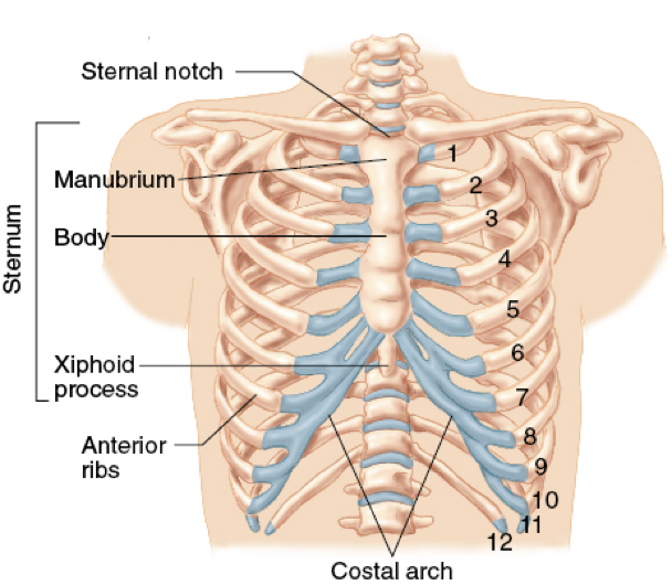
- Thorax
- Formed by 12 thoracic vertebrae and 12 pairs of ribs
- The thoracic cavity contains:
- Heart
- Lungs
- Esophagus
- Great vessels
 The Appendicular Skeleton
- Arms, legs, their connection points, and pelvis
- Occur wherever bones come in contact
- Hinge joint
- Motion restricted to one plane
- Ball-and-socket joint
- Allows rotation and bending
The Upper Extremities
- Composed of arms, forearms, hands, and fingers
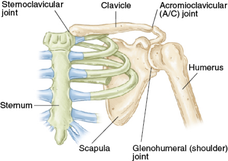
- Shoulder girdle
- Clavicle
- Scapula
- Humerus
- Shoulder girdle
- Arm
- Humerus is the supporting bone
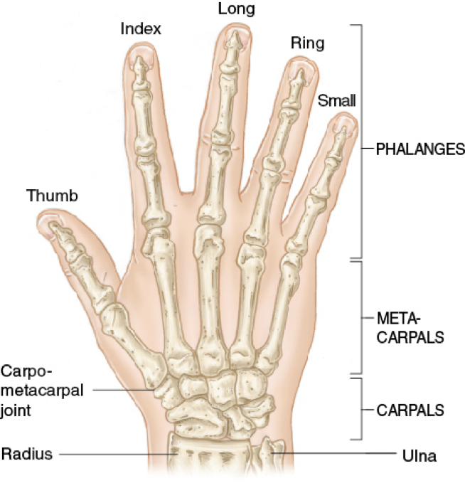
- Forearm
- Radius on the lateral side
- Ulna on the medial side
- Wrist and hand
- Ball-and-socket joint
- Principal bones:
- Carpals
- Metacarpals
- Phalanges
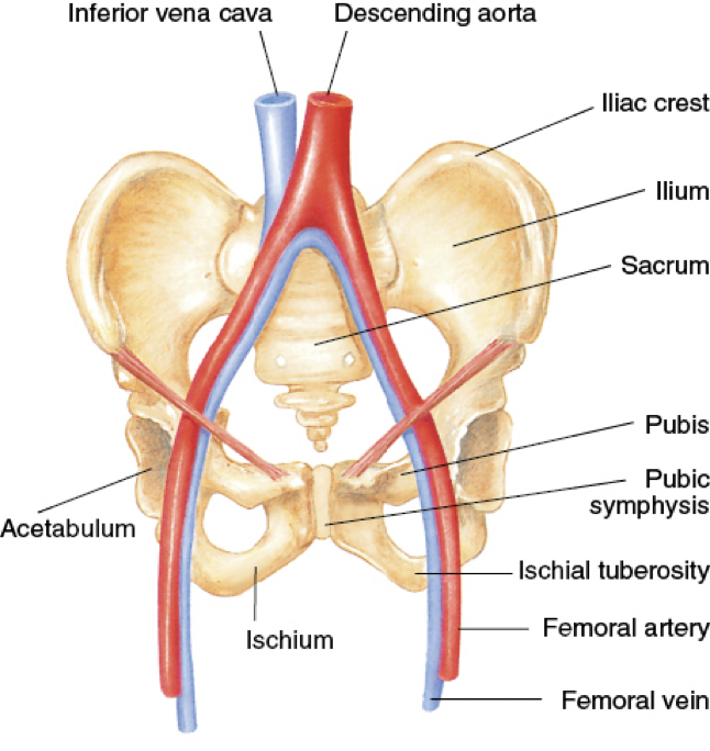
The Pelvis
- Closed bony ring consisting of three bones
- Sacrum
- Two pelvic bones
- Each pelvic bone is formed by the fusion of the ilium, ischium, and pubis.
- Posteriorly, the ilium, ischium, and pubis bones are joined by the sacrum.
- Anteriorly, the pubic symphysis is where the right and left pubis are joined.
The Lower Extremities
- Main components: thigh, leg, and foot
- Femur (thighbone)
- Femur connects to the pelvic girdle by a ball-and-socket joint
- The knee connects the upper leg to the lower leg
- Kneecap (patella)
- Lower leg
- Tibia (shinbone)
- Fibula
- Ankle
- A hinge joint
- Allows flexion/extension of the foot
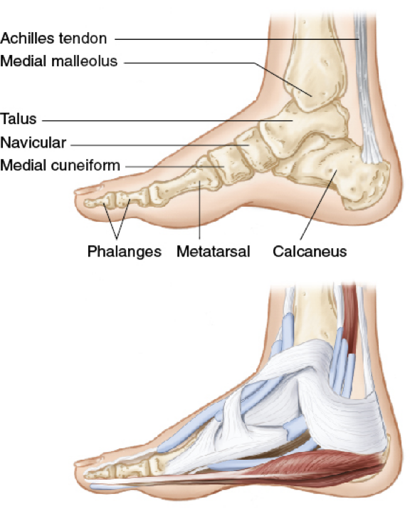
- Foot
- 7 tarsal bones
- 5 metatarsal bones
- Toes are formed by phalanges
The Skeletal System: Physiology
- Functions of the skeletal system
- Gives the body its shape
- Protects fragile organs
- Allows for movement
- Stores calcium
- Helps create blood cells

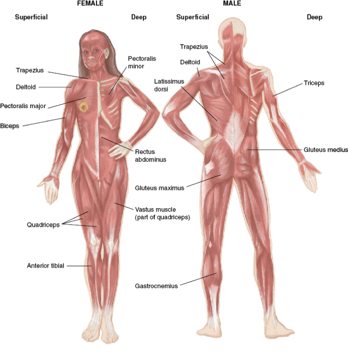
The Musculoskeletal System: Anatomy
The musculoskeletal system provides:
- Form
- Upright posture
- Movement
- Protection for vital internal organs
3 types of muscle
- Skeletal (voluntary) muscle - not striated
- Smooth muscle - digestive system
- Cardiac muscle - the heart
The Musculoskeletal System: Physiology
- Contraction and relaxation of this system make it possible to move and manipulate the environment.
- A by-product of this movement is heat.
- Another function of the muscles is to protect the structures under them.
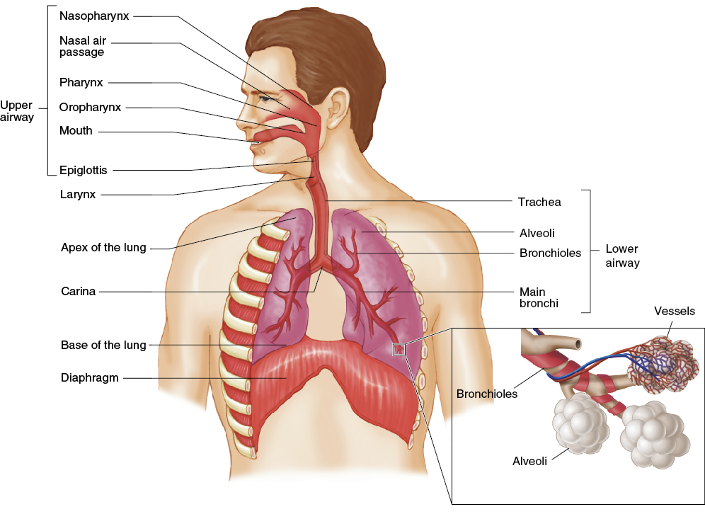
The Respiratory System: Anatomy
- Structures of the body that contribute to respiration (the process of breathing)
| Upper AirwayNoseMouth (oral cavity)TongueJaw (mandible)LarynxPharynxNasopharynxOropharynxLaryngopharynxTracheaEpiglottisEsophagus | Lower AirwayThyroid cartilageAdam’s appleCricoid cartilage: immediately below the thyroid cartilageCricothyroid membrane Trachea Ends at the carina, dividing into right and left bronchi leading to bronchiolesDivided into lobesContain bronchi, bronchioles, and alveoliAllow for gas exchange |
|---|
Muscles of Breathing
- The diaphragm and intercostal muscles are the primary muscles of breathing.
- Other muscles involved in breathing:
- Neck (cervical muscles)
- Abdominal muscles
- Pectoral muscles
The Respiratory System: Physiology
- Functions to provide the body with oxygen and eliminate carbon dioxide
- Ventilation and respiration are two separate, interdependent functions of the respiratory system.
- Respiration: the exchange of oxygen and carbon dioxide in alveoli and tissues
- Provides oxygen to the cells and removes waste carbon dioxide
- Diffusion: the passive process in which oxygen molecules move from an area with a higher concentration of molecules to an area of lower concentration.
- The brainstem controls breathing.
- Ventilation: simple air movement into and out of the lungs
- Tidal volume
- Residual volume
- Dead space
- Minute volume
Characteristics of Normal Breathing
- Normal rate and depth (tidal volume)
- Regular rhythm or pattern of inhalation and exhalation
- Clear, audible breath sounds on both sides of the chest
- Regular rise and fall movement on both sides of the chest
- Movement of the abdomen
- Inadequate breathing patterns in adults:
- Labored breathing
- Muscle retractions
- Pale, cyanotic, cool, damp skin
- Tripod position
- Agonal gasps (gasping breaths)
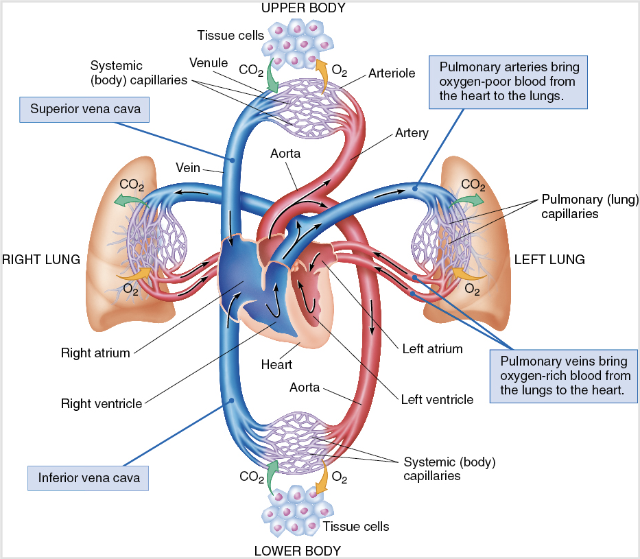
The Circulatory System: Anatomy
- A complex arrangement of connected tubes
- Arteries, arterioles, capillaries, venules, veins
- Two circuits
- Systemic circulation (body)
- Pulmonary circulation (lungs)
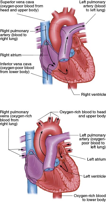
The Heart
- Made of specialized cardiac muscle
- Works as two paired pumps
- The septum divides the right and left sides
- Each side is divided into:
- Atrium (upper chamber)
- Ventricle (lower chamber)
- Circulation
- The heart receives its blood from the aorta.
- The right side receives deoxygenated blood from the veins.
- The left side receives oxygenated blood from the lungs.
- Normal adult resting heart rate (HR): 60–100 beats/min
- Stroke volume (SV)
- Amount of blood moved by one beat
- Cardiac output (CO)
- Amount of blood moved in 1 minute
- HR × SV = CO
- Electrical conduction system
- Causes smooth, coordinated contractions
- Contractions produce the pumping action
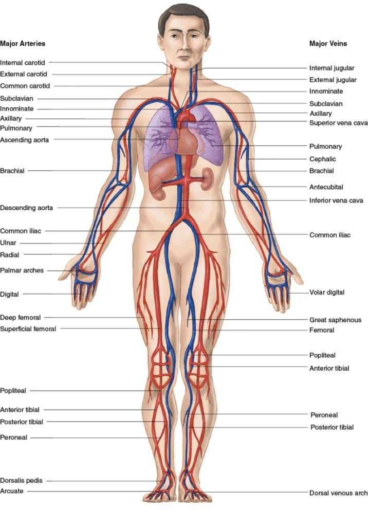
Arteries
- Arteries carry blood from the heart to all body tissues.
- Pulmonary artery
- Carries oxygen-poor blood to the lungs
- Arteries branch into smaller arteries and then into arterioles.
- Arterioles branch into smaller vessels until they connect to the capillaries.
- Pulse
- Created by the forceful pumping of blood out of the left ventricle and into major arteries
- Palpated most easily at the neck, wrist, or groin
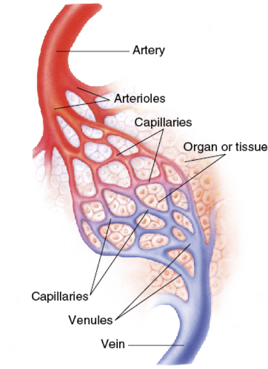
Capillaries & Veins
- Capillaries connect arterioles to venules
- Capillaries allow contact between blood and cells
- Veins return oxygen-depleted blood to the heart
- The superior vena cava carries blood returning from the head, neck, shoulders, and upper extremities
- The inferior vena cava carries blood from the abdomen, pelvis, and lower extremities
Spleen
- A solid organ located under the rib cage
- Filters blood
- Particularly susceptible to injury from blunt trauma
- This can lead to severe internal bleeding
- Solids do not rupture, but hollow organs can
- Solid organs → internal bleeding, infection, sepsis, death
- Hollow organs → rupture of the organ, internal bleeding, sepsis
The Blood
- Blood composition
- Plasma (liquid)
- Red blood cells (erythrocytes)
- White blood cells (leukocytes)
- Platelets
- Blood pressure: the pressure blood exerts against the walls of the arteries
- Systole occurs when the left ventricle contracts
- Diastole occurs when the left ventricle relaxes
- Blood pressure readings
- Systolic blood pressure: high point of wave
- Diastolic blood pressure: low point of wave
- Normal circulation in adults
- Automatically adjusted and controlled
- Perfusion: circulation of blood in an organ or tissue in adequate amounts to meet the needs of its cells
- Blood enters organs and tissues through arteries.
- Blood leaves organs and tissues through veins.
- Inadequate Circulation in Adults
- The system can adjust to small blood loss.
- With a large loss, adjustment fails, and the patient goes into shock.
- Functions of blood
- Perfusion
- Transporting oxygen
- Transporting carbon dioxide
- Transporting wastes and nutrients
- Clotting (coagulation)
The Nervous System
- The nervous system is perhaps the most complex organ in the body
- Divided into two main portions:
- Central nervous system (CNS)
- Peripheral nervous system
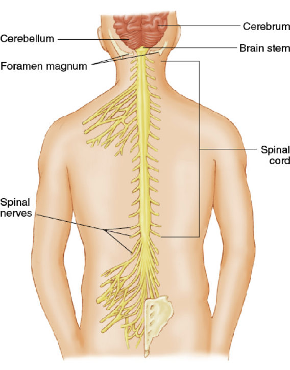
Brain
- Controlling organs of the body
- Subdivisions
- Cerebrum
- Cerebellum
- Brain stem
- Spinal cord
- Continuation of the brain
- Transmits messages between brain and body
Peripheral Nervous System
- Somatic nervous system
- Transmits signals from the brain to voluntary muscles
- Autonomic nervous system
- Involuntary actions
- Split into two areas
- Sympathetic nervous system (fight-or-flight)
- Parasympathetic nervous system (slows body)
The Integumentary System: Physiology 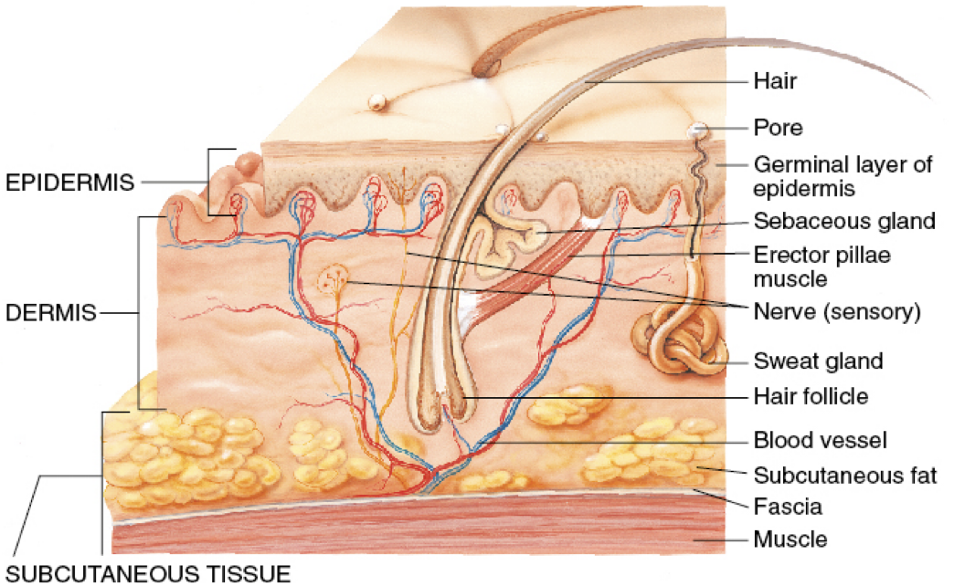
- Two layers
- Epidermis (superficial)
- Dermis (deeper)
- Below the skin lies subcutaneous tissue.
- Fat that insulates and serves as an energy reservoir
- The skin is the largest single organ in the body.
- Three major functions
- Protect the body in the environment
- Regulate body temperature
- Transmit the information from the environment to the brain
The Digestive System: Anatomy 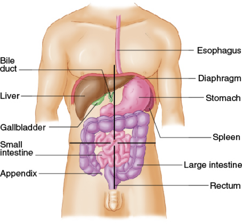
- Digestion: processing of food that nourishes the cells
- Abdomen: the second major body cavity
- Contains major organs of digestion and excretion
- Quadrants are the easiest way to identify areas:
- Right upper
- Lower upper
- Right lower
- Left lower
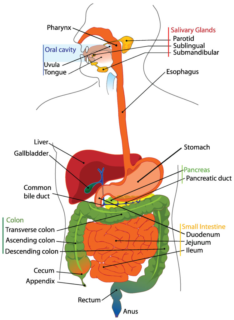
- Mouth
- Lips, cheeks, gums, teeth, tongue
- Salivary glands
- Oropharynx
- Esophagus
- Stomach
- Pancreas
- Liver
- Bile ducts
- Small intestine
- Large intestine
- Appendix
- Rectum
The Digestive System: Physiology
- Enzymes are added to food.
- By salivary glands, stomach, liver, pancreas, and small intestine
- Enzymes convert food into basic sugars, fatty acids, and amino acids.
- Further processed by the liver
- Circulated via blood throughout the body