Biological_Psychology_11_ATAR_Psychology
1. Structure of the Nervous System
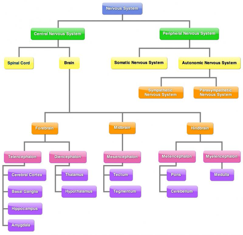
Central Nervous System (CNS)
Composed of the brain and spinal cord
Processes sensory information from PNS and sends motor commands back out
Peripheral Nervous System (PNS)
Comprises nerves outside the CNS
Divided into two main divisions:
Somatic Nervous System
Controls voluntary muscle movements
Carries sensory information to CNS
Autonomic Nervous System (ANS)
Regulates involuntary body functions
Subdivided into:
Sympathetic Nervous System
Prepares body for fight-or-flight response
Parasympathetic Nervous System
Calms the body and maintains homeostasis
2. Features of Neurons
Definition: Neurons are specialized cells responsible for receiving, transmitting, and processing information.
Structure:
Dendrites: Receive signals from other neurons.
Cell Body (Soma): Contains the nucleus and integrates incoming signals.
Axon: Transmits impulses away from the cell body.
Axon Terminals: Release neurotransmitters into the synapse.
Myelin Sheath: Insulates axon, increasing impulse speed.
Types of Neurons:
Motor Neurons: Transmit impulses from CNS to muscles and glands.
Sensory Neurons: Carry sensory information to CNS.
Interneurons: Relay messages between sensory and motor neurons.

3. Neural Transmission
Process:
Action potentials travel in one direction from dendrites to axon terminals.
At axon terminals, neurotransmitters are released into the synaptic cleft.
Electro-Chemical Signal:
Action potentials = electrical signals within neurons.
Neurotransmitters = chemical signals between neurons.
Synapse:
Junction between pre-synaptic and post-synaptic neurons.
Converts electrical impulses into chemical signals and back.
4. Brain Structure and Functions
Hindbrain: Controls basic autonomic functions; includes medulla (heart rate, breathing). Controls voluntary muscle movements; includes cerebellum (balance, coordination, swallowing). At base of brain, back of skull.
Medulla:
At base of brain stem. Front of Cerebellum.
Relays information between spinal cord and brain.
Regulates vital voluntary bodily functions (coughing, sneezing, blood pressure).
Damage = interruptions between brain and spinal cord. Can be fatal, as medulla controls vital organs.
Cerebellum:
Behind brainstem, beneath occiptal and temporal lobes.
Assist in coordinating voluntary movement and balance by relaying motor information to/from cerebral cortex.
Involved in balance, judging distance, coordination of fine motor movements.
Damage = reduced motor control, difficulty in balance, maintaining equilibrium.
Affected by alcohol.
Midbrain: Involved in processing information related to hearing, vision, and movement; contains reticular formation (attention and arousal).
Reticular formation:
Throughout brainstem, from spinal cord to midbrain.
Stimulates brain with bombarding of sensory information - keeps cerebral cortex active and alert.
Controls physiological awareness and muscle tone by regulating function of ANS (sleep/wake cycle).
Damage = disruption to cleep/wake cycle, loss of attention, pain management and balance.
Forebrain: Largest brain region; responsible for complex functions including cognition and emotion; includes cerebral cortex and structures like hypothalamus and thalamus.
Cerebrum:
Largest part of brain, consists of white matter (myelinated axons), cerebral cortex (grey matter; dendrites, unmyelinated axons, cell bodies of neurons).
Outer layer = cerebral cortex. Responsible for higher cognitive functions, voluntary movement, emotions + personality.
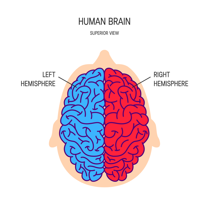
Split into two cerebral hemispheres.
Thalamus:
Two small egg-shaped structures joined together. One at top of brain, one at top of brainstem.
Thalamus receives all information (except smell) on way to cerebral cortex. Information travels up spinal cord and to thalamus.
Conducts motor signals and relays information from brainstem to cortex.
Coordinates shifts in consciousness.
Damage = loss of senses, issues with movement or tremors, attention problems, etc.
Hypothalamus:
Peanut-sized structure.
Located below thalamus.
Maintains homeostasis, regulates hormone release (connected to endocrine system); hunger/thirst.
Controls ‘body-clock’, assists with sleep/wake cycle.
Damage = disruption in body temperature regulation, growth, eating habits, weight control, emotions, sexual behaviour, motivation and sleep cycles.
4.1 Cerebral Cortex
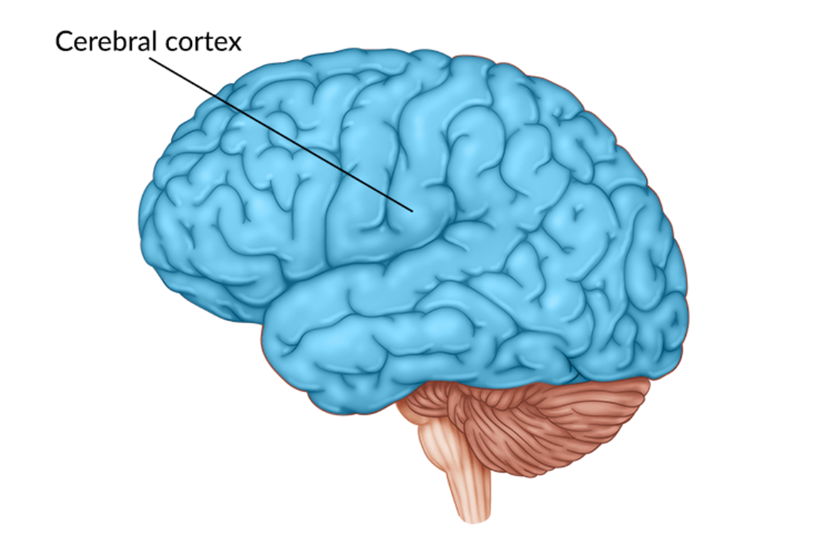
At surface of brain.
Damage = problems with cognition, sensation, movement, behaviour.
Two hemispheres:
Left (intellect) = verbal, analytical functions; writing, speaking, understanding. Mathematics, judging time, rhythm.
Right (creativity) = information processing, detecting/expressing emotion, non-verbal communication; jokes, irony, spacial/visual skills.
Hemispheric specialisation, contralateral control of body.
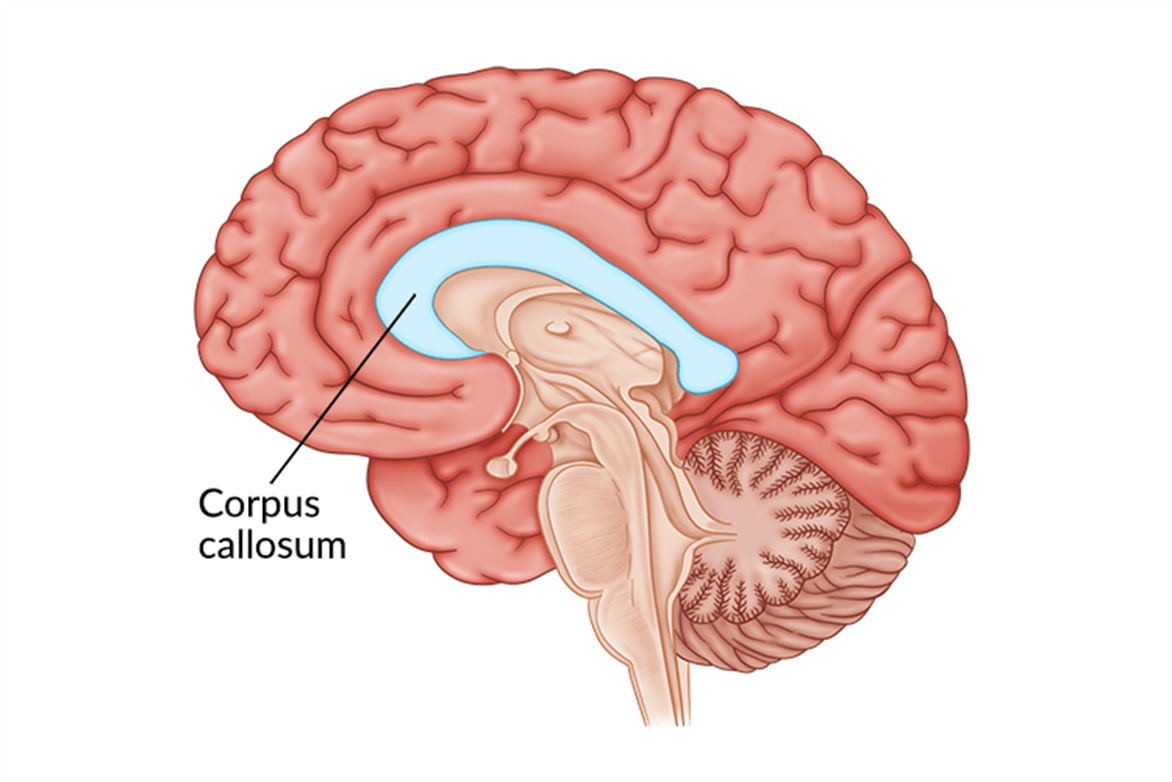
Corpus Callosum:
Longitudinal fissure of a thick band of nerve fibres connecting hemispheres.
Physically connects hemispheres.
Transmits information.
Divided into four lobes:
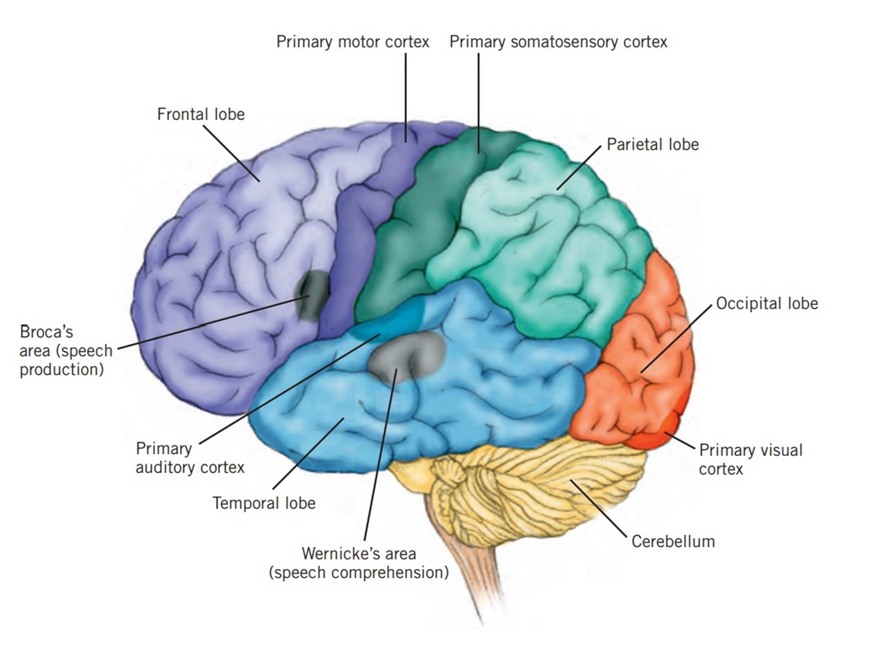
Frontal Lobe: reasoning, planning, movement, emotions, and personality.
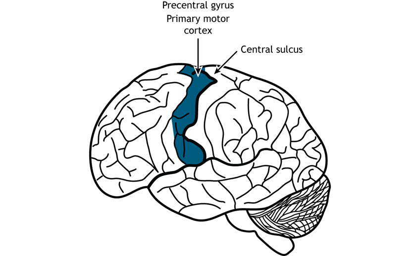
Primary motor cortex:
Rear of frontal lobe.
Directs skeletal muscles ~ voluntary movement (left/right - contralateral control).
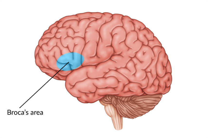
Broca’s area:
Production of articulate speech.
Left frontal lobe, near primary motor cortex.
Controls respiration muscles, cheeks, lips, jaws, tongue.
Sentence structure, grammar.
Related to Wernicke’s area.
Damage = Broca’s aphasia, can understand, can’t produce (Broca’s broken speech).
Parietal Lobe: Processing sensory information; spatial awareness.
Somatic sensations.
Spatial awareness.
Perception of location, movement of body parts.
Integration of sensory language.
Sensory cortex:
Strip of neurons at front of parietal lobe, next to primary motor cortex.
Registers, processes sensations detected by body’s sensory receptors.
Contralateral reception.
Temporal Lobe: Auditory processing; memory formation; understanding language.
Understanding speech.
Audio information interpretation.
Smell processing.
Facial recognition.
Long-term memory formation.
Primary auditory cortex:
Registers, processes auditory information; left is verbal, right is non-verbal.
Contribute to memory via link to hippocampus.
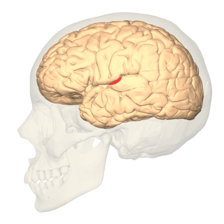
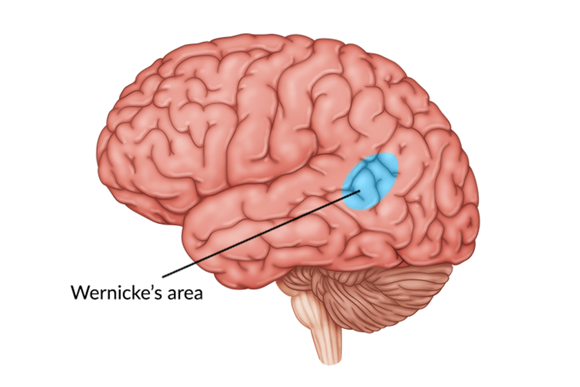
Wernicke’s area:
Left temporal lobe, near primary cortex.
Identifying sounds to comprehend meaning.
Accesses words in memory, controlling comprehension of speech and formulation of meaningful sentences.
Connections to Broca’s area, left temporal lobes receiving sound. Also speech registered in primary auditory cortex of both hemispheres.
Damage = Wernicke’s aphasia; can speak fluently but does not make sense. Difficulty in communication. Can see and pronounce but not understand.
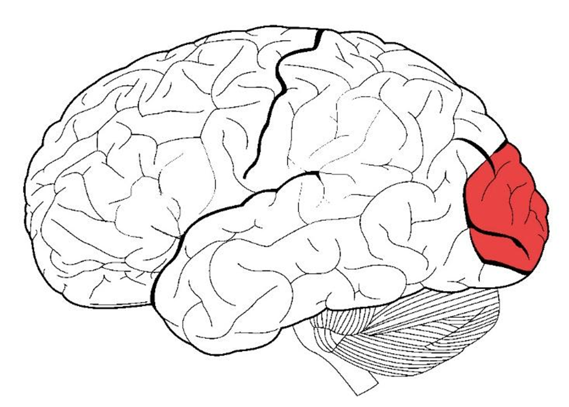
Occipital Lobe: Visual processing, facial recognition, perception of distance/depth.
Information from each eye is transmitted to primary visual cortex in left occiptal lobe, vice versa.
Primary visual cortex:
Processes visual information (into whole image or pattern, given meaning) from retinas via optic nerve, then send to other brain lobes.
Contains variety of neurons (specialised response neurons; specific to colour, shape, motion, etc.).
4.2 Localisation of Functions
Broca’s Area: Speech production.
Wernicke’s Area: Language comprehension.
5. Historical Studies
Phineas Gage:
Case study illustrating the relationship between brain injury and personality changes.
Suffered a severe injury when a metal rod penetrated his skull, damaging the frontal lobe.
Post-accident changes included:
Personality shift: Became impulsive, irritable, and irresponsible.
Inability to hold down a job or maintain social relationships.
Demonstrated the role of the frontal lobe in regulating personality and social behavior.
Roger Sperry:
Conducted studies on split-brain patients to explore the functions of cerebral hemispheres.
Findings indicated lateralization of brain functions, with each hemisphere specializing in certain tasks.
Highlighted how brain function is localized, impacting cognition and behavior.
Walter Freeman:
Pioneered frontal lobotomies as a treatment for mental illness.
His methods raised significant ethical questions regarding neuropsychological treatments.
Conducted thousands of lobotomies, often without proper consent, leading to questions about patient rights and treatment efficacy.
6. Contemporary Methods of Study
EEG (Electroencephalogram)
How it Works: Measures electrical activity in the brain via electrodes attached to the scalp. It detects voltage fluctuations resulting from ionic current flows within the neurons.
Strengths: High temporal resolution allows for the measurement of brain activity in real time, making it effective for tracking dynamic changes in brain activity.
Limitations: Low spatial accuracy; does not pinpoint the exact location of brain activity, limiting detailed brain mapping.
Uses:
Diagnose epilepsy, seizure disorders.
Sleep research
CT (Computed Tomography)
How it Works: Uses X-rays to create detailed cross-sectional images (slices) of the brain. A computer combines these images to provide a comprehensive view of brain structure.
Strengths: Useful for quickly detecting brain injuries, tumors, and bleeding; widely available and fast.
Limitations: Exposure to ionizing radiation can pose risks, and less effective in differentiating between types of soft tissues compared to MRI.
Uses:
Fractures.
Diagnose brain tumours, aneurysms.
Measure size of brain tumour.
Assess brain injury.
MRI (Magnetic Resonance Imaging)
How it Works: Utilizes strong magnetic fields and radio waves to generate detailed images of brain structures by aligning hydrogen atoms in water molecules and detecting their signals.
Strengths: Provides high-resolution images with detailed structures of the brain; no ionizing radiation exposure.
Limitations: More expensive and less accessible than CT; takes longer to conduct, and some patients may find the enclosed space uncomfortable.
Uses:
Diagnose brain tumours, aneurysms.
Measure size of brain tumour.
Assess effects of stroke.
Assess brain injury.
fMRI (Functional Magnetic Resonance Imaging)
How it Works: Measures brain activity by detecting changes in blood flow; areas of the brain that are more active receive more blood, which can be measured using MRI technology.
Strengths: Non-invasive method that provides both structural and functional information; excellent spatial resolution allows for precise localization of brain activity.
Limitations: Lower temporal resolution than EEG, so it cannot capture rapid brain activity changes with the same effectiveness as EEG.
Uses:
Activity in brain when performing a task.
Plan for tumour removal surgery.
Detect brain activity of patients with neurological conditions such as Parkinson’s disease.
Structure of the Nervous System
Central Nervous System (CNS)
Composed of brain and spinal cord
Processes sensory information from PNS and sends motor commands back out
Peripheral Nervous System (PNS)
Comprises nerves outside the CNS
Divided into:
Somatic Nervous System: Controls voluntary muscle movements
Autonomic Nervous System (ANS): Regulates involuntary body functions (divided into Sympathetic and Parasympathetic Systems)
Features of Neurons
Definition: Specialized cells for receiving, transmitting, and processing information
Structure:
Dendrites, Cell Body (Soma), Axon, Axon Terminals, Myelin Sheath
Types of Neurons:
Motor, Sensory, Interneurons
Neural Transmission
Process: Action potentials transmit signals, neurotransmitters released in synapses
Synapse: Junction converting electrical impulses to chemical signals
Brain Structure and Functions
Hindbrain: Basic autonomic functions (medulla & cerebellum)
Midbrain: Processes hearing, vision, movement; contains reticular formation
Forebrain: Largest region responsible for complex functions (cerebral cortex, thalamus, hypothalamus)
Cerebral Cortex
Two Hemispheres:
Left: Analytical, language
Right: Creative, non-verbal
Lobes:
Frontal: Reasoning, movement, Broca's area (speech production)
Parietal: Sensory processing, spatial awareness
Temporal: Auditory processing, Wernicke's area (language comprehension)
Occipital: Visual processing
Historical Studies
Phineas Gage: Brain injury affecting personality
Roger Sperry: Split-brain studies highlighting lateralization
Walter Freeman: Ethical concerns in frontal lobotomies
Contemporary Methods of Study
EEG: Measures electrical activity; good temporal resolution
CT: Structural imaging using X-rays
MRI: High-resolution imaging with no radiation
fMRI: Detects changes in blood flow for brain activity