Copy of Copy of HBS EOC Study Guide.docx
HBS EOC Study Guide
Unit 1: Road to Rehabilitation
1.1 Beginning with Bones
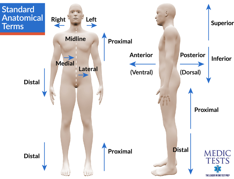
- Anterior is the front part of a human, including face and abdominal region, while posterior is the back part of a human, including shoulder blades and heels.
- Superior refers to something that is higher up on the body, while inferior refers to something lower on the body.
- Medial refers to something closer to the midline, while lateral refers to something further away from the midline.
- Proximal refers to something on an appendage closer to the trunk, while distal refers to something further away from the trunk.
- Superficial refers to something closer to the surface of the skin, while deep refers to something that is further within the body.
- Dorsal refers to something associated with the spinal side of the body, while ventral refers to something associated with the abdominal side of the body. (think of some fins you might know!)
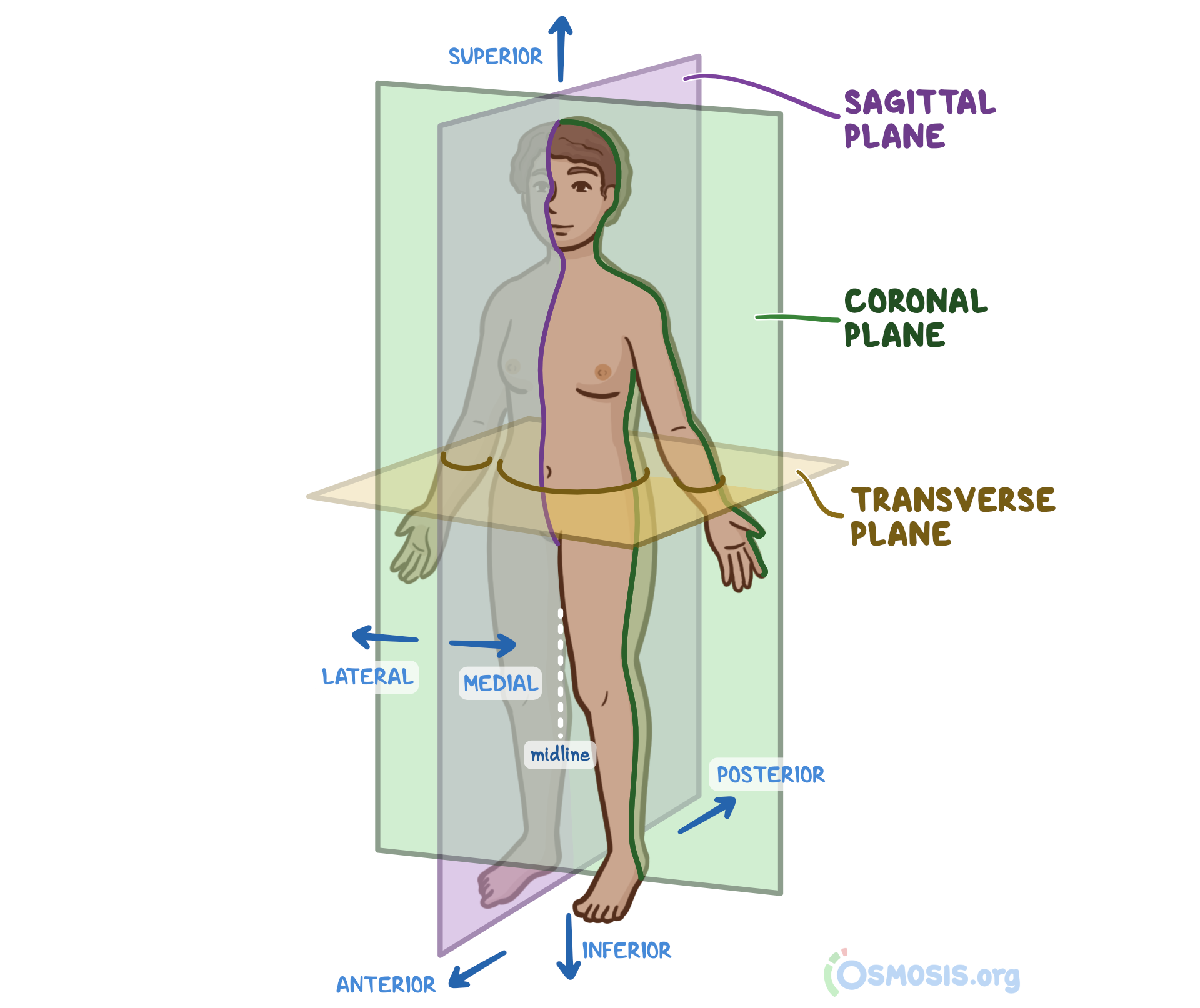
- Here, draw and label each plane. (coronal, sagittal, transverse, and median)

- Match the regional term with its area in layman's terms. ‘
| T eye |
|---|---|
| DD spine |
| CC belly button |
| BB chest cavity |
| AA feet/ankle |
| Z sternum (chest/between left and right ribs) |
| Y shoulder blade |
| X tailbone |
| W back of knee |
| V pelvis |
| U knee |
| S mouth |
| R elbow |
| Q back of the head |
| P nose |
| O lower back |
| N groin |
| M booty |
| L thigh |
| K fingers/toes |
| J upper part of pelvis - part of the hip you can feel |
| I neck |
| H head |
| G hands/wrists |
| F heel |
| E cheek |
| D arm |
| C armpit |
| B where blood is taken from - inside of elbow |
| A tummy |
- Here, label the body cavities. (cranial, dorsal, pelvic, ventral, thoracic, vertebral, abdominal, abdomino-pelvic)
Cranial Vertebral Thoracic Abdominal Pelvic Abdomino-Pelvic Dorsal = belly side of body Ventral = back side of body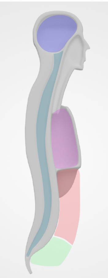
- Cells work together to form tissues,
which work together to form organs,
which work together to make organ systems. These all together make an organism! - Summarize the following different types of tissues below
Nervous Tissue | Connective Tissue | Epithelial Tissue | Muscular Tissue |
|---|---|---|---|
|
|
|
|
- What are the 4 types of bones (think shape)? Give two examples of each type.
Long bone - femur and humorous
Short bone - tarsals and carpals
Flat bone - scapula and ribs
Irregular bone - vertebrae and mandible 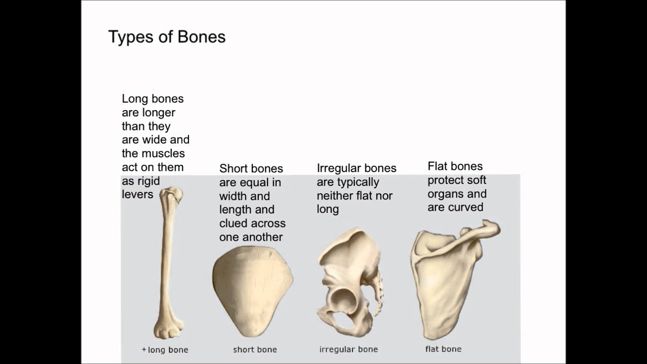
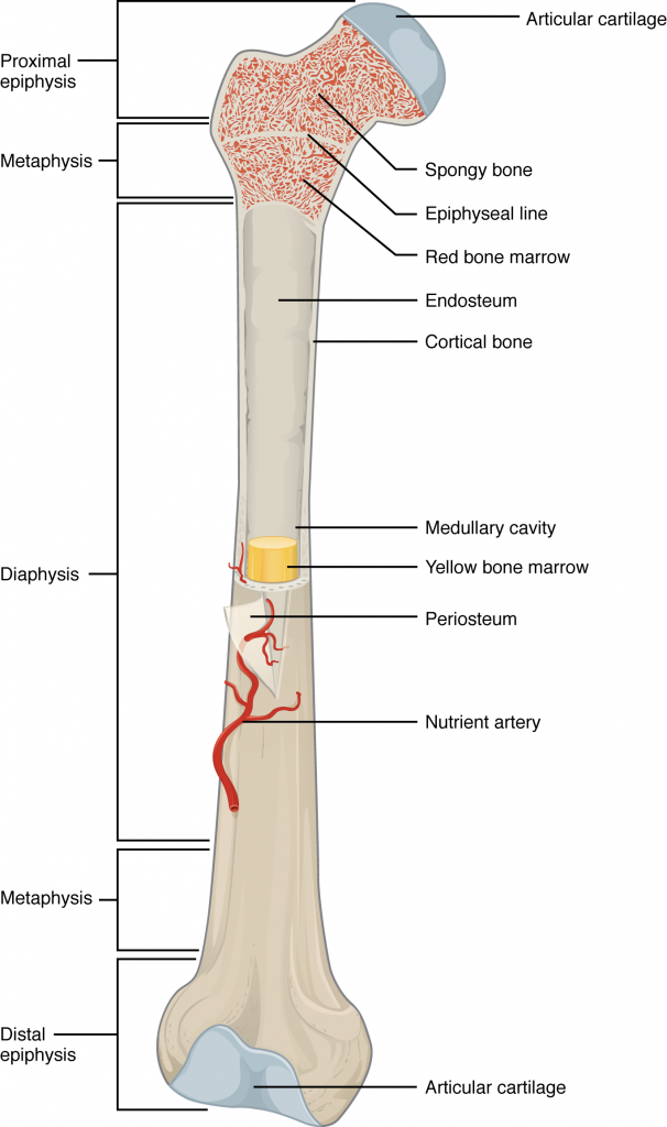
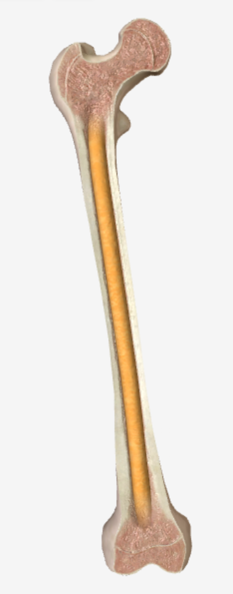
- Label the bone below with the following terms: epiphysis, metaphysis, diaphysis, spongy bone, compact bone, periosteum, medullary cavity, yellow bone marrow
- Label the following bones:
- Carpals
- Clavicle
- Femur
- Fibula
- Frontal Bone
- Humerus
- Mandible
- Maxilla
- Metacarpals
- Metatarsals
- Occipital Bone
- Parietal Bone
- Patella
- Pelvic Girdle
- Phalanges
- Radius
- Rib Cage
- Scapula
- Sphenoid
- Sternum
- Tarsals
- Temporal
- Tibia
- Ulna
- Vertebral Column
- Zygomatic

- Identify the kind of tissue shown below in the table using the options listed.
Adipose tissue 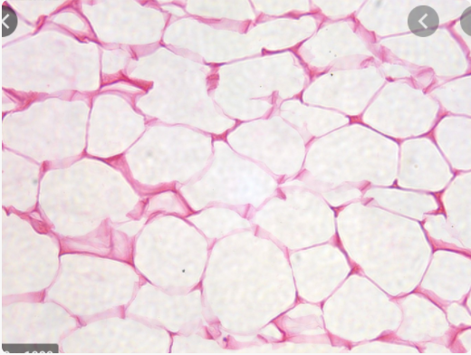 | Skeletal muscle 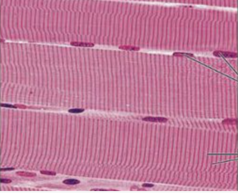 | Epithelial tissue 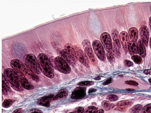 | Compact bone 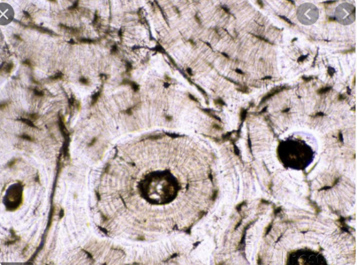 |
|---|
- Osteoblasts are cells that help in the building of bones, while Osteoclasts are cells that help with the breaking down of bone. Excess of either of these types of cells result in weak bones!! Osteoblasts = build Osteoclasts = crash
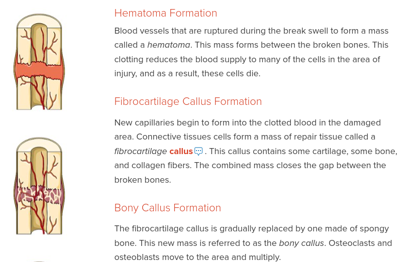
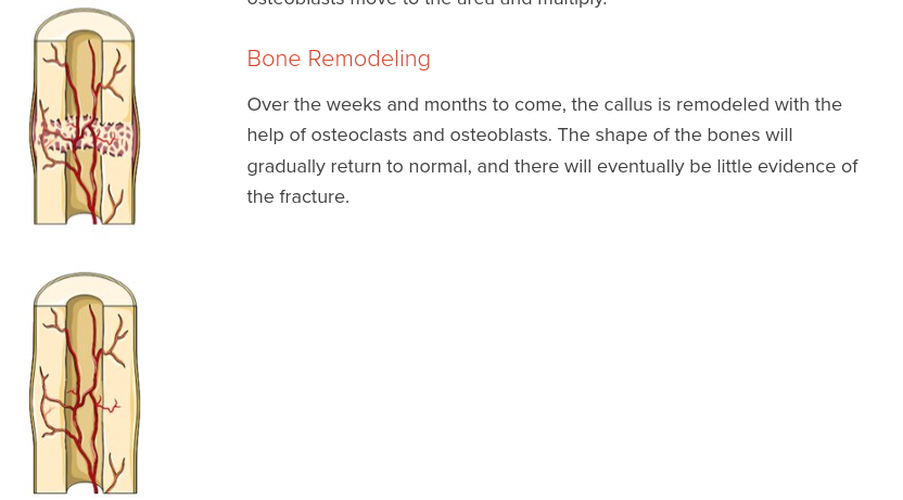
- What are the four stages of fracture repair? Describe them.
- Watch the Designing a Fracture Repair video in 1.1.6 - this helps you understand how to decide on what method to use to help repair a fracture. (plating or nailing). Draw a picture of plating and one of nailing.
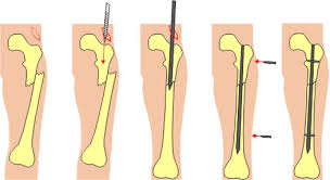
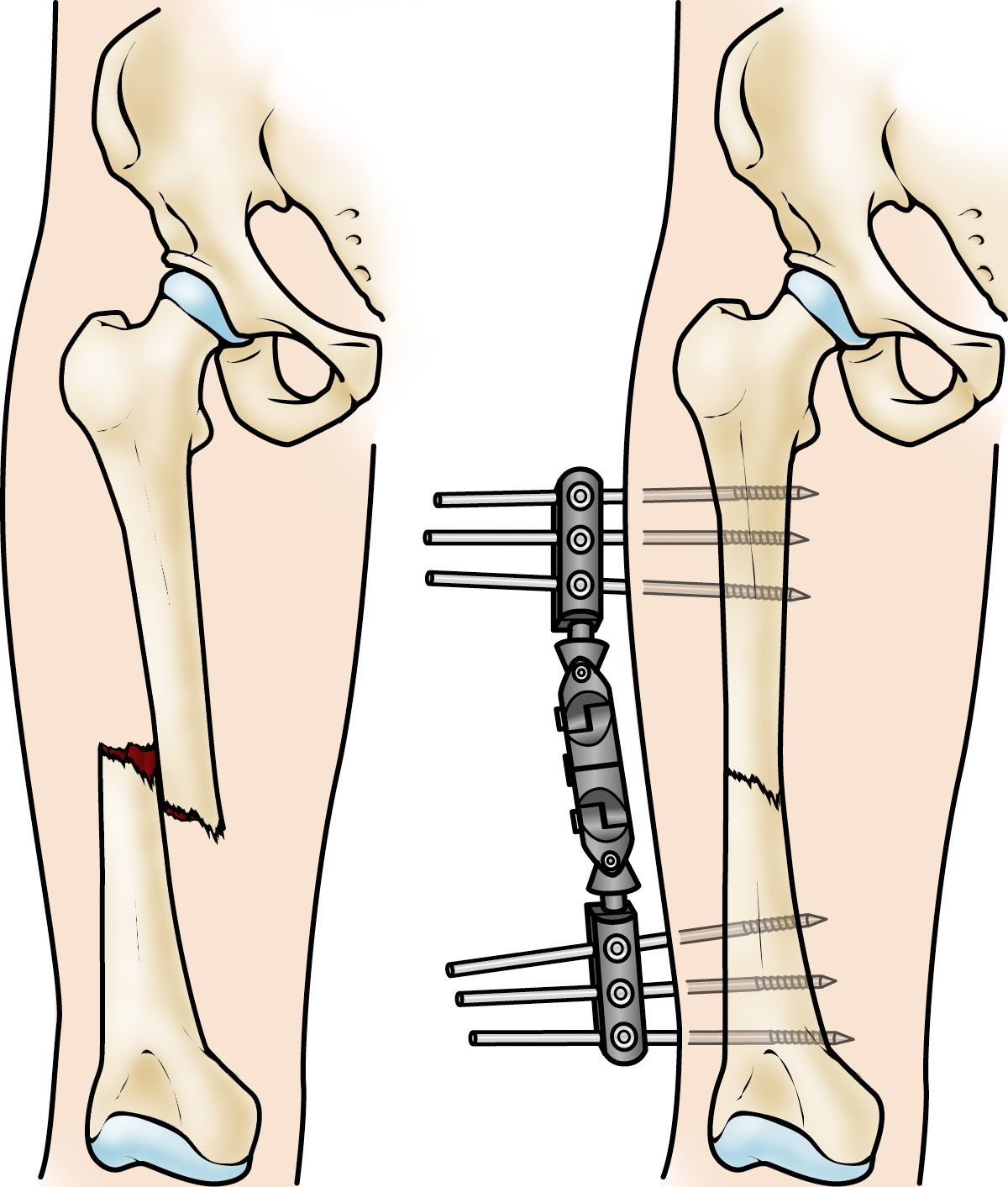 Planting nailing
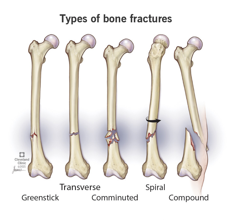
- Draw a picture demonstrating the three types of fractures (transverse, spiral, and impact (aka comminuted)).
1.2 Muscles and Motion
- Complete the following chart about the three types of muscle tissue by using the microscopic image.
Microscopic Image | 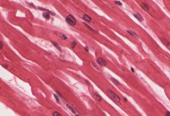 | 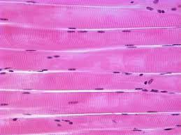 | |
|---|---|---|---|
Type of Muscle Tissue | Cardiac | Smooth | Skeletal |
Striated? | yes | no | yes |
Voluntary? | involuntary | involuntary | voluntary |
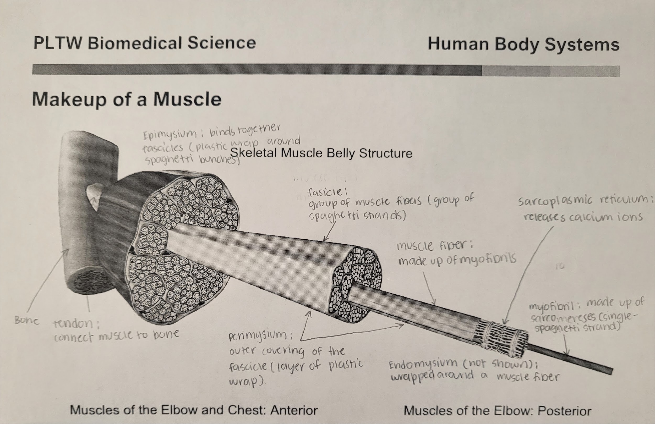
- Label endomysium, epimysium, perimysium, fascicle, tendon, muscle cell, myofibril.
-
- Identify the following labels on the image
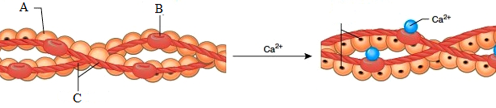
A: actin B: troponin C: tropomyosin

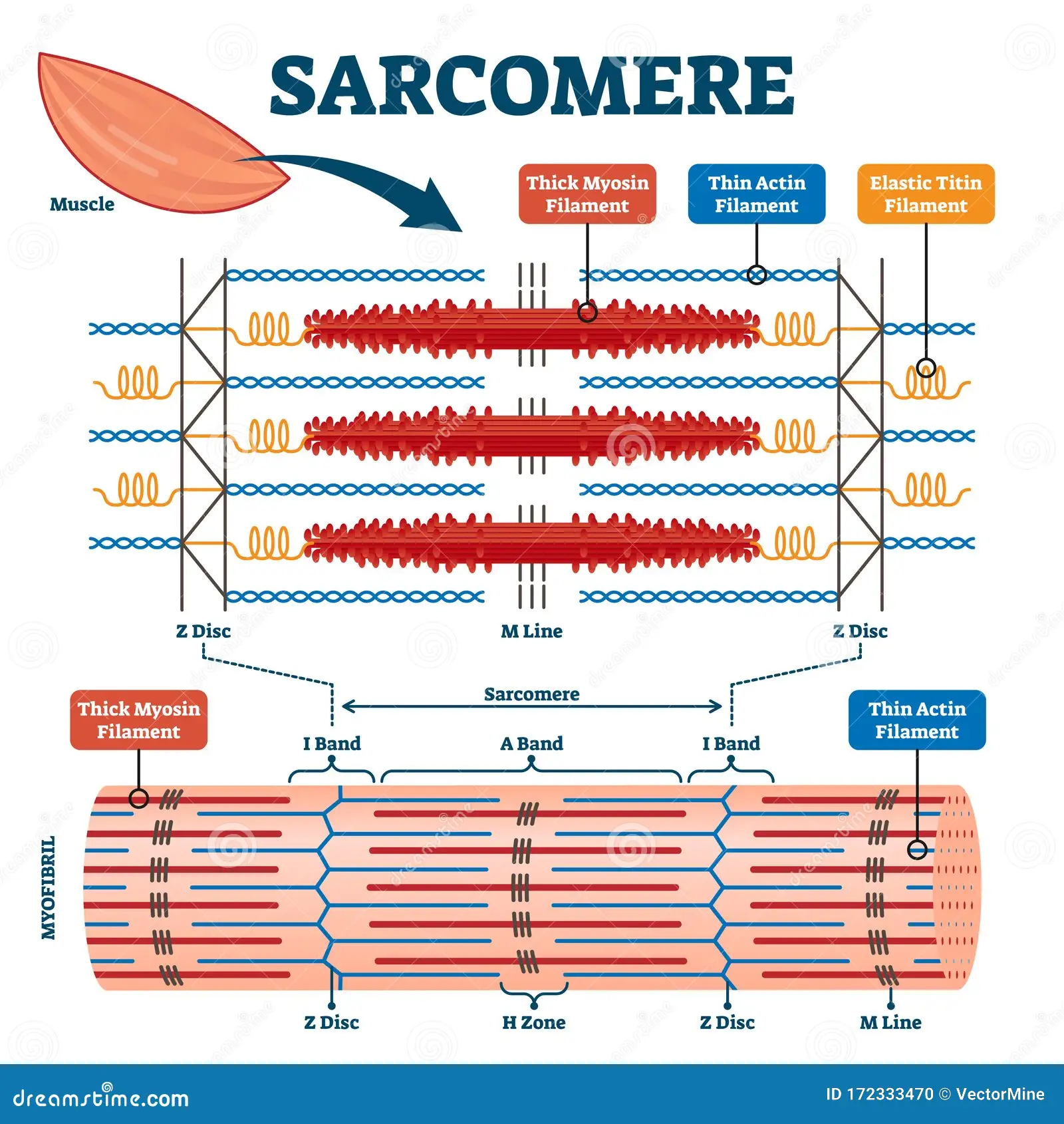
- Label the following CLEARLY: sarcomere, actin, myosin, M line, Z-lines
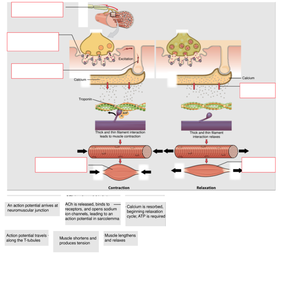
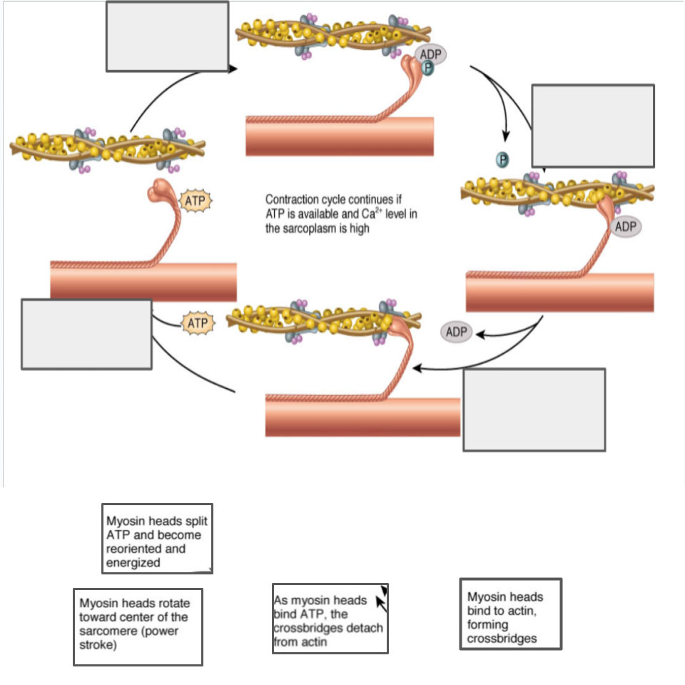
- Fill in the following blanks about muscle contraction using the word bank below.
Actin | ADP / P | ATP (x3) | Calcium (x2) | Contraction |
|---|---|---|---|---|
M-Line | Myosin | Relax | Sarcomere | Sarcoplasmic Reticulum |
Slide | Tropomyosin | Troponin | Weakens (ed) | Z-line |
- Calcium is released from the Sarcoplasmic Reticulum.
- It binds to Troponin which causes Tropomyosin roll away off Actin binding sites.
- Myosin heads that are charged with hydrolyzed ATP “grab” actin. The ADP / P (hydrolyzed ATP) is released from the myosin heads as it pulls, “spending” the energy in that molecule.
- The Sarcomere shortens as the actin filaments slide past myosin filaments. M-Line moves toward the Z-line.
- New ATP then binds, which Weakens the cross-bridge (myosin/actin connection).
- Myosin heads detach and Relax, which causes the actin filaments to Slide back to their resting position.
- The ATP is hydrolyzed, and the myosin heads wait and are ready for a new Contraction cycle to begin when Calcium reveals troponin binding sites once again!
- List all 6 of the muscle rules here.
- Muscles must have at least two attachments and must cross at least one joint.
- Muscles always “pull” and get shorter.
- The attachment that moves is known as the insertion and the attachment that remains stationary is known as the origin.
- Muscles that decrease the angle between ventral surfaces of the body are known as flexors. Muscles that increase the angle between ventral surfaces of the body are known as extensors
- Muscles work in opposing pairs.
- Muscle striations point to the attachments and show the direction of pull.
- In a muscle, the origin refers to the attachment that does not move, while the insertion refers to the attachment that does move.
Unit 2: Research Ready
2.1 Getting Nervous
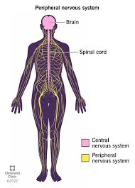
- Differentiate between the central nervous system and the peripheral nervous system.
Central Nervous System | Peripheral Nervous System |
|---|---|
Consists of brain and spinal cord | Consists of sensory receptors, sensory neurons, and motor neurons |
Control/integrating center for the body and receives information from the peripheral nervous system and then tells it what to do | Pass information to the central nervous system and communicate impulses to organs from the central nervous system |
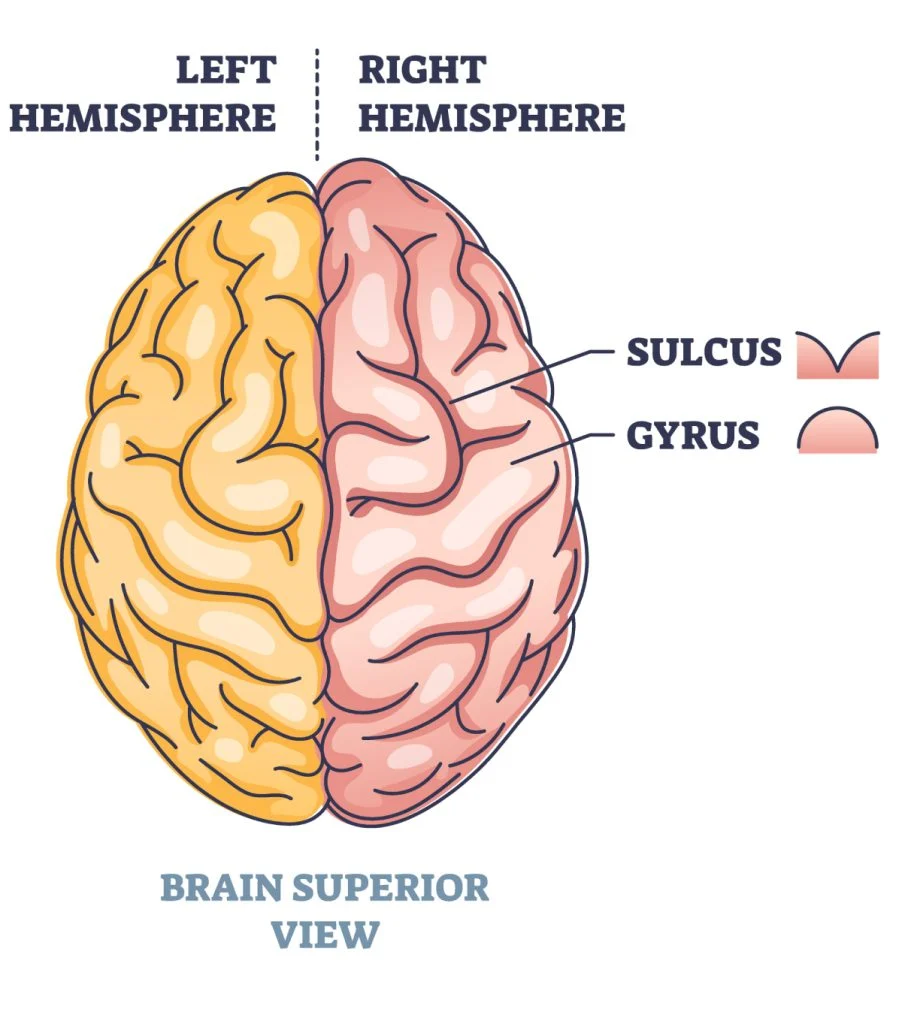
- Cerebrum is the area of the brain containing the four lobes. The four lobes are frontal, temporal, parietal , and occipital .
- The cerebellum is the area of the brain underneath the cerebrum that helps with muscle control and balance.
- The brain stem consists of the medulla oblongata, pons, and midbrain. It helps with breathing, blood pressure regulation, and sleeping. Waking.


List the part of the brain that is associated with each function.
Function | Part of the brain |
|---|---|
Behavior and personality, planning, voluntary muscle movements, mood, emotions, social interactions, and attention | Frontal lobe |
Processing smell and sound, language understanding | Smell: olfactory cortex(cerebral cortex) Sound: auditory cortex(temporal) |
Sensing touch, temperature, pressure, and pain, spatial processing, language, and memory | Parietal lobe |
Visual Perception | |
Muscular coordination and balance | |
Breathing, blood pressure, sleeping and waking | |
Center for pain perception | |
Communication between hemispheres of the brain | |
Coordinates autonomic nervous system, pituitary gland, body temperature, thirst, hunger, sleep, and emotion | |
Long Term memory |
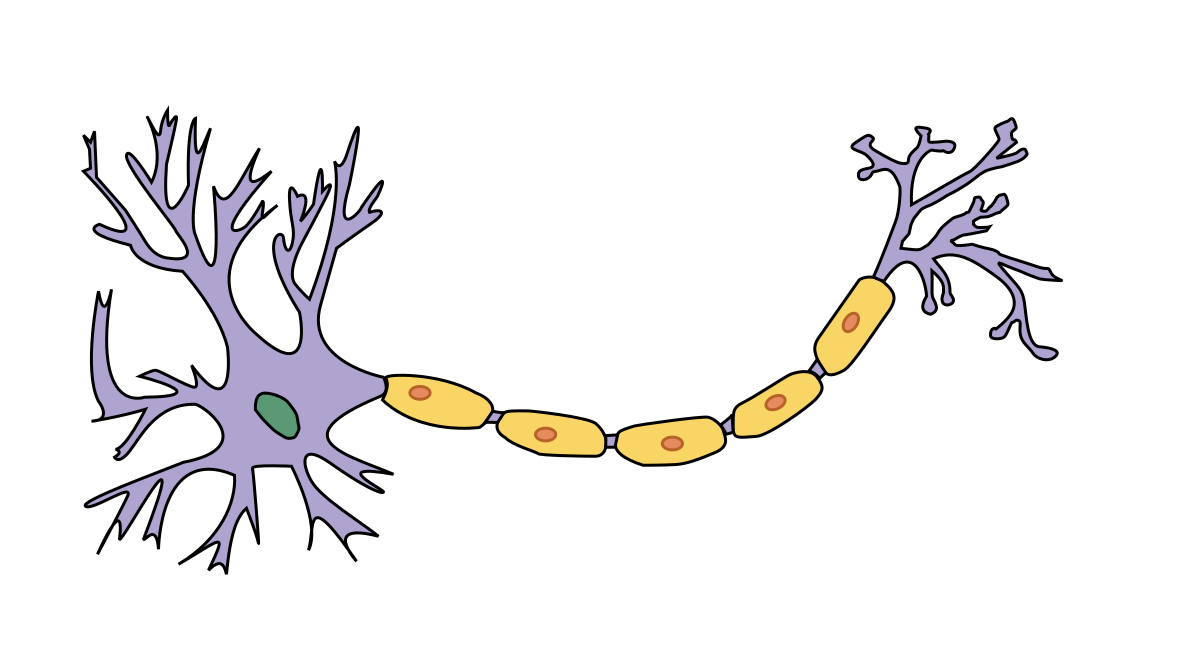
- Label this neuron with these words: dendrite, cell membrane, nucleus, axon, cell body, nodes of ranvier, myelin sheath, schwann cell, axon terminal.
- __________ neurons send signals to the brain, while ____________ send signals within the brain. __________ neurons send signals to muscles.
- ___ Hyperpolarization - ___ Resting Potential - ___ Repolarization -
___ Depolarization ___ K+ closes at -80 & pump ___ - K+ floods in
___ - Na+ floods into the cell ___ - balances back to -70
___ - Na+ closes; K+ opens ___ - Excites Na+ to opens
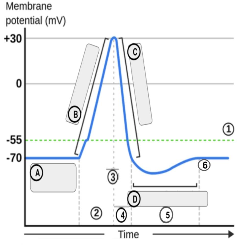
- The ______________ uses ATP to diffuse 3 ________ ions out of the cell and 2 ___________ ions into the cell to maintain resting potential.
- Resting membrane potential is ___________ mV.
- During depolarization, ___________ channels are open and _____________ channels close.
- During repolarization, ___________ channels are open and _____________ channels close.
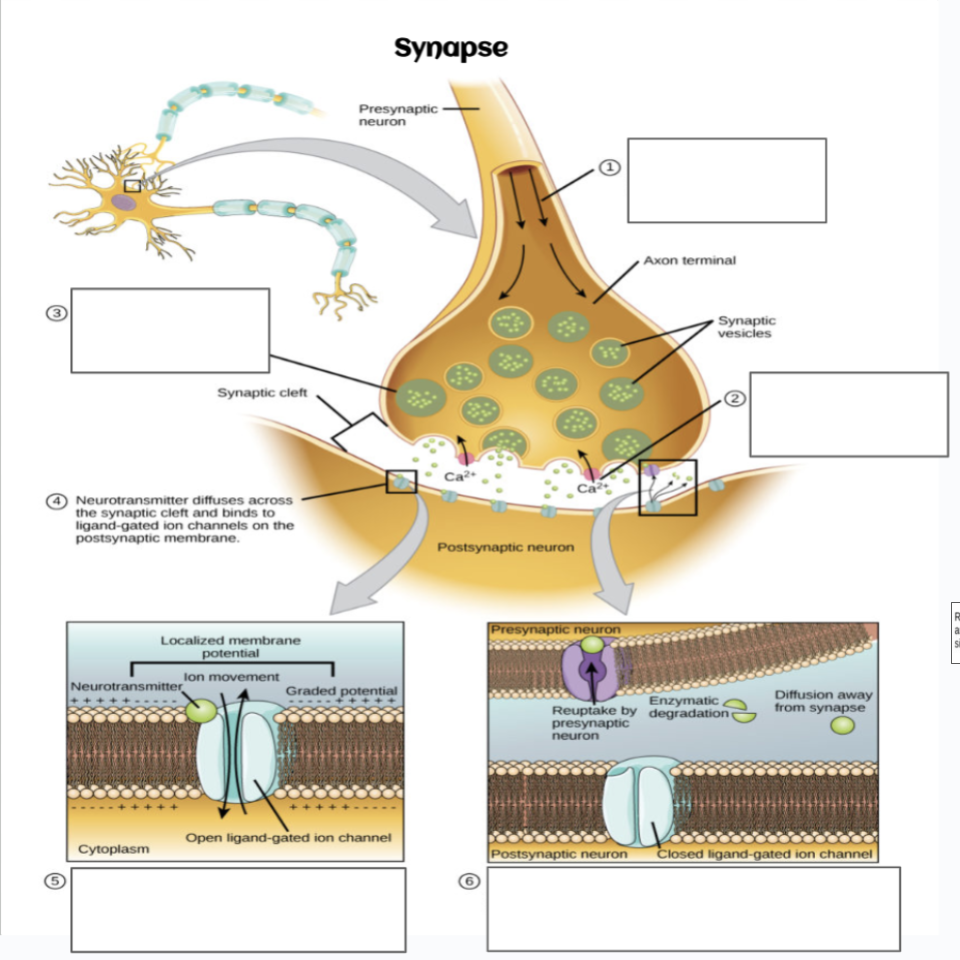
- Describe the steps of what happens when an action potential reaches the end of an axon.
- Match the neurotransmitter with its description.
Neurotransmitter | Function |
|---|---|
F Acetylcholine | a. Mood and sleep regulation; digestion |
E Dopamine | b. Fight or flight |
D GABA | c. Primary excitatory neurotransmitter in the brain |
C Glutamate | d. Primary inhibitory neurotransmitter in the brain |
B Epinephrine and nor-epinephrine | e. Pleasure, motivation, mood, attention, memory, movement |
A Serotonin | f. Muscle contraction, learning, memory |
2.2 Everything Endocrine
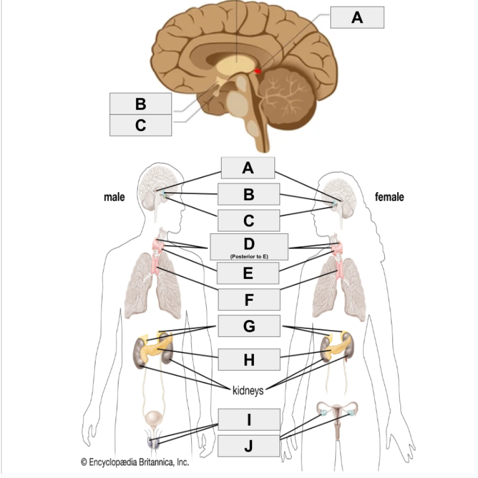
- Identify the endocrine structures
 |
|
|---|
- Match the endocrine gland with its description
Gland | Description |
|---|---|
H Hypothalamus |
|
I Pituitary Gland |
|
J Pineal Gland |
|
G Thyroid Gland |
|
D Thymus |
|
E Adrenal Gland | G. Growth and development and metabolism |
C Pancreas | H. Reproduction, thyroid regulation, growth, emotions, water levels, stress |
B Ovary |
|
A Testes | J. Regulates sleep/wake cycles (melatonin) |
- Describe what happens in your body when you eat lots of sugar. Include hormones, organs, target cells, etc.
- Describe what happens in your body when you haven’t eaten in a while.
- Differentiate between Type 1 and Type 2 Diabetes.
Type 1 doesn’t make enough insulin and type 2 don’t react to the insulin
- For males, estrogen levels stay low, but increase slightly around puberty, then stay the same. For the same hormone in women, levels increase rapidly around puberty, and stay high until menopause.
- For females, progesterone levels start off high, decrease rapidly, then increase and stay high around puberty until around age 50. For the same hormone in men, the level stays very low their whole life.
- For males, testosterone levels change often, but hit a peak around age 25, and slowly decrease slightly. For the same hormone in women, it stays low their whole life.
- Testosterone is the hormone that plays a role in building bone and muscle mass, body hair growth, and development of reproductive tissue.
- Estrogen is the hormone that plays a role in puberty, menstruation, pregnancy, menopause, sperm development, and bone health.
- Progesterone is the hormone that plays a role in menstruation, pregnancy, and testosterone production.
Unit 3: Adventure Awaits
3.1 Cardiopulmonary Connection
- Draw a diagram of blood flow through the heart. (This can be an actual human heart or a heart box)
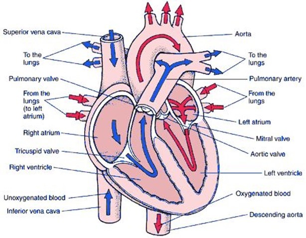
- Describe the role of valves.
Valves prevent backflow of blood into previous chambers of the heart
- Coronary arteries are vessels that bring oxygenated blood to the heart itself.
- The pulmonary vein supplies blood to the left side of the heart muscle.
- Superior vena cava and inferior vena cava supplies blood to the right ventricle, atrium
- Draw a diagram of the structure of arteries, veins, and capillaries.
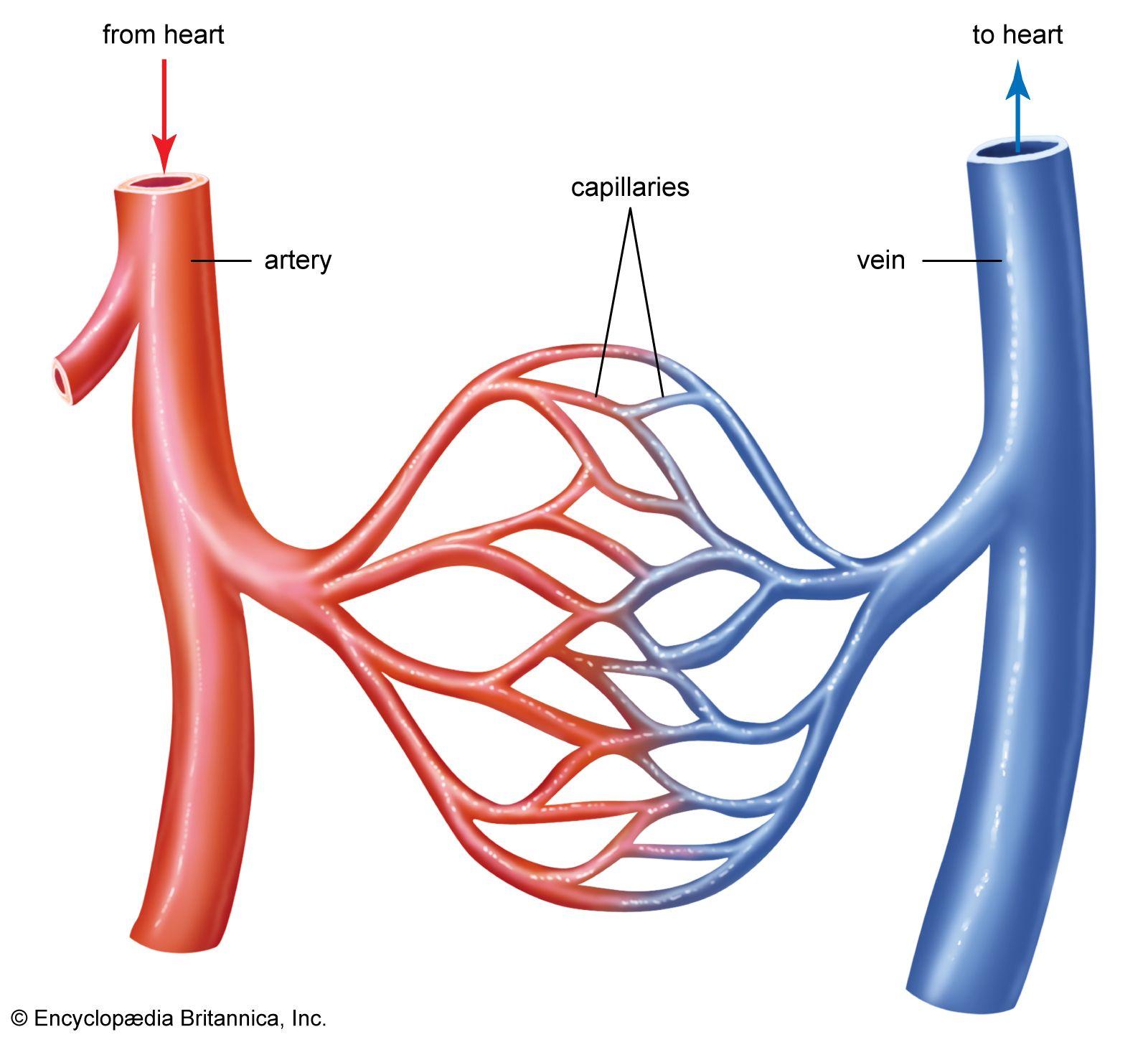
- Arteries and veins are well named. Describe the placements of these vessels. (Each vessel is the name of an artery AND a vein, except for the starred names. ie, in your body, you have brachial artery AND a brachial vein)
Vessel | Location |
|---|---|
Carotid* (just an artery) | |
Jugular* (just a vein) | |
Subclavian | |
Axillary | |
Brachial | |
Aorta* (just an artery) | |
Radial*(just an artery) | |
Ulnar*(just an artery) | |
Brachiocephalic | |
Descending aorta*(just an artery) | |
Iliac | |
Femoral | |
Popliteal | |
Cephalic* (just a vein) | |
Basilic* (just a vein) |
- ___________ pressure is the pressure in the arteries when the heart undergoes systole: the heart ________ to _______ blood through the ____________
- ____________ pressure is the pressure in the arteries when the heart undergoes diastole: the heart is at _____________, allowing the ___________ the fill with ____________.
- Complete this chart:
Blood Pressure Category | Systolic (mm Hg) | Diastolic (mm Hg) | |
|---|---|---|---|
Normal | Less than 120 | and | |
Elevated | 120-129 | and | |
____________ - stage 1 | 80-89 | ||
____________ - stage 2 | 140 or higher | or | |
Hypertensive Crisis | Higher than 120 |
- What are factors that could cause changes to a person’s blood pressure?
- Cardiac output is a measure of how much __________ is pumped by both ___________ in _____ _________. It is measured in _____________.
- Heart rate is a count of how many times the heart ____________ in _____ ____________. It is measured as __________ ______ ________.
- ________ __________ is the amount of blood being pumped out of the heart with each heartbeat. On average, it is stable at ___________ mL/beat.
- What is the formula to calculate cardiac output?
- Calculate the cardiac output of a patient whose heart rate is 185 bpm.
- ABI, or _____ ________ ___________ is calculated by dividing the ankle systolic pressure by the arm systolic pressure.
- To understand this, read through this image below.
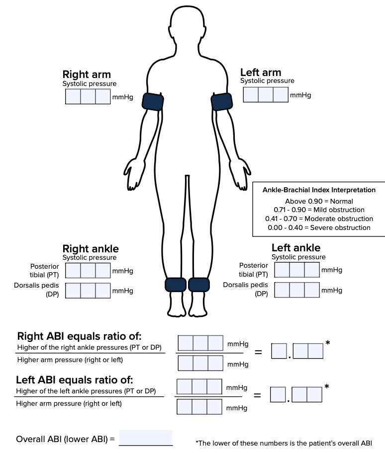
- Peripheral Artery Disease is a cardiovascular condition characterized by the _________________ of blood flow to the _______________. It can increase a person’s risk for _______ ____________. Patients may have pain while _____________, and they may experience a change in ____________. If left untreated, peripheral artery disease may completely __________ blood flow. (critical limb ________).
- _______________ commonly causes peripheral artery disease. It is a buildup of plaque in blood vessels.
- Use 3.1.3 to label this diagram. Label it with these terms: right atrium, right ventricle, left atrium, left ventricle, SA node, AV node, Bundle of His, Purkinje fibers Identify the waves on an EKG diagram
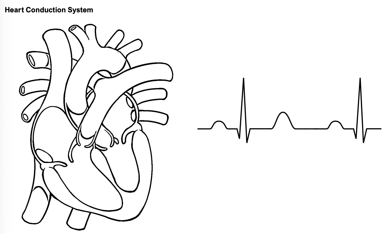
- Describe the steps of heart conduction as listed on #6 on 3.1.3.
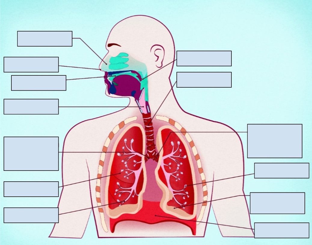
- Label the above diagram with these terms: bronchiole, right lung, right main (primary) bronchus, larynx, oral cavity, nostril, nasal cavity, pharynx, trachea, left main (primary) bronchus, alveoli, base of left lung, diaphragm.
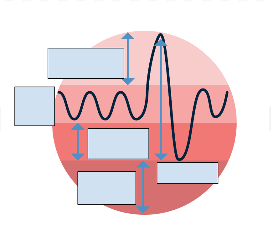
- Fill out the following blanks for what they represent in a Spirometer Graph
Unit 4: Patient Perspectives
4.1 Keeping It Renal
- Describe the four structures of the urinary system
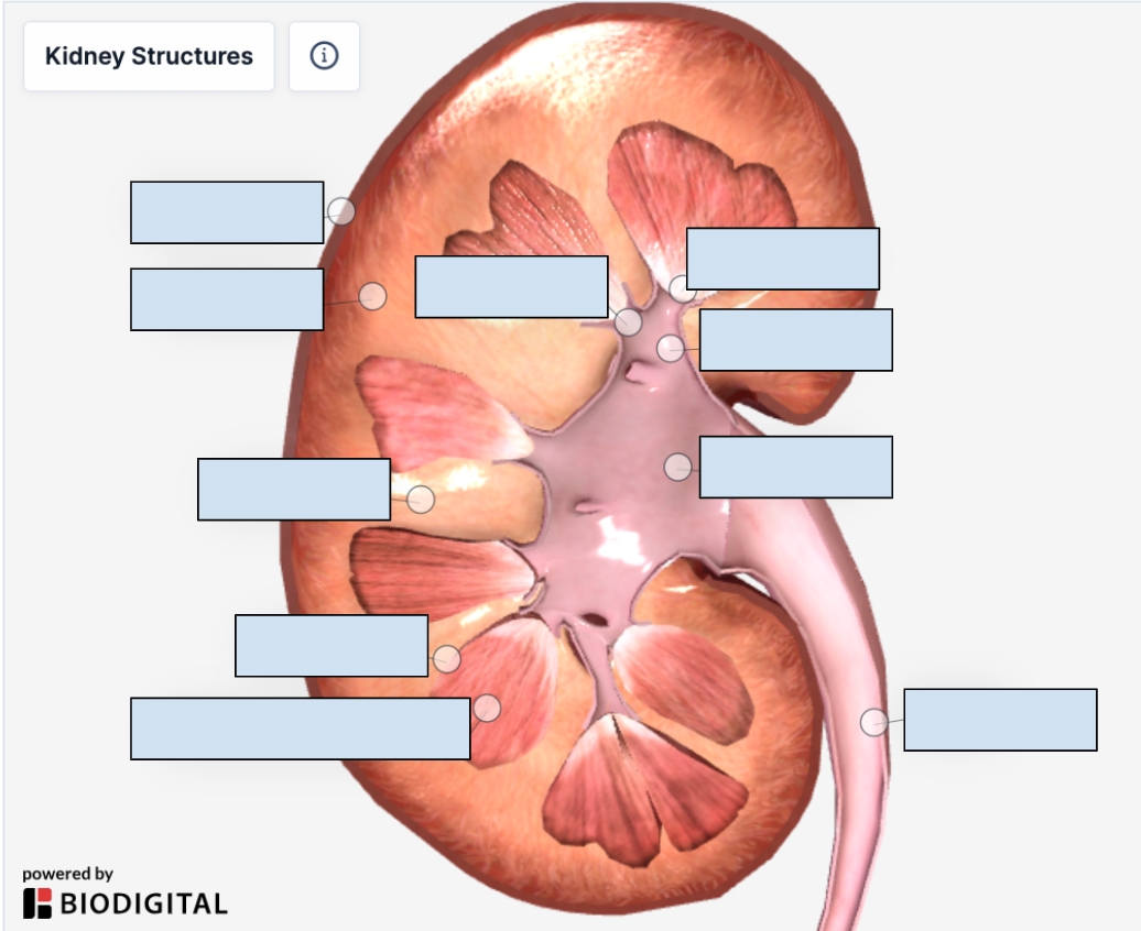
- Label this diagram of a kidney
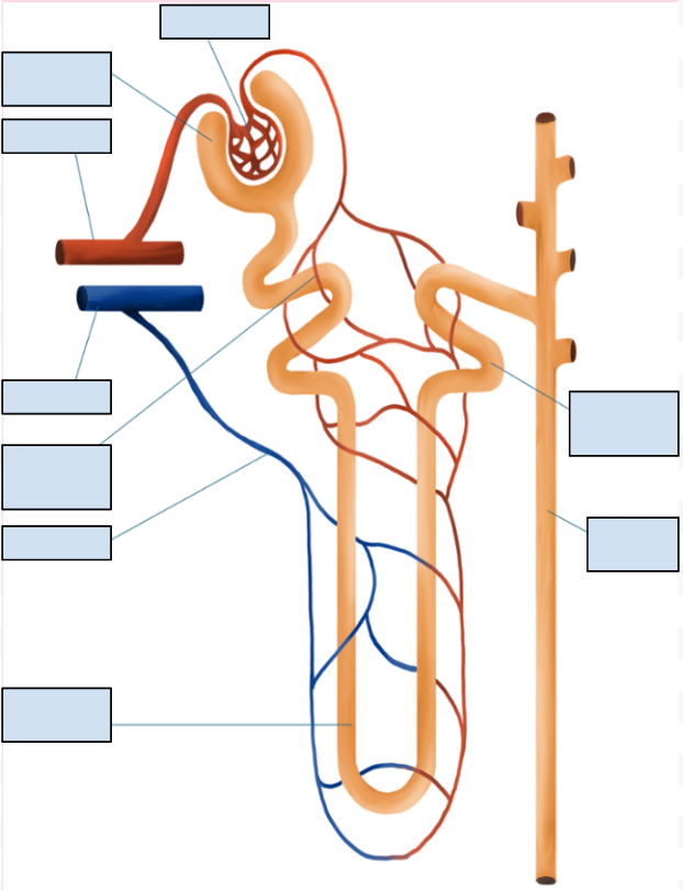
- Label this diagram of a nephron.
- ______________ _____________ ________ represents how rapidly the blood is cleansed of metabolic wastes, how effectively the kidneys carry out both tubular reabsorption and secretion, and how well the kidneys maintain homeostasis in the body.
- Differentiate between filtration, reabsorption and secretion.
- Polycystic Kidney Disease is an __________ disorder of the kidneys. There are two types: ___________ ___________ _____ and _____________ __________ _____. PKD is characterized by hundreds of fluid filled _________ throughout both kidneys. They block the blood vessels and urine-producing tubes within the kidney. As a result, they may lead to ___________ __________ after a few decades. Symptoms include __________ in the urine, ______________ urination, pain in the back or abdomen, and high _________ ___________.PKD can also occur by spontaneous ____________ of the PKD gene.
-