Human Biology Yr11 ATAR
Year 11 ATAR Human Biology content
Cell organelles
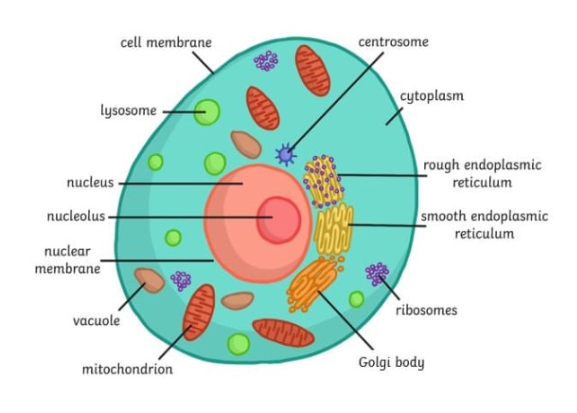
Cell membrane
Composed of phospholipid bilayer
Regulates passage of substances in and out of cell
Contains proteins for transport and cell recognition
Maintains cell shape and structure
Important for cell signalling and communication
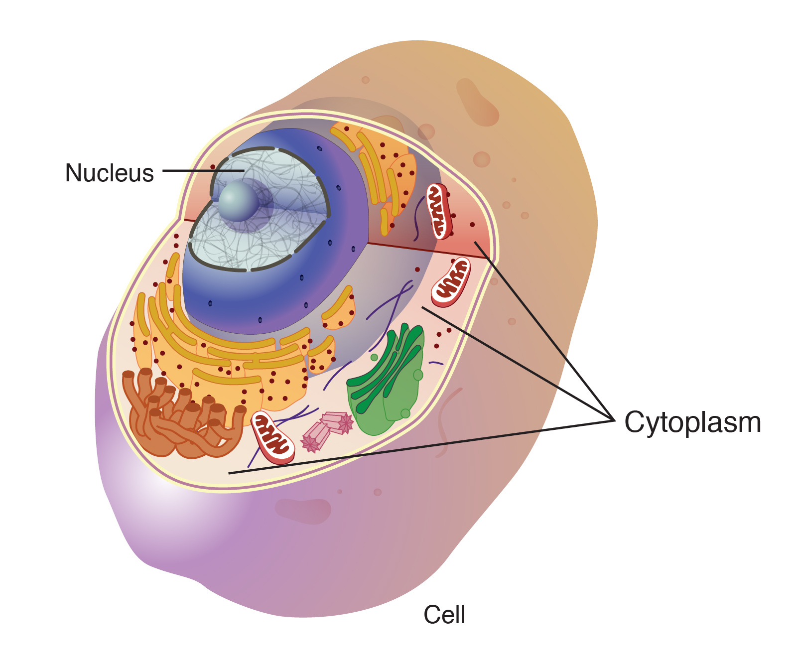
Cytoplasm
Location: Found in all types of cells
Function: Site of many cellular processes
Composition: Mostly water with proteins, salts, and organic molecules
Organelles: Contains various organelles like ribosomes and the cytoskeleton
Movement: Facilitates movement of organelles and materials within the cell
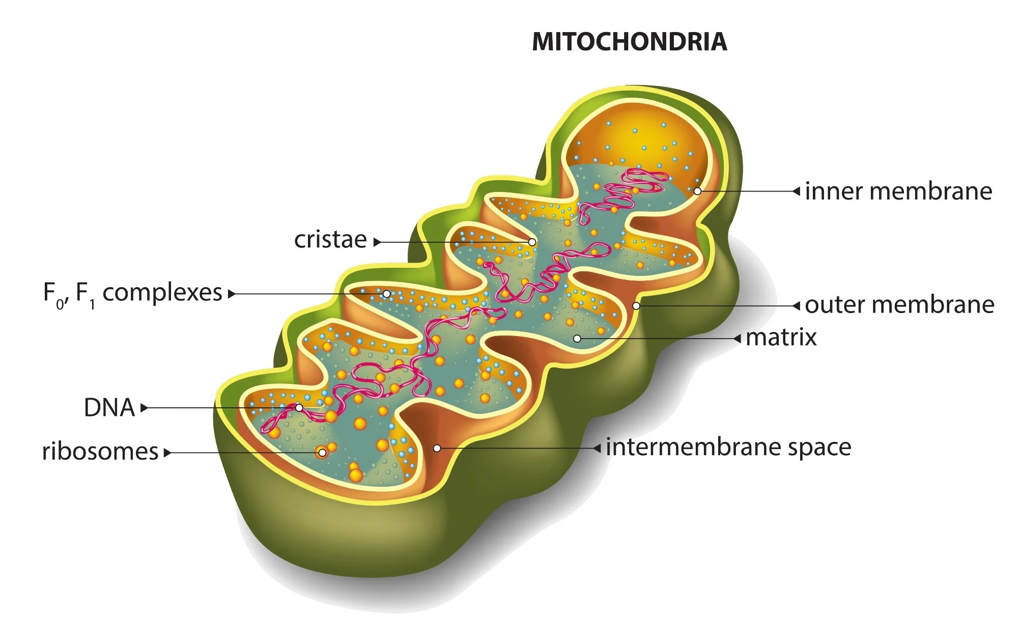
Mitochondria
Function: Powerhouse of the cell
Structure: Double membrane with inner folds (cristae)
ATP production: Through oxidative phosphorylation
DNA: Contains its own circular DNA
Endosymbiotic theory: Originated from bacteria
Diseases: Mitochondrial disorders affect energy production
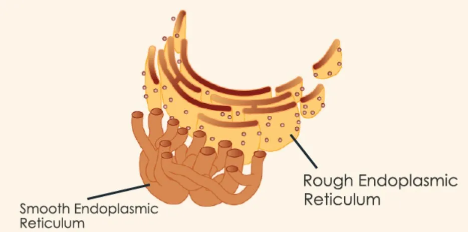
Rough endoplasmic reticulum
Site of protein synthesis: ribosomes attached
Involved in lipid metabolism
Plays a role in detoxification processes
Forms a network of membrane-bound tubules
Important for the synthesis of membrane proteins
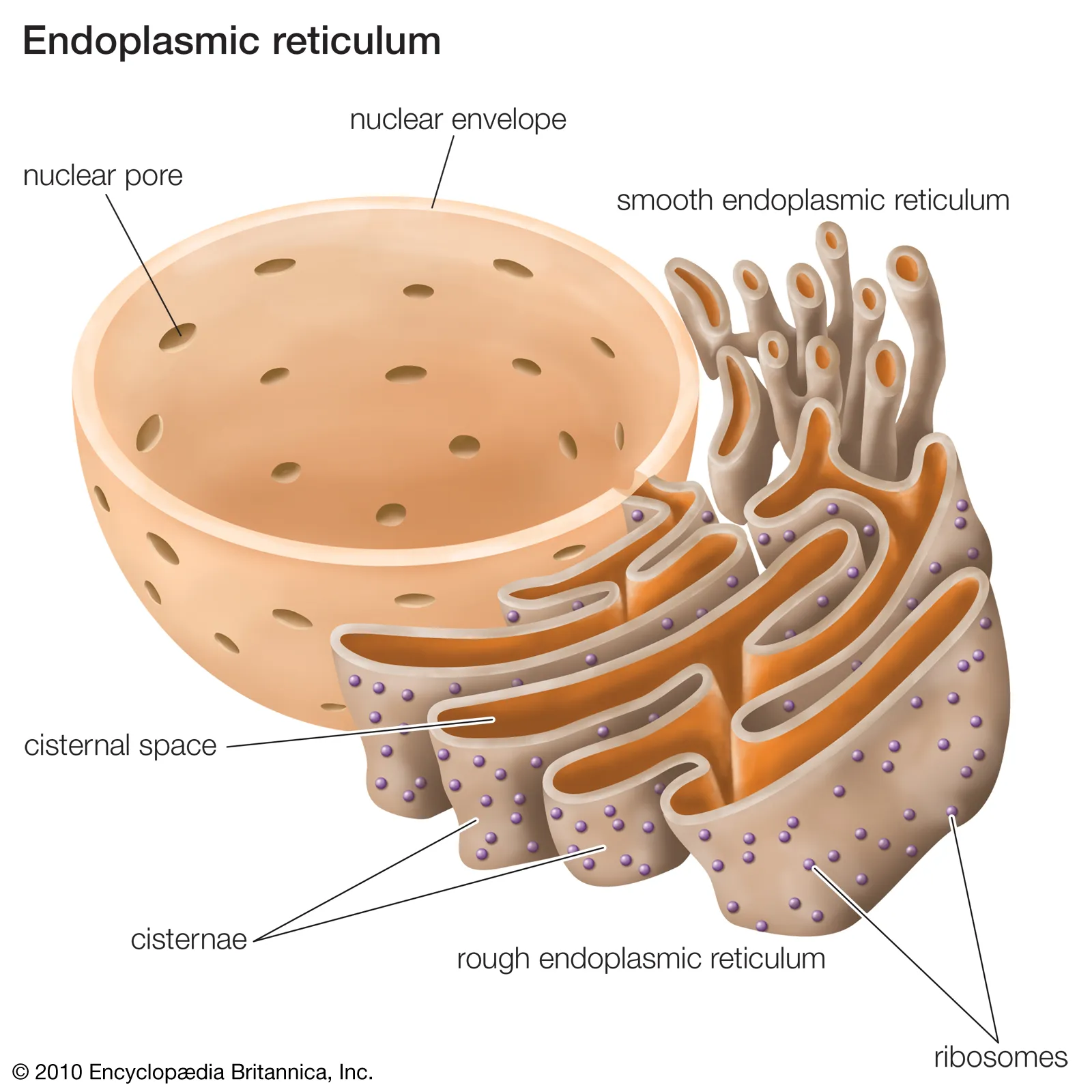
Smooth endoplasmic reticulum
Function: lipid synthesis, detoxification
Structure: tubular network
Location: connected to rough ER
Key enzymes: lipid biosynthetic enzymes, cytochrome P450
Role in cell: storage of calcium ions
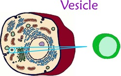
Vesicles
Function: Transport materials within the cell
Types: Endocytic, exocytic, lysosomal
Formation: Budding off from organelles
Composition: Lipids, proteins
Role: Maintain cell homeostasis
Golgi body
Function: Modifies, sorts, and packages proteins
Structure: Consists of flattened sacs called cisternae
Location: Found in eukaryotic cells
Secretory Pathway: Involved in protein trafficking
Post-Translational Modifications: Adds carbohydrates
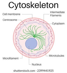
Cytoskeleton
Components: Microfilaments, intermediate filaments, microtubules
Functions: Cell shape, cell motility, intracellular transport
Regulation: Protein kinases, GTPases
Associated proteins: Actin, tubulin, keratin
Diseases: Neurodegenerative disorders, cancer metastasis
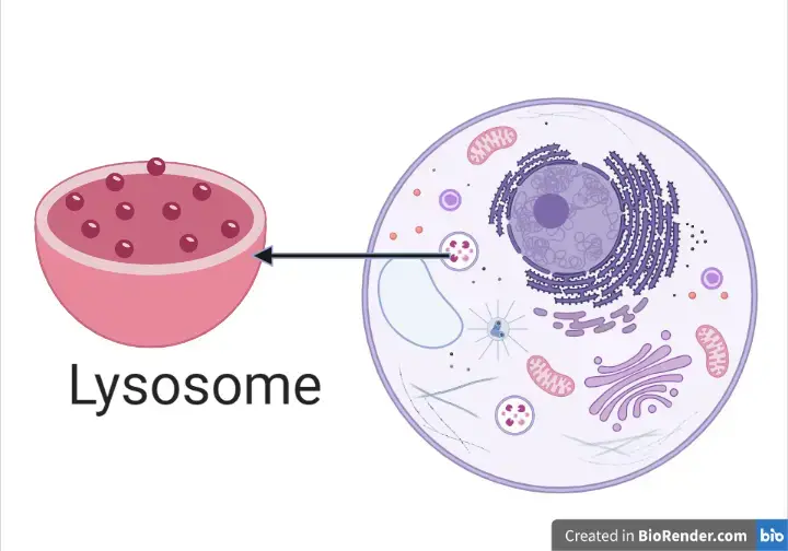
Lysosomes
Function: Digestion and recycling of cellular waste
Structure: Membrane-bound organelles containing hydrolytic enzymes
Formation: Budding from the Golgi apparatus
pH: Acidic environment for enzyme activity
Autophagy: Breakdown of damaged organelles and macromolecules
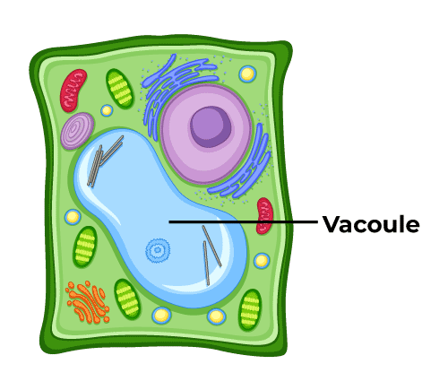
Vacuoles
Function: Storage, waste disposal, structural support
Types: Central vacuole (plants), Contractile vacuole (protists)
Structure: Membrane-bound organelle
Content: Water, enzymes, ions, nutrients
Role in plant cells: Maintains turgor pressure, stores pigments
Nucleus
Contains genetic material: DNA
Regulates cell activities
Surrounded by nuclear envelope
Contains nucleolus
Site of transcription and RNA processing
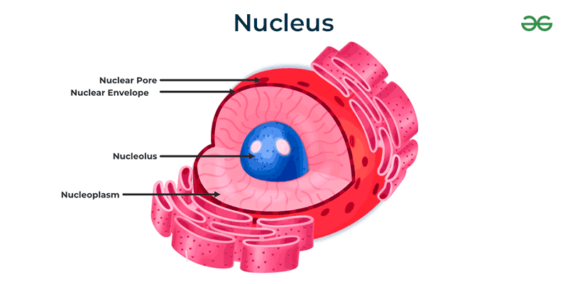
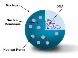
Nuclear membrane
Function: Separates nucleus from cytoplasm
Structure: Double membrane with nuclear pores
Composition: Lipids, proteins, and chromatin
Importance: Regulates passage of molecules in and out of nucleus
Maintenance: Dynamic structure undergoing constant remodelling
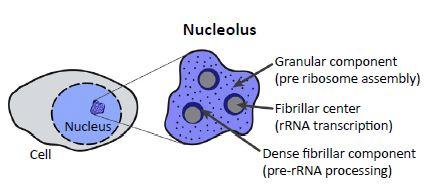
Nucleolus
Function: Ribosome biogenesis
Location: Inside the nucleus
Structure: Contains DNA, RNA, and proteins
Components: Fibrillar centre, dense fibrillar component, granular component
Importance: Essential for protein synthesis
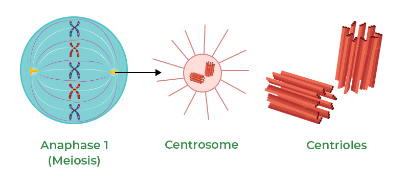
Centrosome/centrioles
Centrosomes are found in animal cells
Consist of a pair of centrioles
Play a role in cell division
Organize microtubules during cell division
Help in formation of spindle fibres
Essential for proper chromosome segregation
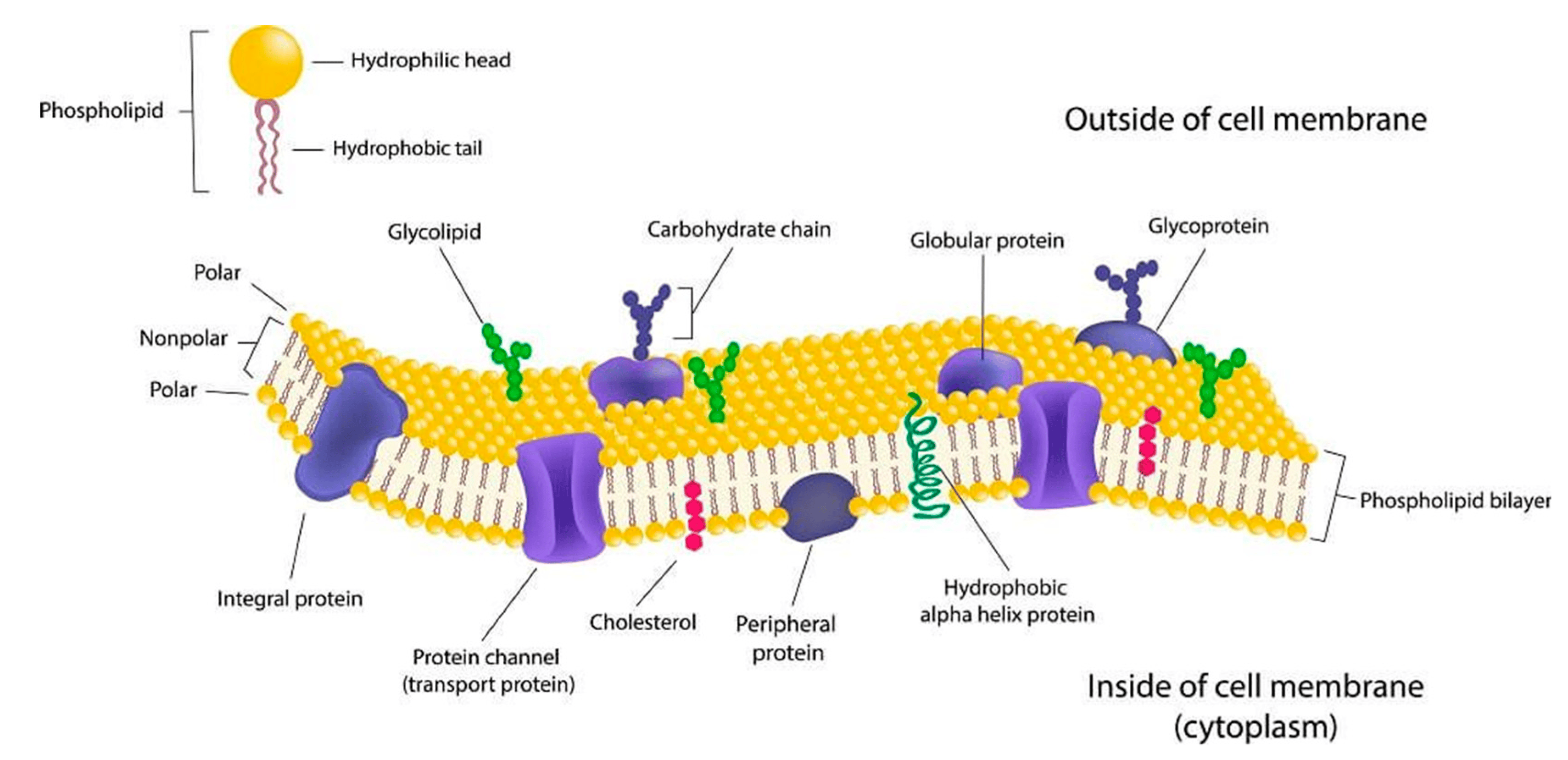
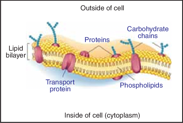
Fluid Mosaic Model
Introduction
Definition of the fluid mosaic model
Proposed by S.J. Singer and G.L. Nicolson in 1972
Structure of Cell Membrane
Phospholipid bilayer
Proteins (integral and peripheral)
Cholesterol
Carbohydrates
Fluidity of the Membrane
Lipid bilayer allows for fluid movement
Proteins can move within the membrane
Mosaic Nature
Proteins and other molecules create a mosaic pattern
Various functions of proteins (transport, receptors, enzymes)
Role of Cholesterol
Helps regulate membrane fluidity
Stabilizes the membrane structure
Dynamic Nature
Constantly changing due to movement of molecules
Allows for cell communication and transport
Conclusion
Importance of the fluid mosaic model in understanding cell membrane structure and function
Substance Movement Across Cell Membranes
Diffusion
Diffusion is a fundamental process where molecules move from an area of high concentration to an area of low concentration. This passive transport mechanism allows substances to naturally spread out to achieve equilibrium within the cell.
Definition: Movement of particles from high to low concentration
Factors affecting rate: Concentration gradient, temperature, surface area
Types: Simple diffusion, facilitated diffusion, osmosis
Importance: Essential for nutrient uptake and waste removal
Examples: Oxygen entering cells, water moving through a semipermeable membrane
Facilitated Diffusion
Facilitated diffusion is a type of passive transport that involves the assistance of specific carrier proteins to transport molecules across the membrane. These proteins help facilitate the movement of substances that may be too large or polar to pass through the lipid bilayer.
Facilitated diffusion
Passive transport
Carrier proteins or channel proteins
Movement of molecules down concentration gradient
No energy input required
Carrier Proteins: Transport molecules across cell membranes by binding to specific substances and undergoing a change in shape.
Channel Proteins: Form pores in cell membranes to allow specific ions or molecules to pass through via facilitated diffusion.
Osmosis
Osmosis is the movement of water molecules across a selectively permeable membrane from an area of low solute concentration to an area of high solute concentration. This process is essential for maintaining the cell's internal environment and preventing dehydration or swelling. It is passive diffusion.
Osmosis:
Definition: Movement of water molecules across a semi-permeable membrane
Direction: Water moves from an area of low solute concentration to high solute concentration
Importance: Crucial for maintaining cell turgidity and regulating cell volume
Active Transport
Active transport is a mechanism that moves molecules against their concentration gradient, from an area of low concentration to an area of high concentration. This process requires energy in the form of adenosine triphosphate (ATP) to pump substances across the membrane.
Energy requirement: high
Movement: against concentration gradient
Examples: sodium-potassium pump, endocytosis, exocytosis
Vesicular Transport
Vesicular transport involves the packaging of large molecules or particles into vesicles for transport across the cell membrane. This process allows cells to engulf and release substances such as proteins, nutrients, or waste materials in membrane-bound vesicles.
By understanding these different mechanisms of substance movement across cell membranes, researchers and healthcare professionals can better grasp the complexities of cellular function and develop targeted interventions for various cellular processes.
Vesicular transport moves large molecules across the cell membrane
Types include endocytosis and exocytosis
Endocytosis brings substances into the cell via vesicles
Exocytosis expels substances out of the cell via vesicles
Endocytosis and Exocytosis
Endocytosis
Definition: Process of a cell taking in molecules by engulfing them in a vesicle
Types:
Phagocytosis: Cell engulfs solid particles
Pinocytosis: Cell engulfs liquid droplets
Receptor-mediated endocytosis: Specific molecules bind to receptors on cell membrane
Steps:
Binding of molecules to receptors
Formation of vesicle
Vesicle detaches and moves into the cell
Vesicle fuses with lysosome for digestion
Exocytosis
Definition: Process of a cell releasing molecules by fusing vesicles with the cell membrane
Types:
Constitutive exocytosis: Continuous release of molecules
Regulated exocytosis: Release of molecules in response to a signal
Steps:
Vesicle containing molecules moves towards cell membrane
Vesicle fuses with cell membrane
Molecules are released outside the cell
Factors affecting the exchange of materials across the cell membrane
SA:V: Surface area to volume ratio is vital in biology for efficient material exchange across cell membranes. Higher ratios enhance nutrient and waste exchange, aiding cells in meeting metabolic needs and maintaining homeostasis, making it crucial in cell biology and physiology.
Concentration Gradient: Movement of materials from high to low concentration.
Size of Molecules: Small molecules pass through easily, while larger ones require transport proteins.
Charge: Charged molecules need specific channels or carriers to cross the membrane.
Lipid Solubility: Lipid-soluble molecules can diffuse through the lipid bilayer.
Temperature: Higher temperature increases kinetic energy, enhancing diffusion.
Pressure: Higher pressure can force molecules through the membrane.
Membrane Permeability: Membranes with more proteins allow for more selective permeability.
pH Gradient: Differences in pH can affect ion movement across the membrane.
Presence of Transport Proteins: Facilitated diffusion and active transport require specific proteins.
Osmotic Pressure: Movement of water across the membrane due to solute concentration differences.
Tissue
Nervous tissue is responsible for transmitting electrical signals in the body.
Muscular tissue enables movement and generates force.
Connective tissue provides support and structure to the body.
Epithelial tissue covers the body's surfaces and lines internal organs.
Nervous Tissue:
Function: Transmit electrical impulses.
Components: Neurons and neuroglia.
Location: Brain, spinal cord, nerves.
Specialized for communication and control.
Muscular Tissue:
Function: Generate force for movement.
Types: Skeletal, smooth, cardiac.
Skeletal: Voluntary movement.
Smooth: Involuntary movement.
Cardiac: Found in the heart.
Connective Tissue:
Function: Support and connect tissues/organs.
Types: Loose, dense, adipose, cartilage, bone, blood.
Provides structural framework.
Epithelial Tissue:
Function: Cover and protect body surfaces.
Types: Simple, stratified, squamous, cuboidal, columnar.
Found in skin, lining of organs.
Avascular, but innervated.
Microscopy Techniques
Light Microscopy
Uses visible light to illuminate the sample
Types:
Bright-field microscopy
Phase-contrast microscopy
Fluorescence microscopy
Electron Microscopy
Uses a beam of electrons to illuminate the sample
Types:
Transmission electron microscopy (TEM)
Scanning electron microscopy (SEM)
Scanning Probe Microscopy
Uses a physical probe to scan the sample surface
Types:
Atomic force microscopy (AFM)
Scanning tunnelling microscopy (STM)
Confocal Microscopy
Uses a pinhole to eliminate out-of-focus light
Provides high-resolution, 3D images
Super-resolution Microscopy
Overcomes the diffraction limit of light microscopy
Types:
Structured illumination microscopy (SIM)
Stochastic optical reconstruction microscopy (STORM)
Stimulated emission depletion microscopy (STED)
Science Understanding for Efficient Metabolism
Metabolism Overview
Definition and importance
Anabolism vs. catabolism
Role of Oxygen in Metabolism
Importance of oxygen for cellular respiration
ATP production through aerobic metabolism
Nutrients Required for Metabolism
Carbohydrates
Energy source for cellular activities
Glucose breakdown in glycolysis
Proteins
Amino acids for growth and repair
Protein breakdown in proteolysis
Lipids
Energy storage and cell membrane structure
Lipid breakdown in beta-oxidation
Vitamins and Minerals
Coenzymes and cofactors for metabolic reactions
Importance of micronutrients in metabolism
Integration of Nutrients in Metabolism
Metabolic pathways and interconversion of nutrients
Regulation of metabolism through hormones and enzymes
Impact of Imbalanced Nutrient Intake
Consequences of nutrient deficiencies on metabolism
Health implications of excess nutrient consumption
Future Research and Applications
Advances in understanding metabolic processes
Personalized nutrition and metabolic health strategies
Biochemical Processes in the Cell
I. Anabolic Reactions
Definition: Building complex molecules from simpler ones, requiring energy.
Examples:
Protein synthesis
DNA replication
Photosynthesis
II. Catabolic Reactions
Definition: Breaking down complex molecules into simpler ones, releasing energy.
Examples:
Cellular respiration
Glycolysis
Lipid metabolism
III. Enzyme Control
Enzymes: Biological catalysts that regulate biochemical reactions.
Specificity: Enzymes are specific to substrates they act upon.
Regulation:
Allosteric regulation
Competitive inhibition
Feedback inhibition
IV. Importance
Energy production: Catabolic reactions release energy for cellular functions.
Biomolecule synthesis: Anabolic reactions build essential molecules for growth and repair.
Homeostasis: Enzyme-controlled processes maintain cellular balance.
Enzyme Function and Factors Affecting it
pH
Enzymes have an optimal pH for activity
pH changes can denature enzymes
Temperature
Enzymes have an optimal temperature for activity
Extreme temperatures can denature enzymes
Inhibitors
Competitive inhibitors compete with substrate for active site
Non-competitive inhibitors bind to allosteric site, altering enzyme shape
Co-enzymes and Co-factors
Co-enzymes are organic molecules that help enzyme function
Co-factors are inorganic molecules that help enzyme function
Concentration of Reactants and Products
High substrate concentration can increase enzyme activity until saturation
High product concentration can inhibit enzyme activity through feedback inhibition
Enzyme States
Saturated State
Enzyme active sites are fully occupied by substrate molecules
Reaction rate reaches maximum, plateauing
Adding more substrate does not increase reaction rate
Denatured State
Enzyme structure is altered, leading to loss of function
Caused by extreme pH, temperature, or chemicals
Active site shape changes, preventing substrate binding
Optimal State
Enzyme functions at its peak efficiency
pH and temperature are within optimal range
Substrate concentration allows for maximum reaction rate
Inactive State
Enzyme is not functioning due to unfavourable conditions
Can be reversed once conditions become favourable
Enzyme remains intact but temporarily non-functional
Cellular Respiration
Central Idea: Cellular respiration is the process by which cells generate energy from glucose.
Main Branches:
Types of Cellular Respiration
Aerobic Respiration
Anaerobic Respiration
Steps of Cellular Respiration
Glycolysis
Krebs Cycle
Electron Transport Chain
Energy Production
ATP Synthesis
NADH and FADH2 Production
Importance of Cellular Respiration
Energy for Cell Functions
Waste Product Removal
Factors Affecting Cellular Respiration
Oxygen Availability
Temperature
pH Levels
Diagram:
Respiratory System and Gas Exchange
Transport of materials within the internal environment
Cell Level:
Gas exchange occurs at the cellular level through the process of diffusion.
Oxygen enters cells while carbon dioxide exits cells.
Tissue Level:
Tissues receive oxygen and release carbon dioxide through capillaries.
Oxygen is utilized for cellular respiration to produce energy.
Organ Level:
Lungs are the primary organs for gas exchange in the respiratory system.
Alveoli in the lungs facilitate the exchange of gases between air and blood.
Oxygen from inhaled air diffuses into the bloodstream, while carbon dioxide diffuses out.
Structure and Function:
The respiratory system includes the nose, trachea, bronchi, and lungs.
Structures like alveoli provide a large surface area for efficient gas exchange.
Diaphragm and intercostal muscles aid in breathing by expanding and contracting the lungs.
Overall:
The respiratory system ensures the body receives oxygen for cellular functions and removes carbon dioxide as a waste product.
Efficient gas exchange at different levels of organization is vital for maintaining homeostasis and sustaining life.
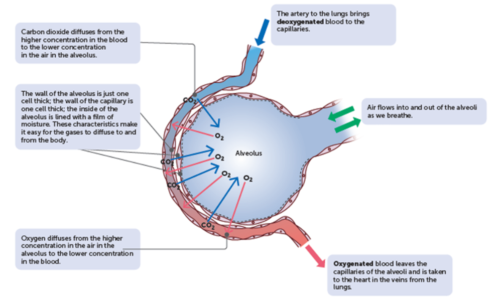
Efficient exchange of gases in the lungs
Breathing:
Involves inhalation and exhalation.
Inhalation: Diaphragm contracts, ribcage expands, air is drawn into the lungs due to low pressure.
Exhalation: Diaphragm relaxes, ribcage contracts, air is pushed out of the lungs due to high pressure.
Blood flow:
Oxygen-poor blood from the body enters the lungs through the pulmonary arteries.
Oxygen-rich blood is carried back to the heart through the pulmonary veins.
Facilitates the exchange of gases between the alveoli and blood vessels.
Structure of the alveoli:
Tiny air sacs in the lungs where gas exchange occurs.
Surrounded by capillaries where oxygen diffuses into the blood and carbon dioxide diffuses out.
Large surface area and thin walls optimize gas exchange efficiency.
Why cells are so small
Higher surface area to volume ratio
Efficient exchange of nutrients and waste
Faster diffusion of molecules
Optimal cellular functions
Easier to maintain homeostasis
Diseases
Emphysema:
What causes it? | Long-term exposure to irritating particles in the air taken into the lungs. |
Who is most at risk of having it? | Smokers, people who work in situations where a lot of dust is produced, people in cities with high air pollution. |
Describe what happens to the alveoli and lungs: | The irritating particles cause damage to the alveoli.
|
What two problems result from the damage to the alveoli? | Lungs are constantly inflated, breathing out no longer is passive, requires voluntary effort. |
Can it be cured? | CANNOT be cured once lungs are damaged. |
Lung cancer:
Lung cancer is similar to most other cancers; a mass of cells divides in an uncontrolled way. Lung cancer can be caused by exposure to asbestos fibres and other pollutants. Smoking Tabacco is the greatest risk for lung cancer. Some chemicals seem to initiate cancerous growths and others, tumour growth. A symptom of lung cancer is excessive production of mucus causing 'smokers cough'. The trapped mucus ruptures the alveoli. It can be treated if on a smaller scale.
Causes Triggers Effects | ||
|
|
|
Blood
Components of blood
Plasma: Liquid component of blood, carries nutrients, hormones, and waste products throughout the body.
Erythrocytes (Red Blood Cells): Contain haemoglobin to transport oxygen from the lungs to tissues and carbon dioxide back to the lungs.
Platelets: Assist in blood clotting by forming clots to stop bleeding when a blood vessel is injured.
Leukocytes (White Blood Cells): Part of the immune system, defend the body against infections and foreign invaders by attacking pathogens.
Blood Clotting
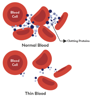
Blood clotting: helps minimise blood loss and prevent infection after an injury that causes damage to blood vessels.
Steps to blood clotting:
Vasoconstriction
Muscles of arteries at the site of damage narrow and tighten immediately to reduce blood flow and loss.
Platelet plug
Platelets within the blood build up as a ‘plug’ at the site of damage, sticking to the rough surface to stop the bleeding. It releases more constricting substances.
These platelets have thrombin receptors on their surfaces that bind serum thrombin molecules which in turn convert soluble fibrinogen in the serum into fibrin at the wound site.
Fibrin forms long strands of tough insoluble protein that are bound to platelets — forms a mesh on top of the platelet plug.
Coagulation
For serious injuries a complex set of reactions results in the formation of an insoluble protein called fibrin, which forms a mesh that traps blood cells, platelets and plasma to for a clot/thrombus and hold the clot into place.
After the clot has formed
Clot retraction
the network of fibrin contracts, becoming denser and stronger, this pulls the edges of the damaged blood vessels together.
Serum
a clear substance (serum) is released, dries and forms a scab that prevents micro-organisms from entering.
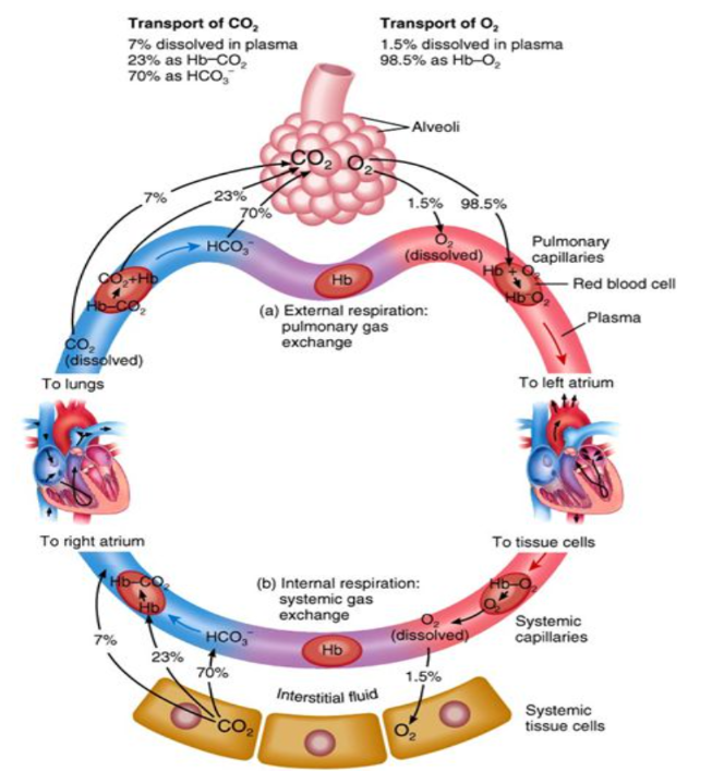
Blood as a transport system
Transport of nutrients and wastes:
100% nutrients and wastes (except CO2) - dissolved and transported in plasma.
Nutrients - essential elements and molecules obtained from food we eat.
Metabolic wastes - substance produced by cells that cannot be used. If built up it would cause harm.
Transport of oxygen:
97% oxygen transported by erythrocytes as haemoglobin.
Erythrocytes are well suited to their function:
Contains haemoglobin, able to combine with oxygen.
Have no nucleus = more room for haemoglobin molecules.
Bi-concave discs = centre increases surface area and volume.
Doesn’t have other organelles to use up oxygen itself.
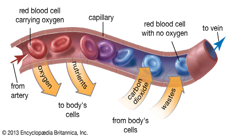
Steps:
Oxygen diffuses from high concentration in alveoli into surrounding capillaries (pulmonary capillaries) and into erythrocytes. Some dissolves in plasma.
→ oxygen in alveoli diffuse into capillaries, erythrocytes and plasma.
Oxygen and haemoglobin combine, creating oxyhaemoglobin inside the erythrocytes to make blood oxygenated and bright.
→ oxygen + haemoglobin = oxyhaemoglobin + bright red.
Oxygenated blood travels to heart to be pumped throughout whole body.
→ oxygenated blood to heart and pumped everywhere.
Oxyhaemoglobin breaks down to release oxygen around body cells.
→ oxyhaemoglobin breaks down = oxygen around cells.
This oxygen diffuses from blood into tissue fluid and into cells to be used for cellular respiration.
→ from blood, oxygen diffuses into tissue fluid and cell walls for cellular respiration.
The blood is now deoxygenated and dark red (haemoglobin without oxygen), and travels back to lungs for more oxygen to repeat the process.
→ blood is deoxygenated and travels to lungs for oxygen. Repeat process.
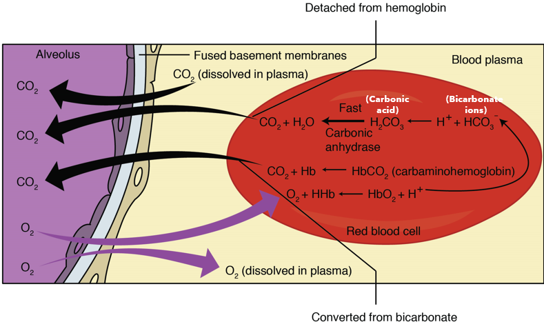
Transport of carbon dioxide:
Carbon dioxide dissolves in plasma (7-8%).
In erythrocytes as carbaminohaemoglobin (22%).
In plasma as bicarbonate ions (70%).
Steps:
Carbon dioxide produced as waste during cellular respiration.
→ cellular respiration creates carbon dioxide.
Carbon dioxide diffuses from high concentration in cells to low concentration in plasma.
→ Carbon dioxide from cells to plasma (descending concentration.
Some dissolves, some enters erythrocytes and combines with haemoglobin, most reacts with water in plasma, forms carbonic acid. The carbonic acid then ionises into hydrogen and bicarbonate ions.
→ Some carbon diffuses in the previous step, some enter erythrocytes and combines with haemoglobin or react with water in plasma = carbonic acid ~ ionises into hydrogen + bicarbonate ions.
Carbon dioxide travels back to capillaries surrounding alveoli with 3 methods.
→ 3 ways for CO2 to go to capillaries on alveoli.
Carbaminohaemoglobin breaks down, releasing carbon dioxide into plasma, hydrogen ions and bicarbonate ions reform into carbonic acid which is broken down into water and carbon dioxide in plasma.
→ carbaminohaemoglobin breaks down, releases CO2 into plasma. Hydrogen + bicarbonate ions reform into carbonic acid - breaks down into H2O and CO2 in plasma.
Carbon dioxide diffuses from high concentration in plasma to low concentration in the alveoli to be breathed out.
→ Carbon dioxide diffuses from plasma to alveoli (descending concentration).
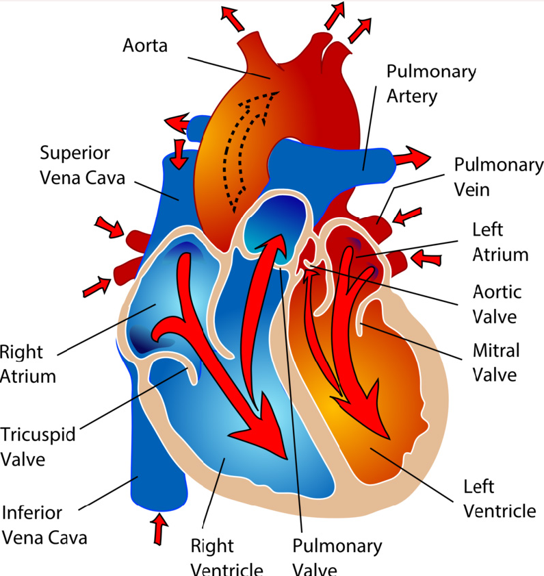
The heart structures
Visual 3D heart
Do it yourself:
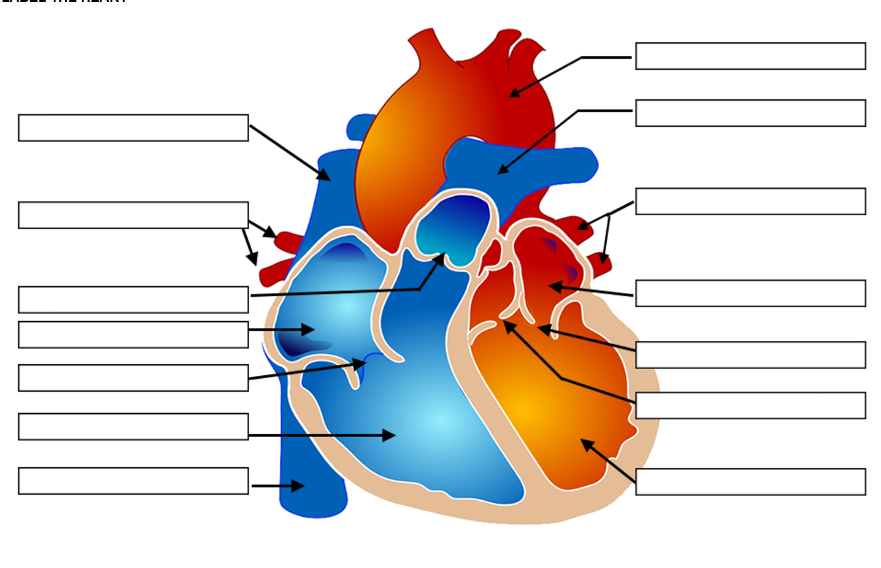
Blood vessels
Aorta - takes oxygenated blood from left side of the heart to the body.
Superior vena cava - blood from upper body to heart.
Inferior vena cava - blood from lower body to heart.
Pulmonary vein - oxygenated blood from lungs to left side of heart.
Pulmonary artery - takes deoxygenated blood from the right of the heart to the lungs
Blood Vessels Direction of Flow Pressure of Blood Flow Thickness of Walls Elasticity of Walls Size of Lumen Valves | ||||||
Arteries | Away from heart | High to pump blood to whole body | Thick for high pressure | Elastic Contains smooth and elastic fibres | Small, narrow lumens | No valves |
Veins | Toward heart | Low pressure | Thin due to low pressure | Less elastic | Large due to lack of muscles | Yes to prevent blood from flowing backwards due to low pressure |
Capillaries | Both directions The connection between arteries and veins | Low | Single cell layer | Not elastic | Very small | No valves |
The cardiac cycle and cardiac output
Diastole - blood fills the ventricles.
Diastole Process: Diastole is the phase of the cardiac cycle when the heart muscle relaxes and the chambers fill with blood. It involves the atria and ventricles expanding to allow blood to flow in. This phase is crucial for the heart to receive oxygenated blood and prepare for the next contraction (systole).
Systole - blood pumps out of ventricles.
Systole is the phase of the cardiac cycle when the heart muscle contracts, pumping blood out of the chambers. It involves the contraction of the atria and ventricles to push blood through the circulatory system.
Events | Description |
Atrial and ventricular diastole | Atrium fills with blood and ventricles also receive blood as the valves between them are open |
Atrial systole and ventricular diastole | Atria contract, pushing blood into the ventricles |
Atrial diastole and ventricular systole | After the atria relax, the ventricles contract, pushing blood out of the heart. |
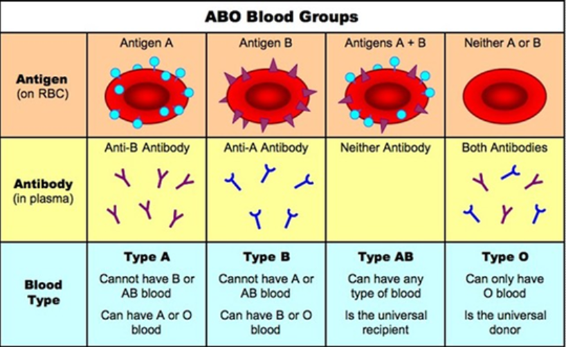
Blood groups and transfusions
Blood group of individual | Antigen/s on surface of RBC of individual | Antibodies in blood plasma Of individual |
A | A | B |
B | B | A |
AB | AB | None |
O | None | A+B |
Antigen present on surface of RBC? | Makes the blood be called? |
Yes | + |
No | - |
Rh antigen on RBC? | Antibodies produced | Therefore can receive blood from |
Yes/positive | None | Both positive and negative |
No/negative | Anti-Rh | Negative only |
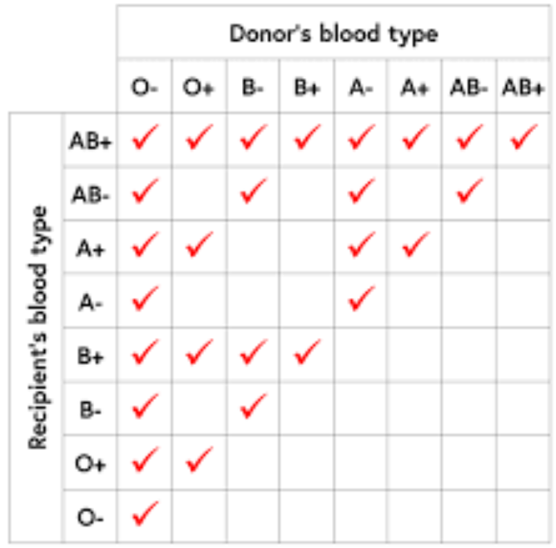
Transfusions:
A transfusion is given to a person suffering excessive blood loss.
If the blood donated is not compatible with the receiver, their blood will agglutinate/clump.
The most widely used type of transfusion is the Red Blood Cell transfusion. Spinning blood at a very high speed in a centrifuge. Components of blood separate and different cell components can be taken.
Examples:
Donor is blood type A, recipient is blood type B - blood will agglutinate.
Donor is blood type A, recipient is blood type O - blood will agglutinate.
Donor is blood type O, recipient is blood type AB - blood transfusion will be successful.
Lymphatic system
The lymphatic system functions to return tissue fluid to the circulatory system and assist in protecting the body from disease.
The lymphatic system consists of:
Capillaries joined to larger lymph vessels (also called lymphatic vessels or lymphatics).
Lymph nodes, which are located along the length of some lymph vessels
The image above displays the lymphatic capillaries, vessels and nodes from the circulatory capillaries to the heart.
Fluid tends to leak out of the arterial end of a blood capillary due to high pressure in the vessel.
Some of this fluid returns to the capillary at the venous end. The excess fluid in the tissues is returned to the blood by the lymphatic system. The fluid returned this way is called lymph.
The lymph vessels originate as blind-ended tubes in the spaces between the cells of most tissues. These are lymphatic capillaries.
→ They are very permeable; proteins and disease causing organisms in the intercellular fluid can easily pass through the walls of the lymph capillaries.
The network of lymph vessels joins to form two lymphatic ducts that empty the lymph into large veins in the upper chest.
Lymph is moved through the lymphatic vessels as a result of smooth muscle, skeletal muscle and valves.
→ The smooth muscle layer of the vessels contract to push the lymph along the vessel.
→ The skeletal muscle muscles surrounding the vessels are also able to contract, providing additional force.
→ There is no pump or force driving the direction of flow therefore the larger lymph vessels have valves that closes when the pressure drops, preventing backflow of lymph.
The image above displays the lymphatic nodes.
Lymph nodes are most numerous in the neck, armpits, groin, and around the alimentary canal.
Lymph enters through vessels on the convex side of the node.
The lymph passes through multiple nodes before entering the circulatory system.
As it passes the nodes, large particles such as bacteria are tapped by fibre network.
The macrophages engulf them.
Filtered lymph now leaves through the lymph vessel on the other side with less disease causing microorganisms.
Nodes are bean-shaped and range from 1mm to 25mm. Each are surrounded by a capsule of connective tissue containing lymphocytes, macrophages, and plasma cells.
→ Macrophages engulf the particles by phagocytosis to destroy them with enzymes.
→ Lymphocytes are white blood cells produced in greater number when infected - this causes swelling and increase in the body’s production of antibodies/macrophage action.
Spaces between the cells of the lymphoid tissue are criss-crossed by a network of fibres.
Alveoli are tiny air sacs in the lungs crucial for gas exchange
Thin walls allow efficient exchange of oxygen and carbon dioxide
Emphysema damages alveoli, reducing elasticity and surface area
Impaired breathing efficiency and decreased oxygen intake occur
Symptoms include shortness of breath, coughing, and wheezing
Limits physical activities and daily tasks
Progressive nature leads to deteriorating lung function
Early detection and management are crucial for emphysema
Healthy Alveoli:
The walls of healthy alveoli are elastic, enabling them to stretch and recoil during inhalation and exhalation, ensuring optimal functioning.
Alveoli with Emphysema:
The walls of the alveoli become damaged and lose their elasticity, leading to a reduction in the surface area available for gas exchange.
Individual may experience difficulty breathing and reduced oxygen intake. Emphysema is often associated with long-term smoking or exposure to harmful chemicals, highlighting the importance of maintaining healthy lungs through lifestyle choices and environmental awareness.
Healthy
Elastic
Stretch and recoil during inhalation and exhalation
Damaged
Lose elasticity
Reduced surface area available for gas exchange
difficulty breathing - reduced oxygen intake
Components of Blood/Circulatory System:
Erythrocytes - rbc - carry oxygen in their biconcave organelle-less disc
Leucocytes/lymphocytes - wbc - locate and attack foreign cells to protect your body of diseases
Thrombocytes - clotting - bind together when blood vessels are damaged - form blood clots to stop excessive damage
Plasma - liquid component of blood
How They Work:
The heart, a muscular organ, functions as the central pump of the circulatory system, propelling blood through arteries to reach various tissues and organs.
Capillaries, the smallest blood vessels, facilitate the exchange of nutrients, oxygen, and waste products between the blood and tissues.
Veins play a vital role in returning blood from the body's tissues back to the heart, completing the circulatory loop necessary for maintaining proper oxygen and nutrient distribution.
Arteries are blood vessels that carry oxygenated blood away from the heart.
random summarised stuff idk…:
DEOXYGENATED blood from superior/inferior vena cava → right atrium tricuspid valve → right ventricle pulmonary artery → lungs for gas exchange in the alveoli
OXYGENATED blood pulmonary veins → left atrium mitral valve → left ventricle aortic valve → blood leaves aorta to whole body
Diastole heart muscle relaxes and allows the chambers to fill with blood
Systole heart muscle contracts and pumps blood out of the chambers. It is followed by diastole.
lungs
air nasal cavity → pharynx → larynx → trachea → bronchi → bronchioles → terminal bronchioles.
diaphragm contracts → less volume → more pressure → intercostal muscles retract/relax → exhale
diaphragm relaxes → more volume → less pressure → intercostal muscles expand → inhale
air in → alveoli → o2 diffuses into blood stream via haemoglobin, co2 diffuses across alveolar membrane from capillaries in alveoli (when exhaled co2 is expelled)
Digestive and excretory system
For extra understanding visit Khan Academy
Digestion
Types of digestion:
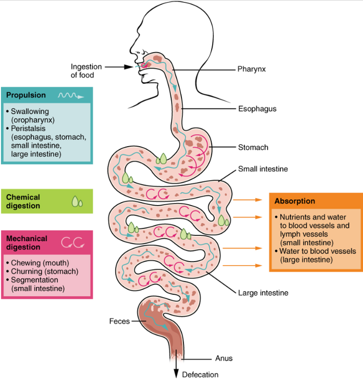
Mechanical digestion
The physical breakdown of food particles into smaller pieces to increase surface area.
Allows for more effective chemical digestion.
Mouth: teeth/cut, grind, tear.
Small intestine: gall bladder releases bile into small intestine. Bile salts emulsify the fat into smaller droplets.
Chemical digestion
The chemical break down of large, complex molecules into smaller, simpler molecules - small enough to be absorbed by into the blood stream.
Enzymes are used - biological catalysts.
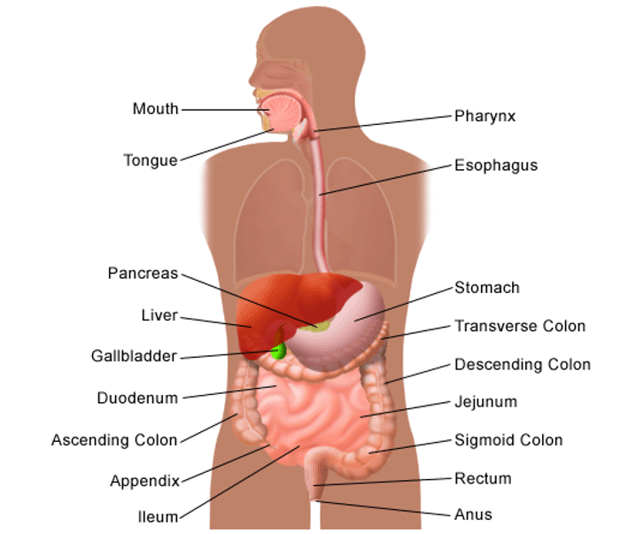
The alimentary canal
The alimentary canal is one continuous tube from the mouth to the anus that food travels through.
Accessory organs help with digestion but food doesn’t enter them.
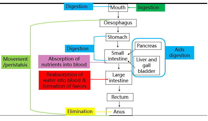
Digestion:
Mouth:
Mastication/chewing by teeth.
Salivary amylase starts carbohydrate digestion (starches).
Stomach:
Churning of stomach muscles.
Gastric protease/pepsin starts protein digestion.
Small intestine:
Segmentation by intestinal walls. Emulsification of fats by bile salts.
Pancreatic and intestinal amylase, protease, and lipase complete carbohydrate, protein and fat digestion into nutrients.
Chemical digestion specifically in the small intestine
Pancreatic amylase turns starch into disaccharides.
Intestinal amylases turns disaccharides into monosaccharides to be absorbed into the blood. Example: sucrase breaks sucrose into glucose.
Pancreatic protease/trypsin turns proteins into peptides.
Intestinal protease/peptidase turns peptides into amino acids to be absorbed into blood.
Pancreatic lipase turns lipids/fats into fatty acid and glycerol for absorption into blood.
Pancreatic ribonuclease and deoxyribonuclease digests DNA and RNA.
Peristalsis
The alimentary canal is lined with circular and longitudinal muscles.
The food moves along the canal via a wave of circular contractions (peristalsis) assisted by mucus (lubricates the lining).
Order of movement by peristalsis:
Bolus in mouth swallowed down to oesophagus to stomach.
Stomach chyme pushed through pyloric sphincter into small intestine.
Sphincters are thicker circular muscles that is constricted between peristalsis which prevents movement from one organ to another too early.
Chyme moves along the small intestine in the order of duodenum, jejunum, to ilium and nutrients are absorbed.
Undigested food material enters the large intestine and moves slowly along it and into the rectum to be stored.
The faeces exit the rectum through the anus.
See diagram:
Absorption of nutrients in the small intestine (150-151):
Nutrients in the small intestine are now small enough to move through the walls into the blood (called absorption).
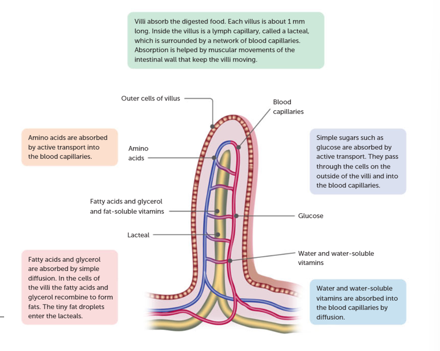
The villi:
Absorption of water in the large intestine and elimination of faeces at the rectum (152):
Carbohydrates:
The enzyme, amylase from the salivary glands mixes with our food.
The enzyme begins to work on breaking starch molecules into smaller pieces.
When the acid concentration in the stomach rises, the amylase stops working.
Absorption: cells lining small intestine have transport proteins, grabbing sugars into cells.
Nucleic acids:
Pancreatic juices released into the small intestine contains enzymes called nucleases.
These enzymes break nucleic acid chains into individual building blocks.
Absorption: transport proteins pick up pieces into cells.
Proteins:
Glands in stomach release and enzyme called pepsin.
Pepsin clips proteins near amino acids, tyrosine and phenylalanine, cutting protein into smaller pieces.
Absorption: transport proteins take amino acids (proteins), individually/in clusters (2-3).
Fats:
Do not dissolve on water; forms floating droplets on top.
Bile salts made by liver is delivered to small intestine, acts like detergent and breaks fat into smaller pieces.
Absorption: micelles brush against cells lining intestine, fatty acids and monoglycerides diffuse into their membranes.
Flow chart of carbs, proteins, lipids:
Carbohydrate --> glucose/fructose (monosaccharides – they're small enough to go through the cell membrane) -> oral cavity -> salivary amylase -> pancreatic amylase in small intestine (duodenum)
Protein --> peptides into amino acids -> stomach + pepsin (protease) -> trypsin in small intestine (duodenum) (pancreatic protease)
Lipid -> fatty acids and glycerol -> bile salts break down -> pancreatic lipase in small intestine
The digestive system in relation to the other systems:
Cell Membrane: The digestive system connects to the cell membrane by absorbing nutrients through transport proteins, facilitating the transfer of molecules between the digestive tract and cells.
Cellular Respiration: It provides glucose and molecules from food breakdown to produce ATP, supporting the energy needs of cellular respiration and ensuring metabolic processes are fuelled efficiently.
Circulatory System: Nutrients absorbed from the digestive tract are transported via the bloodstream to cells throughout the body, ensuring a continuous supply of essential substances for cellular functions.
Respiratory System: By supplying oxygen for cellular respiration and removing carbon dioxide, the respiratory system supports the digestive system in maintaining optimal conditions for nutrient breakdown and energy production.
Lymphatic System: The absorption of fats and fat-soluble vitamins into lymphatic vessels in the small intestine connects the digestive system to the lymphatic system, enabling the transport and processing of these essential nutrients throughout the body.
Peristalsis moves substances along the alimentary canal
Organs and their ezymes:
Mouth - salivary amylase in salivary glands digests carbs/starch (chemical digestion).
Stomach - pepsin digests proteins (chemical digestion).
Small intestine, duodenum - pancreatic lipase digests lipids (chemical digestion).
Absorption of nutrients in small intestine
Main components of the villi:
Lacteal
fatty acids, glycerol and fat soluble vitamins
Amino acids
Outer cells of villus
Blood capillaries
Glucose
Water, water soluble vitamins
The villi:
Back to this diagram…(summarised:
Pink box:
Fatty acids + glycerol absorbed by simple diffusion.
Fatty acids + glycerol recombine = form fats.
Tiny fat droplets enter lacteals.
Peach box:
Amino acids absorbed by active transport into blood capillaries.
Green box:
Undigested food absorbed.
On the villus there is a lymph capillary called lacteal which is surrounded by a network of blood capillaries.
Absorption is helped by muscular movements of the intestinal wall keeping the villi moving.
Purple box:
Glucose (simple sugars) absorbed by active transport.
They pass through outside cells of villi into blood capillaries.
Blue box:
Water and vitamins absorbed into blood capillaries by diffusion.
Absorption of nutrients:
Nutrient | Absorbed into | By method |
Simple sugars/glucose | Blood capillaries | Active transport
(low concentration to high concentration across carrier protein) |
Amino acids | Blood capillaries | Active transport |
Fatty acids and glycerol | Lacteal | Simple diffusion |
Water and water soluble vitamins | Blood capillaries | Simple diffusion – osmosis |
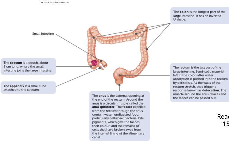
The large intestine
Organs:
Large intestine |
Faeces are created Bacterial creates vitamins |
Rectum | Last part of the large intestine When stretched from faeces deposited will trigger defection |
Anus | The anus is the external opening at the end of the rectum |
Appendix | Small tube attached to the caecum stores bacteria |
Movement of material through large intestine, rectum, and anus:
Material enters the caecum pouch.
It travels up ascending colon.
Then through the transverse colon.
Then down the descending colon.
Key terms:
Elimination - removal of indigestible material, bacteria and bile bile pigments from the body.
Faeces - material passed out of the rectum.
Digestive disorders:
Diarrhoea:
Frequent defecation of watery faeces.
Caused by irritation of small/large intestine which increases peristalsis (contents move through before water can be absorbed adequately).
May be the result of:
Bacteria
Viruses
Parasites
Cancer (bowel)
Coeliac disease
Lactose intolerance
Constipation:
Occurs when large intestine movements reduced and contents are stationary for a long time.
As water is absorbed, the faeces become drier and harder.
Defecation becomes difficult (possibly painful).
May be caused by:
Lack of roughage in diet (cellulose/fibrous material).
Humans have no enzymes to digest cellulose, but still important to stimulate movements of alimentary canal.
Lack of exercise
Emotional issues
Bowel cancer:
Also known as colorectal cancer.
An uncontrolled growth of cells in wall of large intestine.
May be linked to:
Diet
High alcohol consumption
Smoking
May increase risk:
Diet high in red/processed meat
Diet low in fibre (fruit/vegetables)
Overweightness/obesity
Physical inactivity
Coeliac disease:
Unable to tolerate protein called gluten (gluten free people).
If gluten is consumed, their immune system responds by damaging/destroying the villi in small intestine.
Without healthy villi, nutrients cannot be absorbed and person becomes malnourished.
Symptoms vary from person to person.
Some people may not have symptoms but are still endangered to malnourishment.
Coeliac disease is difficult to diagnose.
It is inherited, no cure. The only solution is to follow a gluten free diet.
Recap point
Organ | Role in waste removal |
Lungs | Removal of carbon dioxide from respiration |
Skin – sweat glands | Removal of mostly water via sweat and other wastes such as urea & salts |
Liver | Processing of substances for excretion |
Kidneys | Principal excretory organ, mainly urea, uric acid and creatine. |
Alimentary canal | Passes out bile pigments after breakdown of haemoglobin products |
Excretory system
The excretory system regulations the chemical composition of body fluids by removing metabolic wastes and retaining the proper amount of water, salts, and nutrients. The organs included in this are:
Kidneys
Liver
Lungs
Skin functioning at organ level
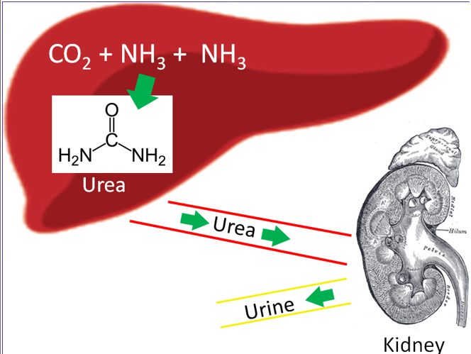
Deamination of amino acids in the liver produces urea → transported to kidneys for removal.
The liver
The liver is located in the upper abdominal cavity.
Anything with the mentioning of “hepatic” refers to the liver.
Its function:
All blood from stomach and small intestine passes through the liver to be processed and metabolised.
**metabolised means to be absorbed and used by the body
Memorise diagrams and images by:
Look, cover, speak/write, check method.
Draw them
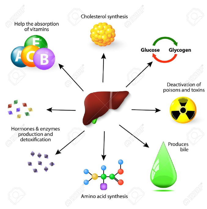

The liver plays a major role in processing materials for excretion.
Excretion is the removal of metabolic wastes (by-products of cellular chemical processes).
Deamination helps process excess amino acids from protein digestion to later be excreted from the kidneys.
Why deamination needs to take place
Proteins we eat are digested by protease enzymes into amino acids (proteins + protease → amino acids).
Amino acids are transported by the hepatic portal vein to liver.
Other proteins in body (such as worn out RBCs) are also broken down into amino acids and taken to liver.
Most amino acids are used to make new proteins (basically proteins are broken down to amino acids to be made into new proteins).
SOMETIMES proteins used as back up energy source. This causes toxic ammonia which needs to be converted to urea for excretion.
Deamination process in liver (169)
Amino acids are made of an amine group and carbon skeleton.
In deamination, the amine group (NH2) is removed from the amino acid, assisted by enzymes.
The amine group (NH2) is converted to ammonia and the carbon group is converted to carbohydrates.
The ammonia (NH2, now NH3) is soluble and highly toxic = needs to be rapidly converted to urea (CH4N2O), which is transported to kidney to be excreted (some in sweat).
Carbohydrates are used in cellular respiration for energy.
The whole process in equations:
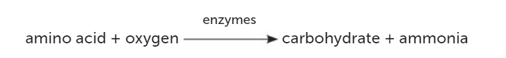
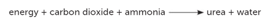
Image:
Recap point:
“Explain the process of deamination and its role in excretion.” - 8-10 points
Most amino acids from digestion of proteins are used to make new proteins in the body.
Sometimes excess amino acids are needed as a back-up energy source.
Amino acids are made of an amine group and a carbon group.
Deamination occurs in the liver to process excess amino acids.
In deamination the amino (NH2) is removed from the amino acid – with the aid of enzymes.
The amino group is converted to ammonia – and the carbon group is converted to carbohydrate.
The ammonia is soluble and highly toxic and needs to rapidly converted to urea (CH4N2O).
Which is transported to the kidney to be excreted (and some in sweat)
The carbohydrates are used in cellular respiration for energy.
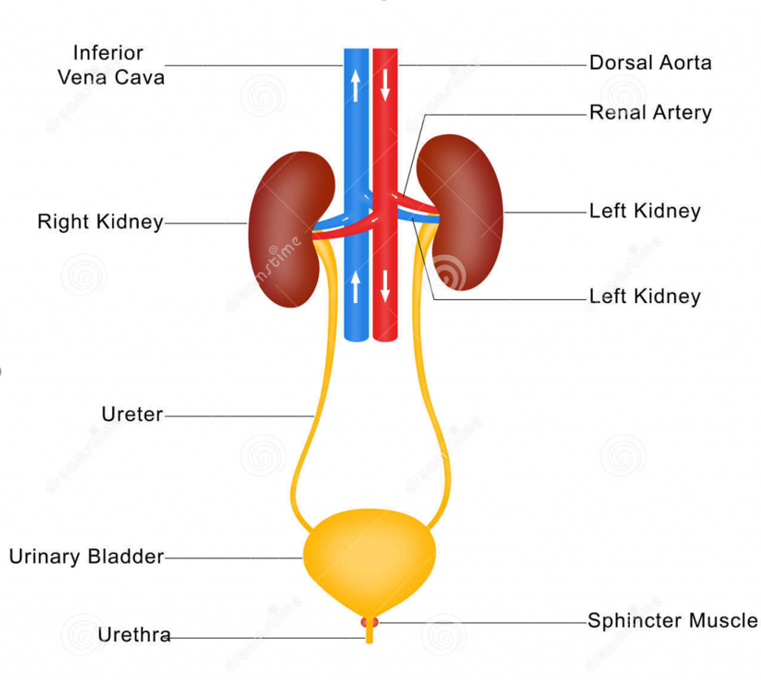
The urinary/renal system
The nephrons in the kidney facilitate three basic processes:
Filtration
Reabsorption
Secretion
Structure Function | |
Renal vein | Takes blood away from kidneys |
Renal artery | Takes blood towards kidneys |
Kidney | Produces urine |
Ureter | Tube that drains urine away from kidney to bladder |
Bladder | Muscular bag which stores urine until passed out body |
Urethra | Tube that allows urine to pass out body |
The kidney:
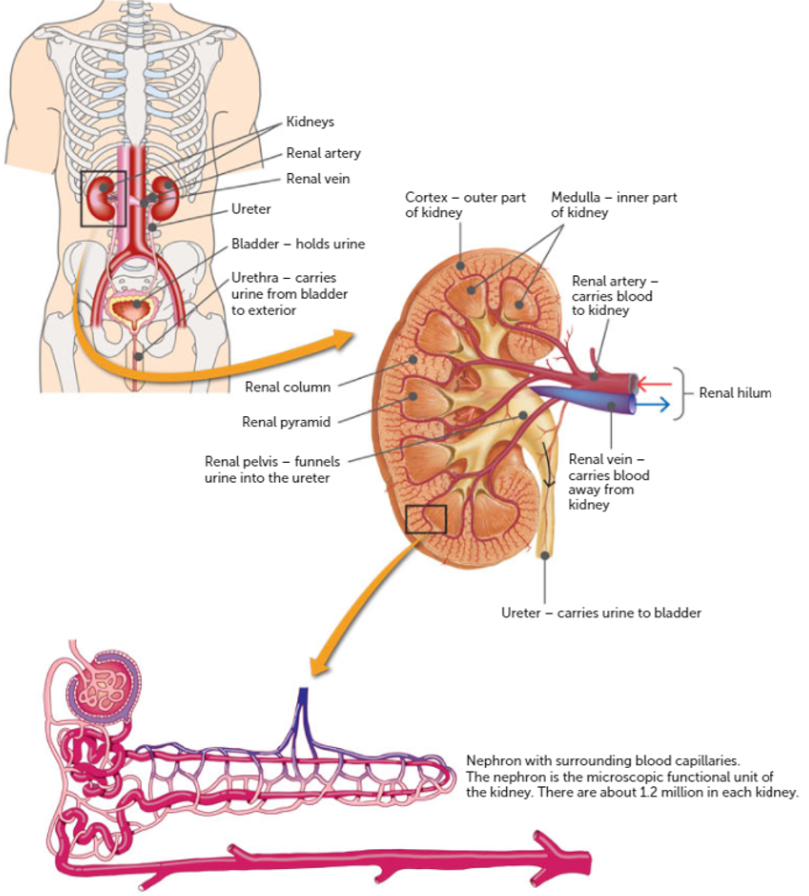
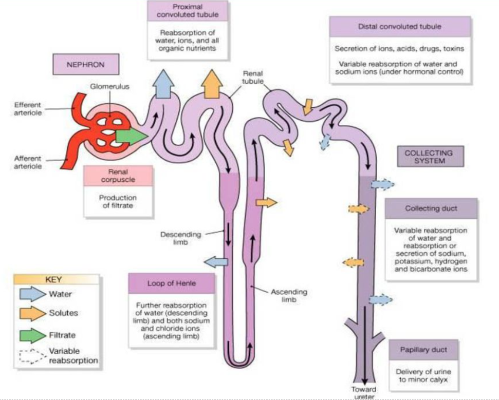
The nephron:
Formation of urine
Visit this site!
Glomerular filtration
occurs at renal corpuscle.
Blood is filtered by the glomerular capillary network due to high pressure of blood entering through the wide afferent arteriole.
~20% of fluid is forced out of blood through the glomerular capillary walls and into the glomerular capsule.
This fluid is known as “filtrate” and includes:
Water
Glucose, amino acids, fatty acids
Salts (sodium ions, potassium ions), ions, hormones
Urea, uric acid, creatine, toxins
Filtrate enters proximal tubules of nephron
Other 80% blood, exits via thinner efferent arteriole.
Reabsorption
99% filtrate reabsorbed.
Useful substances are removed from filtrate through tubular walls, reabsorbed back into peritubular capillaries/blood.
The proximal con. tube, loop of Henle, and distal con. tube are long and folded (+SA).
Movement across the membrane can be passive/active.
Substances move away from
Substances
Active or passive
Proximal con. tubule and loop of Henle
Ions (potassium, chloride and bicarbonate) and water
Passive
Proximal convoluted tubule and loop of Henle
Sodium ions and glucose
Active
Distal convoluted tubule and collecting duct
Water and sodium under hormonal control
Active
Tubular secretion
Maintains pH of blood and urine; removes excess hydrogen and ammonium; keeps blood neutral, and urine slightly acidic.
Substances are moved from the peritubular capillaries into the filtrate in the nephron tubules to be excreted in the urine.
Can be active/passive.
Can be secreted back into filtrate:
Excess hydrogen and ammonium ions
Potassium ions
Creatine and drugs
Some urea and uric acid
Excretion
Remaining filtrate drains from collecting ducts to renal pelvis.
Now called urine.
Will move via muscular contraction through ureter, stored in bladder.
When bladder full, contracts, push urine out body through urethra.
Components of urine:
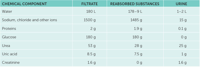
Recap point
The 3 steps to urine formation?
Filtration: Blood pressure forces fluid and solutes out of the glomerulus into the Bowman's capsule.
Reabsorption: Useful substances are reabsorbed back into the blood.
Secretion: Additional waste products are actively transported into the urine.
What is reabsorbed?
Water
Glucose
Amino acids
Blood in urine?
If red blood cells were in urine, it could indicate damage to the kidneys or urinary tract, such as the glomeruli or tubules in the kidneys.
Tubular secretion purpose?
Tubular secretion in the nephron is the process of moving substances from the blood into the renal tubules to regulate pH balance, eliminate waste, and control electrolyte levels.
Disorders of the excretory system
KIDNEY STONES
Causes:
Drinking too little water
The oesophagus is a muscular tube connecting the throat to the stomach.
It moves food by peristalsis.
It has mucous membrane lining for protection.
No digestion occurs in the oesophagus.
It has upper and lower oesophageal sphincters.
A) Gastric juice: Contains hydrochloric acid and enzymes to break down proteins.
B) Bile: Aids in fat digestion and absorption by emulsifying fats.
C) Pancreatic juice: Contains enzymes to digest carbohydrates, proteins, and fats.
D) Intestinal juice: Helps with further digestion and absorption of nutrients.
Filtration: Nephrons filter blood to remove waste products and excess substances.
Reabsorption: Essential substances like water and ions are reabsorbed back into the blood.
Secretion: Additional waste products are actively secreted into the urine.
Musculoskeletal system
The skeletal system is made up of bones and tendons + ligaments + cartilage.
It provides a scaffold to support the weight of the body.
It facilitates movement by being points of attachment for muscles.
It protects vital organs.
It produces red blood cells.
It stores and releases minerals and fats.
Bones:
Collagen-rich fibres
Minerals
Hard and strong
Bone consists of osteocytes in a matrix of calcium. There is compact and spongy bone.
Cartilage is thin, lacks blood vessels and is resistant to compression.
Bone marrow: in the very centre of bones – it is a soft, flexible tissue that produces blood cells and stores fat.
Spongy bone: is a matrix of intercrossing, connecting bars of bone. The spaces are filled with bone marrow and blood vessels. The matrix contains minerals, including calcium, phosphate and collagen. These make the bone hard and strong and prevents them from being brittle.
Compact bone: consists of a solid matrix, making it dense, rigid and strong. It’s made up of the same calcified matrix as the spongy bone, but arranged in layers.
Cartilage:
Collagen-rich fibres
No minerals
Soft and flexible
Cartilage consists of a collagen-rich matrix but does not contain the minerals that make bones hard and strong, nor nerves or blood vessels. Instead, the cells found in cartilage rely on diffusion for any nutrients and oxygen, from blood vessels in close proximity.
Ligaments:
Fibrous connective tissue
Connect bones to other bones
Found at joints
There are two subgroups that make up the skeleton:
The axial skeleton:
Centre axis
Support for posture
Protection of organs
The appendicular skeleton:
Upper and lower limbs
Shoulder and hip girdle
Movement
a = axial, b = appendicular
Type of bone | Description | Examples |
Long | Longer than wide & growth plates. For strength, structure and stability Lots of movement | Femur |
Short | Phalanges | Carpals and tarsals |
Flat bones | Plates/thin Protect vital organs & provide muscle attachment | Sternum |
Irregular | Hip girdle | Vertebrae, sacrum |
Sesamoid | Seed like bones embedded in a tendon | Patella |
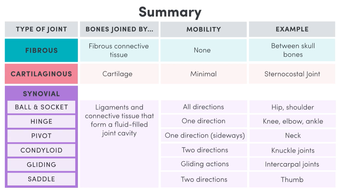
Joints
There are 3 types of joints; fibrous, cartilaginous and synovial. The movement of joints can be classified as immobile/fixed, slightly moveable or freely moveable.
Fibrous
Strong, flexible fibrous tissue.
No mobility.
E.g. skull.
Cartilaginous
Flexible, connective.
Slightly moveable; possible, but minimal - not as strong and rigid as fibrous tissue.
E.g. first sternocostal joint. The end of the first rib is cartilage and attaches straight onto the sternum.
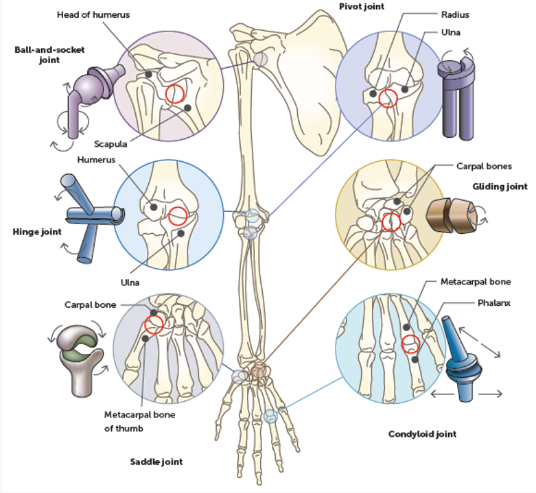
Synovial
Bones don’t touch - ligaments and tendons form a joint cavity that is filled with synovial fluid.
Freely moveable - synovial joints can move in more than one direction.
E.g. ball and socket joint.
There is a socket in which a ball shaped end sits. This is the most mobile type of synovial joint. Specific examples include the hip, shoulder…
E.g. hinge joint.
Has bone, and hinge point. Specific examples include knee, ankle and elbow.
E.g. pivot point.
Bones pivoting around each other. Specific examples include the atlas and axis bones. The atlas pivots on the axis, which allows us to rotate our neck.
E.g. condyloid joint (condylar/ellipsoid).
Oval-shaped end of bone fitting into an oval-shaped hollow in another bone. Similar to ball ack socket joint but has less mobility. Specific examples include knuckle joints (between metacarpals and finger phalanges).
E.g. gliding joint (planar/plane).
Two bones with flat or s lightly curved surfaces glide on top of each other. Specific examples include the intercarpal.
E.g. saddle joint.
Two bones which resemble a rider sitting on a saddle. Has rounded end in another saddle-like end and hollow. It is more stable than other joints.
Bone cells
Osteocyte:
Bone cells maintaining bone tissue. Found in the lacunae.
Osteoblast:
Forms bone tissue (osteocytes). Found in the lacunae for synthesis.
Osteoclast:
Resorption/destruction of damaged bone. Present in bone diseases/broken bones at sites of destruction/repair.
Practice question: describe structure of the cartilage in the bronchi and trachea:
Hyaline cartilage
C-shaped
Very fine protein fibres
Smooth and fine
One-directional collagen fibres
A synovial joint, the knee (hinge)
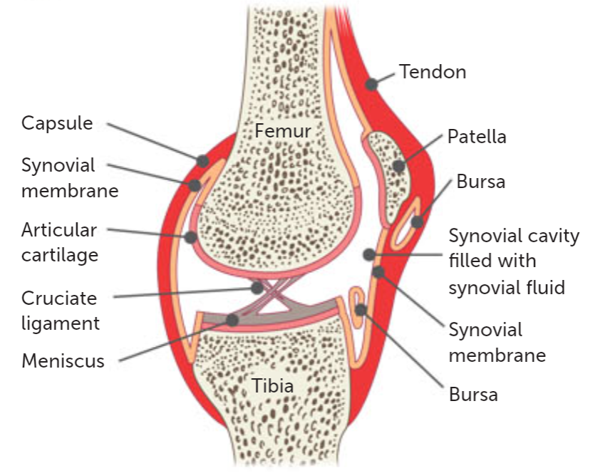
Structure | Description and function |
Fibrous capsule | The fibrous capsule in a synovial joint is a tough outer layer that encloses the joint cavity, providing structural support and stability to the joint. It helps prevent dislocation and contains the synovial fluid, which lubricates the joint and nourishes the cartilage. |
Synovial membrane | The synovial membrane in a synovial joint secretes synovial fluid, which lubricates the joint, reduces friction, and provides nutrients to the cartilage. |
Synovial fluid | Synovial fluid is a viscous, transparent fluid that lubricates and nourishes the joints. It reduces friction between the articular cartilage surfaces during movement and acts as a shock absorber, providing nutrients and removing waste from the avascular cartilage. |
Articular cartilage | The articular cartilage in a synovial joint is a smooth, slippery tissue that covers the ends of bones. Its function is to reduce friction and absorb shock during joint movement. |
Articular discs/menisci | Articular discs/menisci are fibrocartilage structures that improve joint congruence, absorb shock, distribute load, and enhance stability in synovial joints. |
Bursae | Bursae are fluid-filled sacs located near joints, acting as cushions to reduce friction between bones, tendons, and muscles. They help in smooth movement and provide lubrication in synovial joints. |
Ligaments | Ligaments are tough bands of connective tissue that connect bones to other bones in a synovial joint. Ligaments provide stability, support, and limit excessive movement in the joint. |
Genetics+
DNA
DNA - deoxyribonucleic acid, a molecule found in the cells of all organisms.
Contains genetic information that determines the structure of the cell and the way it functions.
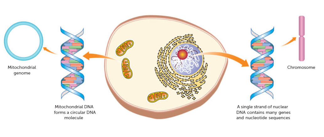
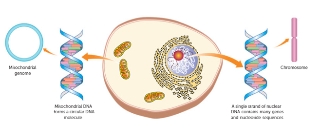
Most DNA molecules are found in the nucleus of each cell, and are therefore called nuclear DNA. There is also a small amount of DNA found in the mitochondria; it is called mitochondrial DNA, or mtDNA, and makes up less than 1% of the total DNA in humans.
It is important for the functioning of the mitochondria → cell.
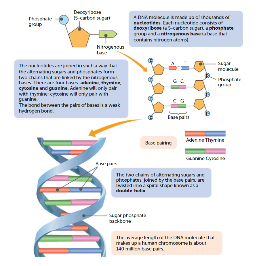
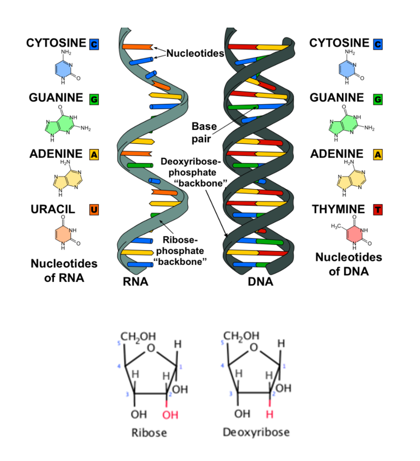
Structure of DNA:
DNA is an example of polymer; a molecule made up of many repeating small units. These repeating units in DNA are called nucleotides - each nucleotide is made up of a sugar molecule, a phosphate group, and a nitrogenous base. There are four nitrogenous bases:
Adenine
Thymine
Cytosine
Guanine
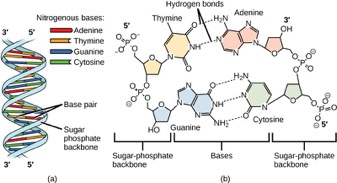
The sugar molecule of one nucleotide bonds of the phosphate group of another one. This forms a long chain of alternating sugars and phosphates, with side chains of bases.
In DNA, two strand join by specific bases being attracted to one another by weak hydrogen bonds.
Adenine + thymine
Cytosine + guanine
The two strands of DNA twist into a spiral shape called a double helix.
The order in which the nitrogenous bases occur in the DNA molecule determines the genetic code. Each gene consists up to 2 million pairs of bases.
The estimated length of the DNA molecules in a human cell is between 2-3 metres, but the width is only 2-millionths of a millimetre. The nuclei of human cells have 46 DNA molecules.
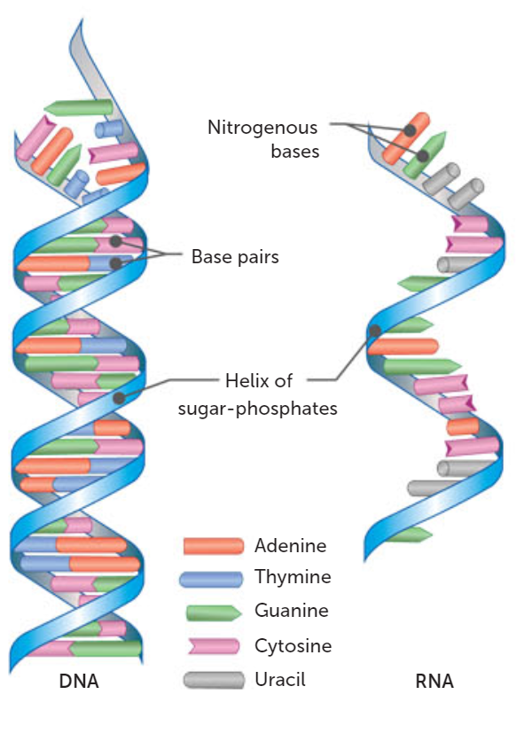
RNA vs DNA
Characteristic DNA RNA | ||
Structure | Double stranded | Single stranded |
Sugar | Deoxyribose sugar | Ribose sugar |
Base pairs | Adenine, thymine, cytosine, guanine | Adenine, uracil, cytosin, guanine |
Synthesises | RNA | Proteins |
Length | DNA longer than RNA | RNA shorter than DNA |
Function | Genetic information ~ cell instructions | Convertis genetic information within DNA to build proteins ~ protein synthesis |
DNA has a double helix structure.
DNA replicates itself.
DNA has deoxyribose sugar.
DNA have the bases: adenine, thymine, cytosine, guanine.
RNA is single stranded.
RNA cannot replicate itself.
RNA has ribose sugar.
RNA have the bases: adenine, uracil, cytosine, guanine.
Chromosomes and mitochondrial DNA
Chromosomes Mitochondrial DNA | |
|
|
 | 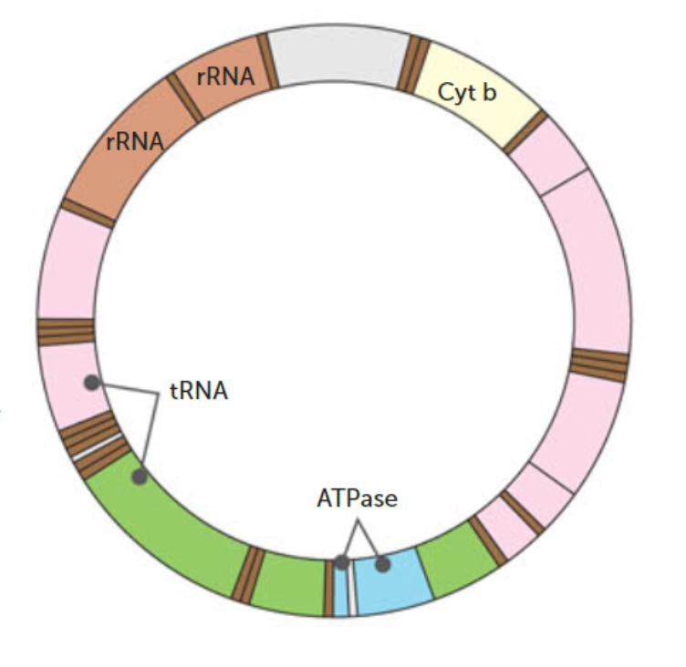 |
DNA determines the structure and function of cells
DNA is bound to proteins in chromosomes in the nucleus. In the mitochondria, DNA is unbound.
DNA stores information fro the production of proteins which determins the structure and function of cells (cell blueprint).
The structural properties of Helical DNA molecule:
Double stranded nucleotide composition of weak bonds involved in base pairing between the complementary strands, allow for replication.
Genetic code
Each gene that determines consists of up to 2 million pairs of bases = variety.
Humans have 46 DNA molecules in the nuclei of all their cells (called chromosomes).
DNA, genes and chromosomes
Protein synthesis
What is protein synthesis:
Synthesis - building ~ synthesising.
Protein synthesis - building proteins.
Involves adding amino acids together at ribosomes.
The instructions for which amino acids to add and in the correct order are found along segments of DNA in the nucleus (genes).
Cell uses enzymes and RNA to help get the instructions to the ribosomes.
Watch this:
Video summary notes:
In a cell there is a nucleus.
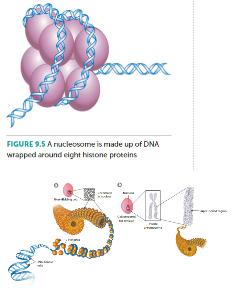
The nucleus contains the genome.
The genome is split between 23 pairs of chromosomes.
Each chromosome contains a long strand of DNA, tightly packaged around proteins called histones.
Sections of the DNA are called genes.
These genes contain instructions for making proteins.
[TRANSCRIPTION]
When a gene is switched on, an enzyme called RNA polymerase attaches to the start of the gene. → The RNA polymerase moves along the DNA, making a strand of mRNA out of free bases in the nucleus. (RNA poly attaches to gene, moves on DNA = strand of mRNA)
The DNA code determines the order in which the free bases are added to the mRNA.
[TRANSLATION]
Before the messenger RNA can be used as a template for protein synthesis, it needs to be processed. The process = removing and adding sections of RNA.
The mRNA then moves out the nucleus, into the cytoplasm. (RNA moves out nucleus)
Protein factories in the cytoplasm; ribosomes, bind to mRNA. The ribosome reads the code in the mRNA to produce a chain made up of amino acids. (Ribo bind to mRNA, reads codes = amino acid chain)
tRNA molecules carry amino acids to ribosome. (tRNA carry amino acids to ribo)
mRNA is read three bases (triplet) at a time.
As each triplet is read, a tRNA delivers the corresponding amino acid which adds onto the growing chain (polypeptide chain) of amino acids. (the amino acid chain/polypeptide chain is made as the triplets are read and amino acids a brought by tRNA)
Once the last amino acid has been added, the polypeptide chain folds into a complex 3d shape = protein. (folds)
Steps to protein synthesis:
Transcription in the nucleus - DNA is copying its code to be taken to out of the nucleus.
Chemical messengers enter the nucleus from the cytosol and bind to DNA. (chem messenger enter, bind to DNA)
DNA helicase breaks weak hydrogen bonds between bases to unzip the two DNA strands for that gene. (DNA helicase breaks bonds between bases, unzips DNA strands)
RNA polymerase moves along DNA template strand + starts synthesising complimentary messenger RNA strand, one base at a time. (RNA polymerase moves along DNA, starts synthesising compl. mRNA strand)
RNA stops copying when it reaches a stop sequence of bases (E.g. UAG). (RNA stop at stop sequence)
m RNA released and moves through nuclear pore to cytoplasm. (mRNA released to cyto)
Translation at the ribosome - code is read to make the protein.
Ribosome in the cytosol attaches to the messenger RNA at start codon (AUG)
Ribosome moves along mRNA 3 codons at a time. (Ribo attaches to mRNA at start codon)
Codon corresponds to a specific amino acid that makes up the protein which is carried by the tRNA with the 3 bases that correspond to the codon/anticodon. (Codons are read, corresponds to a spec. amino acid, carried by tRNA)
The anticodon on the tRNA binds with the complimentary codon on the mRNA, bringing the correct amino acid with it. (anticodon on tRNA binds with compl. codons on mRNA, bringing amino acid)
The next codon on the mRNA is read by the ribosome and its complimentary tRNA carries over the next amino acid which is joined to the previous amino acid in a polypeptide bond. (process repeats = build up of amino acids)
tRNA’s detach and collect another amino acid from the cytosol. (tRNA deatch, process repeated)
This continues until the whole mRNA is read and therefore all amino acid are joined. A protein has now been synthesised which will express the gene that was transcribed. (Until all mRNA is read)
Protein synthesis
Transcription
Translation
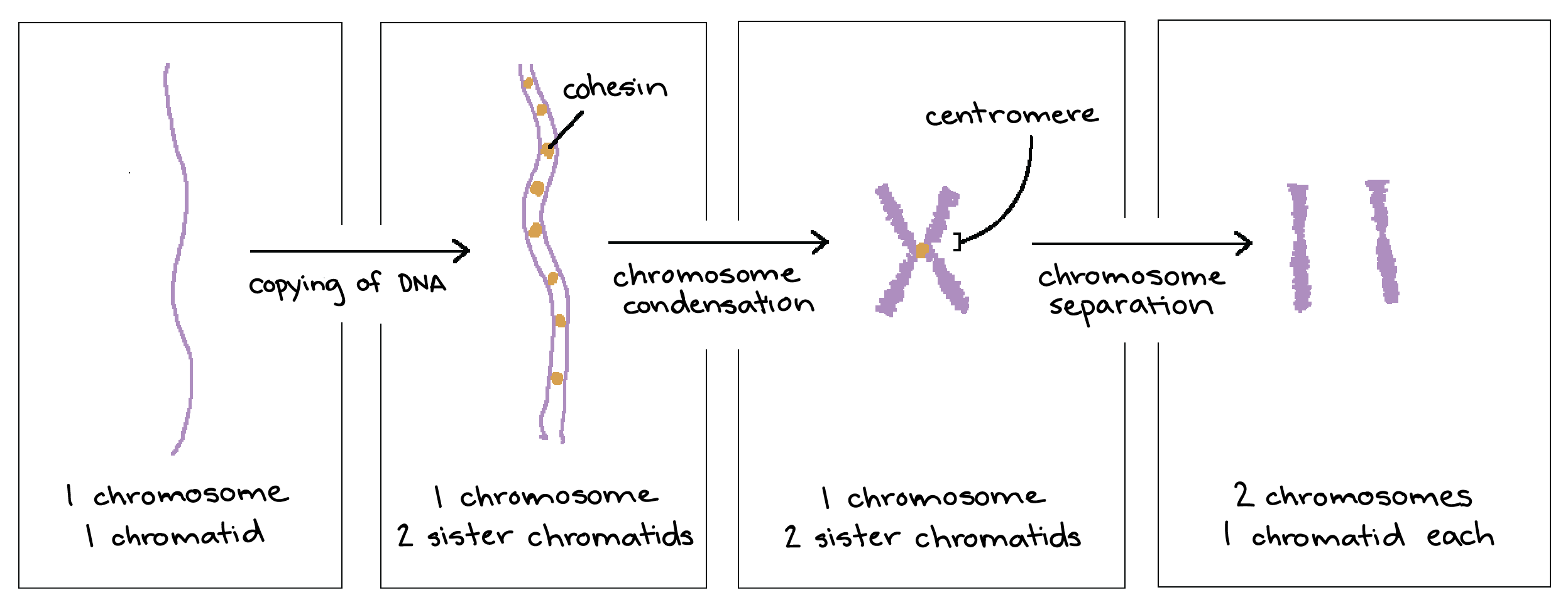
Chromosome, chromatid, chromatin
More in-depth information here!
Chromatin is a long chain of DNA.
Chromosomes is rolled up DNA when it is going through cell division.
Sister chromatids are the branches of the same chromosome.
Chromatin begins to form into chromosomes at the beginning of mitosis/meiosis; metaphase, anaphase. A chromosome can be composed of two chromatids or even one.
Chromosomes and chromatids contain chromatin. Chromatin is of extremely long strands of DNA.
Chromatin contains histone (chromosome histones).
This diagram may help:
Mitosis
I.P.M.A.T.C
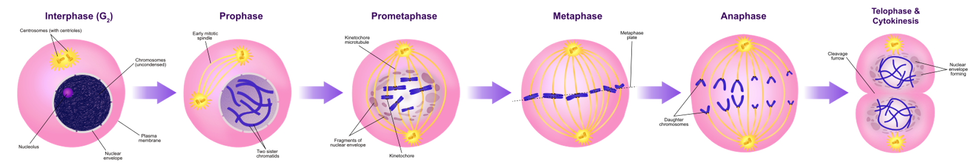
Interphase:
DNA replicates/doubles.
G1, S, G2 stages of cell cycle.
DNA still appears as chromatin/chromosomes not visible yet.
I = identical.
Prophase:
Chromosomes become visible, consisting of 2 sister/identical chromatids joined at a centromere.
Nuclear membrane breaksdown.
Nucleolus disappears.
Centrioles move to opposite poles of cell.
Microtubules radiate from centrioles. Spindle fibres form.
P = Pair, prepare.
Metaphase:
Chromosomes align at equator.
Spindle fibres attach to chromatids at centromere.
M = middle.
Anaphase:
Spindle fibres contract + pull individual chromosomes apart/chromosomes are separated at the centromere.
Individual chromatids are now called chromosomes - they move away from each other to opposite poles of the cell = each side has copy of DNA.
A = apart, away.
Telophase:
Chromosomes at each end form tight groups.
Nuclear membrane forms around each group.
Nucleolus appears in each new nucleus.
= two new nuclei with idnetical sets of DNA.
Chromosomes uncoil into chromatins again.
T = tight groups, two identical nuclei formed.
Cytokinesis:
Cytoplasm divides into two by a furrow forming between two cells, deepening the cuts into two.
Forms two identical daughter cells, identical to original cell (same number, type of chromosomes).
C = cut cytoplasm.
Summarised mitosis:
Interphase - identical: DNA replicates, cell cycle stages G1, S, G2, chromatin visible.
Prophase - unpackage: Chromosomes visible, centromere, breakdown of nuclear membrane, centrioles move.
Metaphase - middle: Chromosomes align at equator, spindle fibres attach.
Anaphase - away: Spindle fibres pull back, chromosomes separate.
Telophase - package: Chromosomes group, nuclear membrane forms, nucleolus appears.
Cytokinesis - cut: Cytoplasm divides, forms two daughter cells.
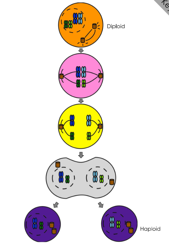
Meiosis
Summarised meiosis:
Prophase I - crossover: Chromosomes condense, homologous chromosomes pair up, crossing over occurs.
Metaphase I - middle: Homologous pairs align at the metaphase plate.
Anaphase I - away: Homologous chromosomes separate and move to opposite poles.
Telophase I - package: Chromosomes arrive at the poles, nuclear membrane reforms.
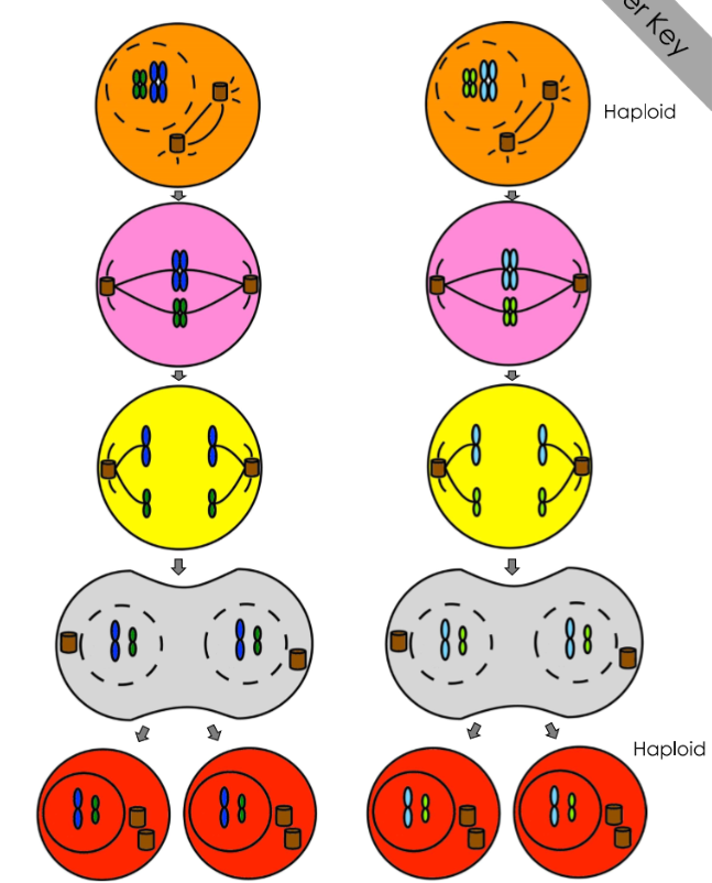
Prophase II - no crossover: Chromosomes condense again.
Metaphase II - middle: Chromosomes align at the metaphase plate.
Anaphase II - sister’s away: Sister chromatids separate and move to opposite poles.
Telophase II - package: Chromatids arrive at poles, nuclei form, and cytokinesis (cut) occurs.
Practise extended response questions:
Compare and contrast DNA replication and Transcription (10 MARKS)
DNA replication | Transcription | Marks |
DNA polymerase builds complementary strand | RNA polymerase builds complementary strand | 2 |
Bi-directional/ occurs on both strands | Occurs on template/non-coding strand only | 2 |
DNA helicase breaks hydrogen bonds | RNA polymerase breaks hydrogen bonds | 2 |
ATCG bases are used | AUCG bases are used | 2 |
Nucleotides contain deoxyribose sugar | Nucleotides contain ribose sugar | 2 |
DNA is proof read | mRNA is processed | 2 |
Similarities
Polymerase creates new strand 5' to 3' | 1 |
Occurs in the nucleus | 1 |
Hydrogen bonds broken between complementary nucleotides | 1 |
DNA uncoiled in a region/bubble | 1 |
RNA primers are used | 1 |
Compare and contrast the structure of DNA with the structure of RNA (10 marks)
DNA | RNA | Marks |
Double stranded | Single stranded | 1 |
Contains deoxyribose sugar | Contains ribose sugar | 1 |
ATCG | AUCG | 1 |
Backbone made of phosphate and deoxyribose sugar | Backbone made of phosphate and ribose sugar | 1 |
Found in nucleus and mitochondria | Found in nucleus and cytoplasm | 1 |
Supercoiled around histones | Found as uncoiled strands or folded structures | 1 |
Contains many genes | Copy of 1 gene | 1 |
Similarities | Marks |
Negatively charged | 1 |
Both can be found in nucleus | 1 |
Nucleotides made of phosphate, sugar and nitrogenous base | 1 |
When cells enter G0 they are either destroyed, stay stagnant or rejoin the cell cycle. Explain the process that results in cells entering G0 and what happens if they rejoin the cell cycle. (12 marks)
Description | Marks |
Cell cycle is made of Interphase (G1, S, G2) prophase, metaphase, anaphase, telophase, cytokinesis | 2 |
End of G1 is a checkpoint, if the cell has not grown correctly/ errors in DNA enter G0 | 1 |
Interphase/S phase- DNA replication occurs | 1 |
Prophase- chromosomes condense into chromosomes, centrioles move to poles of cell and nucleus disappears | 2 |
Metaphase- chromosomes line up along the equator of the cell, spindle fibres attach to centromere | 2 |
Anaphase- spindle fibres contract separating sister chromatids to either pole of the cell | 2 |
Telophase- chromosomes uncoil into 2 nuclei at poles of the cell | 1 |
Cytokinesis- cytoplasm is split into 2 new cells | 1 |
Using your understanding of epigenetics, explain why identical twins are preferred subjects for research than fraternal twins and that twins are chosen over a large span of ages. (2 marks)
Identical twins share identical genotype/ genome so any difference may be attributed to epigenetic factors [1]
A large span of ages are chosen for subject to study the cumulative effect/effect over time of epigenetic factors and more time given in terms of exposure to environmental effects. [1]
Use examples to explain how epigenetic inheritance is different from genetic inheritance? (3 marks)
Genetic inheritance happens through the DNA code that passes from the parent to offspring (1) while in epigenetic inheritance, epigenetic tags can be passed down to future generations without any change in the DNA code as just chemicals attached to DNA /This epigenetic tags are influenced by environmental factors. [1].
Example (1) For example with identical twins an acetyl complex can make gene expression occur rapidly creating something like a tumour in one twin but if the other has not experiences the same environment they may remain healthy despite having the same genes. (any suitable example)
Studies of identical twins have shown that during their life they can develop genetic conditions such as short sightedness, obesity and cancer. The development of these types of conditions has been linked to lifestyle and diet and can be reversed over time. In comparison, the babies born to the Dutch mothers during the famine continue to have small babies and be more susceptible to certain disease regardless of their lifestyle and diet.
Using your knowledge of epigenetic factors, discuss the differences between these two groups of people. (6 marks)
Reproduction
Male reproductive system
Structure | Function |
Testicles |
|
Vas deferens |
|
Prostate gland |
|
Urethra |
|
Seminal vesicles |
|
Scrotum |
|
Epididymis |
|
Erectile tissue | |

Oogenesis
Oogonia is egg mother cells.
Develop in ovaries before birth.
Diploid and divide by mitosis to produce cells that can develop into ova.
Several hundred thousand at birth that have undergone growth.
Primary oocyte.
Begins prophase of first meitotic division and stops (2N).
Each surrounded by single layer of cells - primary follicle.
Follicle growth and maturation
Primary oocyte complete stages of meiosis I to make 2 haploid cells.
Unequal in size.
Larger: secondary oocyte: receives half chromosomes and cytoplasm.
Smaller: polar body: has half chromosomes but no cytoplasm.
Unable to be fertilised.
Follicle: protects and nourishes oocyte until release.
Release oestrogen.
Secondary oocyte
Meiosis II stops at metaphase.
Ovulation occurs: folicle ruptures and expels secondary oocyte and polar body.
Secnondary oocyte enters fallopian tube.
If fertilised meiosis is completed.
Larger: mature egg.
Smaller: second polar body.
Hormones
Hormone | Female | Male |
Luteinising hormone |
|
|
Follicle stimulating hormone |
|
|
Oestrogen |
| |
Progesterone |
| |
Testosterone |
Ovarian Cycle Overview
The ovarian cycle consists of three main phases:
Follicular Phase:
Duration: Day 1 to Day 14 (approx.)
Description: Follicles in the ovaries mature; estrogen levels rise.
Ovulation:
Duration: Around Day 14
Description: A mature egg is released from the ovary, triggered by a surge in luteinizing hormone (LH).
Luteal Phase:
Duration: Day 15 to Day 28 (approx.)
Description: The ruptured follicle transforms into the corpus luteum, producing progesterone to prepare the uterus for potential pregnancy.
The three structures in the fetus that close up at birth are:
Ductus Arteriosus: Connects the pulmonary artery to the aorta, allowing blood to bypass the non-functioning lungs. It closes to ensure blood flows to the lungs for oxygenation after birth.
Foramen Ovale: An opening between the right and left atria of the heart, enabling blood to bypass the lungs. It closes after birth as the pressure changes in the heart.
Ductus Venosus: A vessel that allows blood to bypass the liver, directing it to the heart. It closes after birth, ensuring that blood flows through the liver for processing.
Reproductive organs
The female reproductive organs include:
Ovaries: Produce eggs and hormones (estrogen and progesterone).
Fallopian Tubes: Transport eggs from the ovaries to the uterus; site of fertilization.
Uterus: Houses and nourishes the developing fetus.
Cervix: The lower part of the uterus that opens into the vagina.
Vagina: The canal leading from the external genitals to the uterus; serves as the birth canal and receives the penis during intercourse.
External Genitalia (Vulva): Includes the labia, clitoris, and vaginal opening.
The male reproductive organs include:
Testes: Produce sperm and hormones like testosterone.
Epididymis: Stores and matures sperm.
Vas deferens: Transports sperm from the epididymis to the ejaculatory duct.
Seminal vesicles: Produce seminal fluid that nourishes sperm.
Prostate gland: Secretes fluid that helps protect and energize sperm.
Bulbourethral glands: Produce pre-ejaculatory fluid.
Penis: Delivers sperm to the female reproductive tract.
Oogenesis
Oogonia:
Egg mother cells develop in ovaries before birth.
Diploid and divide by mitosis to produce cells that can develop into ova.
At birth, there are several hundred thousand primary oocytes that have undergone growth.
Primary Oocyte:
Begins prophase of the first meiotic division and stops at this stage (2N).
Each primary oocyte is surrounded by a single layer of cells, forming a primary follicle.
Follicle Growth and Maturation:
The primary oocyte completes stages of meiosis I to make two haploid cells that are unequal in size:
Larger: Secondary oocyte – receives half the chromosomes and more cytoplasm.
Smaller: Polar body – has half the chromosomes but minimal cytoplasm, cannot be fertilized.
The follicle protects and nourishes the oocyte until release and secretes estrogen.
Secondary Oocyte:
Meiosis II stops at metaphase II.
Ovulation occurs: the follicle ruptures, expelling the secondary oocyte and polar body into the fallopian tube.
If fertilized, meiosis is completed, resulting in a mature egg and another polar body.
Spermatogenesis
Spermatogenesis refers to the process of sperm cell development and maturation, occurring in the male reproductive system, specifically within the seminiferous tubules of the testes. Here are the key steps involved in spermatogenesis:
Spermatogonia:
These are the diploid stem cells that are present in the seminiferous tubules. They undergo mitosis to maintain a continual supply of new cells.
Primary Spermatocyte:
Some spermatogonia differentiate into primary spermatocytes, which are also diploid (2N). Each primary spermatocyte will undergo meiosis.
Meiosis I:
The primary spermatocyte undergoes the first meiotic division, producing two secondary spermatocytes, which are haploid (1N) because they contain half the number of chromosomes.
Meiosis II:
Each secondary spermatocyte undergoes the second meiotic division, resulting in a total of four haploid spermatids from each primary spermatocyte.
Spermiogenesis:
The spermatids mature into spermatozoa (sperm cells). This process involves the development of a tail (flagellum) for motility, condensation of the nucleus, and formation of an acrosome (a cap-like structure containing enzymes necessary for penetrating the egg).
Mature Spermatozoa:
The final product, spermatozoa, are released into the lumen of the seminiferous tubules, where they will be stored in the epididymis for further maturation and eventual transport during ejaculation.