Unit 2: Cell Structure and Function
Chapter 4: A Tour of the Cell
The Fundamental Units of Life
All organisms are made up of cell(s)
The cell is the simplest collection of matter that can be alive
All cells are related by their descent from earlier pre-existing cells
Though cells can differ substantially from one another, they share common features
Most cells are too small to be seen by the unaided eye
Concept 4.1: Microscopy
Light microscope (LM) light is passed through a speciment and then through glass lenses
Lenses refract (bend) light and image is magnified
LMs can magnify effectively to about 1,000 times the size
Three important parameters of microscopy
Magnification - ratio of an object’s image size to its real size
Resolution - the measure of the clarity
Contrast - the difference in brightness between the light and dark areas of the image
Most subcellular structures, including organelles are too small to be seen with LMs
Electron Microscopes (EMs) are used
2 Types:
Scanning Electron Microscope (SEMs) - focus a beam of electrons onto the surface of specimen and produce images that look 3D
Transmission Electron Microscope (TEMs) focus a beam of electrons through a specimen
Used mainly to study the internal structure of cells
Concept 4.2: Comparing Prokaryotic and Eukaryotic Cells
The basic structural and functional unit of every organism is one of two types of cells: prokaryotic or eukarytoic
Domains Archaea and Bacteria are prokaryotic cells
Domain Eukarya including the kingdoms protist, fungi, plant, and animal kingdoms are eukaryotic cells
Basic features of all cells:
Plasma membrane (phospholipid bilayer)
Semifluid substance called cytosol
DNA (chromosomes) - genetic information
Ribosomes (make proteins)
Comparing Prokaryotic and Eukaryotic Cells
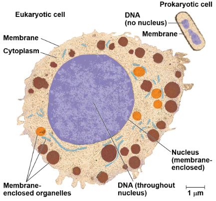
In a eukaryotic cell, DNA is enclosed in a membrane bound nucleus and have numerous membrane bound organelles
Prokaryotic cells are characterized by having
No nucleus
DNA is located in unbound region called the nucleus
No membrane bound organelle
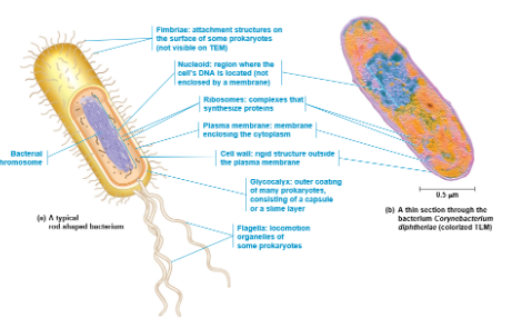
Eukaryotic cell size much larger than prokaryotic cells 10-100 μm in diameter
Typical bacteria are 1-5 μm in diameter
Cell Size
Metabolic requirements set upper limits on the size of cells
Cell size is limited by the relationship of the cell’s outer surface area to its volume (surface area-to-volume ratio) this is critical to cell function
As a cell grows, its volume increases much faster than its surface area
Small cells have a greater surface area to volume ration and can exchange nutrients and waste more readily than large cells
Concept 4.3: The Eukaryotic Cell’s Genetic Instructions Are Housed in the Nucleus and Carried out by the RIbosomes
The nucleus contains most of the DNA in a eukaryotic cell
Ribosomes use the information from the DNA to make proteins
The Nucleus: Information Central
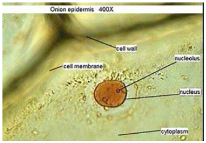
The nucleus contains most of the cell’s genes and is usually the most conspicuous organelle
The nuclear envelope encloses the nucleus, separating it from the cytoplasm
The nuclear membrane is a double membrane; each membrane consists of a lipid bilayer
Nuclear pores regulate the entry and exit of molecules
The shape of the nucleus is maintained by the nuclear lamina, which is composed of protein filaments
In the nucleus, DNA is organized into units called chromosomes
Each chromosome is one long DNA molecule associated with proteins
The DNA and proteins of chromosomes together are called chromatin
Chromatin condenses to form discrete chromosomes as a cell prepares to divide
The nucleolus is located within the nucleus and is the site of ribosomal RNA (rRNA) synthesis
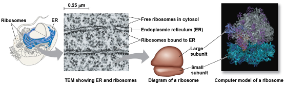
Ribosomes: Protein Factories
RIbosomes are complexes of ribosomal RNA and protein
Ribosomes carry out protein synthesis in two locations":
In the cytosol (free ribosomes)
On the outside of the endoplasmic reticulum or the nuclear envelope (bound ribosomes)
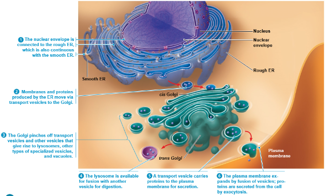
Concept 4.4: The Endomembrane System Regulates Protein Traffic and Performs Metabolic Functions
Compontents of the endomembrane system:
Nuclear envelope
Endoplasmic reticulum
Golgi apparatus
Lysosomes
Vacuoles
Plasma membrane
These components are either continuous or connected through transfer by vesicles
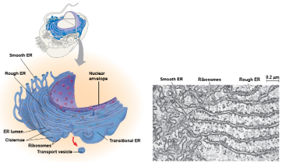
The Endoplasmic Reticulum: Biosynthetic Factory
The endoplasmic reticulum (ER) accounts for more than half of the total membrane in eukaryotic cells
The ER membrane is continuous with the nuclear envelope
There are two distinct regions of ER
Smooth ER: lacks ribosomes
Rough ER: surface is studded with ribosomes
Smooth ER functions include:
Synthesizing lipids
Metabolizing carbohydrates
Detoxifies drugs and poisons
Stores calcium ions
Rough ER functions include:
Has bound ribosomes, which secrete glycoproteins (proteins covalently bonded to carbohydrates
Distributes transport vesicles, proteins surrounded by membranes
Is a membrane factory for the cell
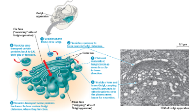
The Golgi Apparatus: Shipping and Receiving Center
The Golgi apparatus consists of flattened membranous sacs called cisternae
Functions of the Golgi apparatus include:
Modifies products of the ER
Manufactures certain macromolecules
Sorts and packages materials into transport vesicles
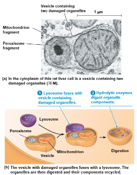
Lysosomes: Digestive Compartments
A lysosome is a membranous sac of hydrolytic enzymes that can digest macromolecules
Lysosomal enzymes work best in the acidic environment inside the lysosome
The three-dimensional shape of lysosomal proteins protects them from digestion of lysosomal enzymes
Some types of cells can engulf another cell by phagocytosis; this forms a food vacuole
A lysosome fuses with the food vacuole and its enzymes digest the molecules
Lysosomes also use enzymes to recycle the cell’s own organelles and macromolecules, a process called autophagy
Phagocytosis:
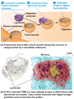
Autophagy:
Vacuoles: Diverse Compartments
Vacuoles are large vesicles derived from the endoplasmic reticulum and Golgi apparatus
The solution inside a vacuole differs in composition from the cytosol

Types of vacuoles:
Food vacuoles are formed by phagocytosis
Contractile vacuoles, found in many freshwater protists, pump excess water out of cells
Certain vacuoles in plants and fungi carry out enzymatic hydrolysis like lysosomes in plants may hold reserves of organic compounds
Central vacuoles, found in many mature plant cells, serve as a repositiory for inorganic ions, including potassium and chloride
Concept 4.5: Mitochondria and Chloroplasts Change Energy from one Form to Another
Mitochondria are the sites of cellular respiration, a metabolic process that uses oxygen to generate ATP (adenosine triphosphate)
Chloroplasts, found in plants and algae, are the sites of photosynthesis
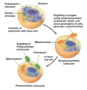
The Evolutionary Origins of Mitochrondria and Chloroplasts
Mitochondria and chloroplasts display the following similarities with bacteria that led to the endosymbiont theory:
Enveloped by a double membrane
Contain ribosomes and multiple circular DNA molecules
Grow and reproduce somewhat independently in cells
The Endosymbiont Theory is widely accepted:
An early ancestor of eukaryotic cells engulfed a nonphotosynthetic prokaryotic cell, which formed a relationship with its host
The host cell and endosymbiont merged into a single organism, a eukaryotic cell with a mitochondrion
At least one of these cells may have then taken up a photosynthetic prokaryote becoming the ancestor of cells that contain chloroplasts
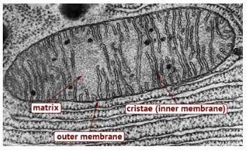
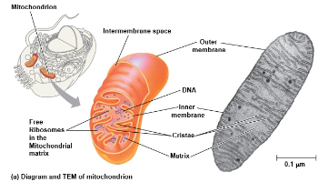
Mitochondria: Chemical Energy Conversion
Mitochondria are in nearly all eukaryotic cells
They have a smooth outer membrane and an inner membrane folded into a cristae
The inner membrane creates two compartments: the intermembrane space and the mitochondrial matrix
Some metabolic steps of cellular respiration are catalyzed in the mitochondrial matrix
Cristae present a large surface area for enzymes that synthesize ATP
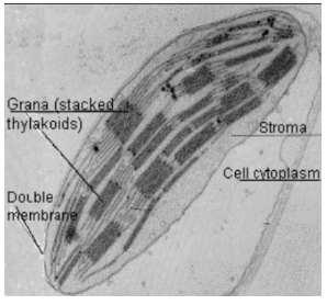
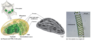
Chloroplasts: Capture of Light Energy
Chloroplasts contain the green pigment chlorophyll, as well as enzymes and other molecules that function in photosynthesis
They are found in leaves and other green organs of plants and in algae
Chloroplasts structure includes:
Thylakoids, membranous sacs, stacked to form a granum
Stroma, the internal fluid
The chloroplast is one of a group of plant organelles called plastids
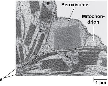
Peroxisomes: Oxidation
Peroxisomes are specialized metabolic compartments bounded by a single membrane
Peroxisomes produce hydrogen peroxide and then convert it to water
Peroxisomes perform reactions with many different functions
Example: Perozisomes in the liver detoxify alcohol and other harmful compounds
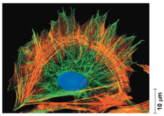
Concept 4.6: The Cytoskeleton Is a Network of Fibers That Organizes Structures and Activities in the Cell
The cytoskeleton is a network of fibers extending throughout the cytoplasm
It organizes the cell’s structures and activities
Roles of the Cytoskeleton Support and Motility
The cytoskeleton helps to support the cell and maintain its shape
It provides anchorage for many organelles and molecules, and is very dynamic
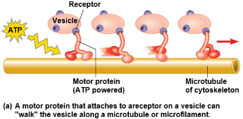
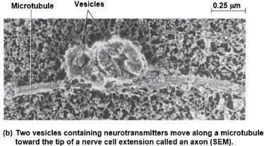
It interacts with motor proteins to produce motility
Inside the cell, vesicles and other organelles can use motor protein “feet” to “walk” along the tracks provided by the cytoskeleton
Three main types of fibers make up the cytoskeleton
Microtubules are the thickest of the three components of the cytoskeleton
Microfilaments, also called actin filaments, are the thinnest components
Intermediate filaments are fibers with diameters in a middle range

Microtubules
Microtubules are hollow rods constructed from globilar protein dimers called tubulin
Functions of microtubules:
Shape and support the cell
Guide movement of organelles
Separate chromosomes during cell division
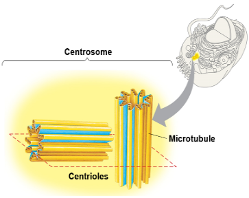
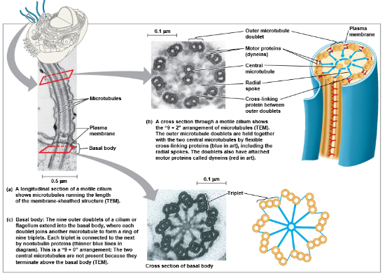
Centrosomes and Centrioles
In animal cells, microtubules grow out from a centrosome near the nucleus
The centrosome is a “microtubule-organizing center”
The centrosome has a pair of centrioles, each with nine triplets of microtubules arranged in a ring
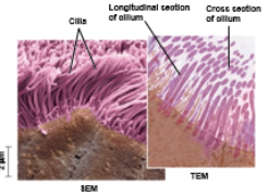
Cilia and Flagella
Microtubiles control the beating of cilia and flagella, microtubule-containing extensions projecting from some cells
Flagella are limited to one or a few per cell, while cilia occur in large numbers on cell surfaces
Cilia and flagella also differ in their beating patterns
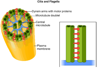
Cilia and flagella share a common structure
A group of microtubules sheathed by the plasma membrane
A basal body (protein structure) that anchors the cilium or flagellum
A motor protein called dynein, which drives the bending movements of a cilium or flagellum
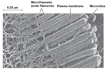
Microfilaments (Actin Filaments)
Microfilaments are thin solid rods, built from molecules of global actin subunits
The structural role of microfilaments is to bear tension, resisting pulling forces within the cell
Bundles of microfilaments make up the core of microvilli of intestinal cells
Microfilaments that function in cellular motility interact with the motor protein myosin
Example: actin and myosin interact to cause muscle contractions, amoeboid movement of white blood cells, and cytoplasic streaming in plant cells
Intermediate Filaments
Intermediate filaments are larger than microfilaments but smaller than microtubules
Intermediate filaments are only found in the cells of some animals, including vertebrates
They reinforce cell shape and fix organelles in place
Intermediate filaments are more permanent cytoskeleton elements than the other two classes
Concept 4.7: Extracellular Components and Connections Between Cells Help Coordinate Cellular Activities
Most cells synthesize and secrete materials that are external to the plasma membrane
These extracellular materials are involved in many essential cellular functions
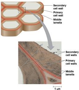
Cell Walls of Plants
The cell wall protects the plant cell, maintains its shape, and prevents polysaccharides and protein
Plant cell walls are made of cellulose microfibrils embedded in other polysaccharides and protein
Plant cell walls may have multiple layers
Primary cell wall: relatively thin and flexable
Middle lamella: thin layer between primary walls of adjacent cells
Secondary cell wall (in some cells): added between the plasma membrane and the primary cell wall
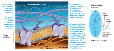
The Extracellular Matrix (ECM) of Animal Cells
Animal cells lack cell walls but are covered by an elaborate extracellular matrix (ECM)
The ECM is made up of glycoproteins such as collagen, proteoglycans, and fibronectin
ECM proteins bind to cell-surface receptor proteins in the plasma membrane called integrins
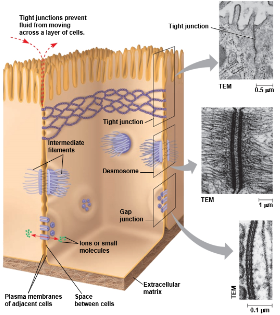
Cell Junctions
Neighboring cells in an animal or plant often adhere, interact, and communicate through direct physical contact
There are several types of intercellular junctions that facilitate this
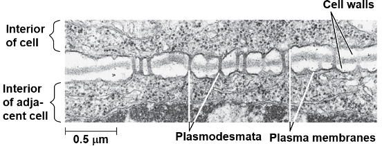
Plasmodesmata
Tight junctions
Desmosomes
Gap junctions
Plasmodesmata is in plants
Plasmodesmata are channels that perforate plant cell walls
Through plasmodesmata, water and small solutes (and sometimes protein and RNA) can pass from cell to cell
Animal cells have three main types of cell junctions
Tight junctions - limit passage of molecules and ions through the space between cells
Desmosomes - maintains cohesion with adjacent cell
Gap junctions - channel that allows for ions and small molecules to pass
All are especially common in the epithelial tissue
Concept 4.8: A Cell is Greater Than the Sum of Its Parts
None of the components of a cell work alone
Cellular functions arise from cellular order
Example: a macrophage’s ability to destroy bacteria involves the whole cell, coordinating components such as the cytoskeleton, lysosomes, and plasma membrane

Chapter 5: Membrane Transport and Cell Signaling
Concept 5.1: Cellular Membranes Are Fluid Mosaics of Lipids and Protein
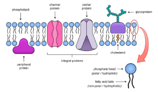
Phospholipids - most abundant lipids in most membranes
Phospholipids are amphipathic molecules, containing hydrophobic and hydrophilic regions
A phospholipid bilayer can exist as a stable boundary between two aqueous compartments
The fluid mosaic model states that the membrane is a mosaic of protein molecules bobbing in a fluid bilayer of phospholipids
Groups of certain proteins or certain lipids may associate in long-lasting, specialized patches
The Fluidity of Membranes
Most of the lipids and some proteins in a membrane can shift about sideways
This movement of phospholipids is rapid; proteins move more slowly
Some proteins move in a directed manner; others seem to be anchored in place
Some proteins simply drift in the membrane
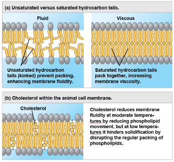
Membranes must be fluid to work properly; they are usually about as fluid as olive oil
As temperature cool, membrane switch from a fluid state to a solid state
The temperature at which a membrane solidifies depends on the types of lipids it contains
A membrane remains fluid to a lower temperature if it is right in phospholipids with unsaturated hydrocarbon tails
The steroid cholesterol has different effects on membrane fluidity at different temperatures
At warm temperatures (such as 37° C), cholesterol restrains movement of phospholipids
At cool temperatures, it maintains fluidity by preventing tight packing
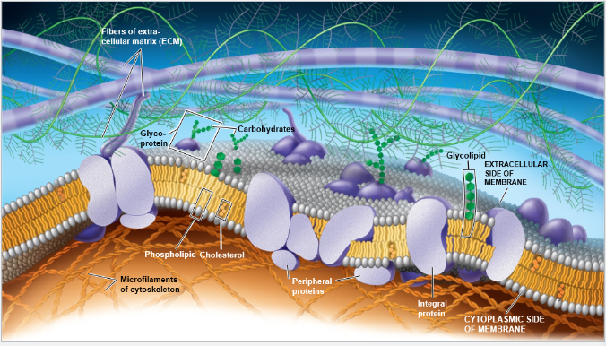
Membrane Proteins and Their Functions
A membrane is a collage of different proteins embedded in the fluid matrix of the lipid bilayer
Proteins determine most of the membrane’s specific functions
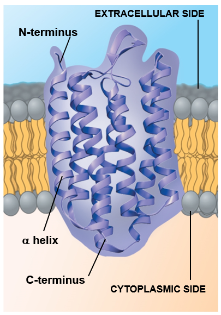
Integral proteins penetrate the hydrophobic interior of the lipid bilayer
The majority of these span the membrane and are called transmembrane proteins
The hydrophobic regions of an integral protein consist of one or more stretches of nonpolar amino acids, often coiled into α helices
Peripheral proteins are loosely bound to the surface of the membrane
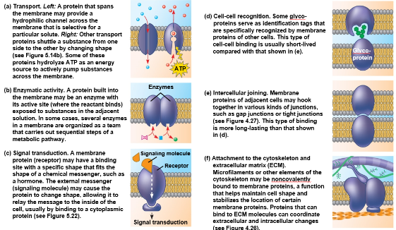
Functions:
Transport
Enzymatic activity
Signal transfuction
Cell-cell recognition
Intercellular joining
Attachment to the cytoskeleton and extracellular matrix (ECM)
The Role of Membrane Carbohydrates in Cell-Cell Recognition
Cells recognize each other by binding to surface molecules, often containing carbohydrates, on the extracellular surface of the plasma membrane
Membrane carbohydrates may be covalently bonded to lipids (forming glycolipids) or, more commonly, to proteins (forming glycoproteins)
Carbohydrates on the external side of the plasma membrane vary among species, individuals, and even cell types in an individual
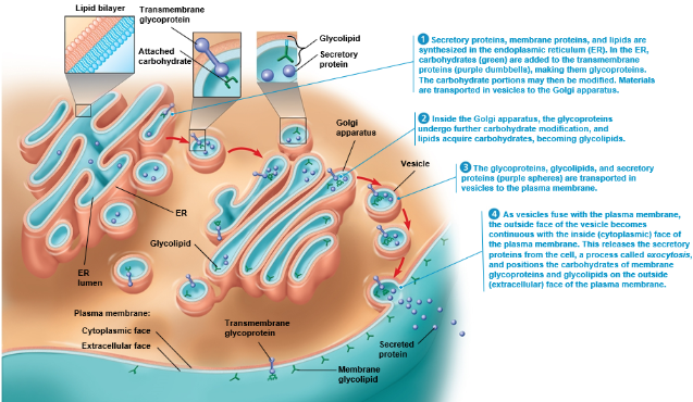
Synthesis and Sidedness of Membranes
Membranes have distinct inside and outside faces
The asymmetrical arrangement of proteins, lipids, and associated carbohydrates in the plasma membrane is determined as the membrane is built by the ER and Golgi apparatus
Concept 5.2: Membrane Structure Results in Selective Permeability
A cell must regulate transport of substances across cellular boundaries
Plasma membranes are selectively permeable, regulating the cell’s molecular traffic
The Permeability of the Lipid Bilayer
Hydrophobic (nonpolar) molecules, such as hydrocarbons, can dissolve in the lipid bilayer of the membrane and cross it easily
Polar molecules, such as sugars, do not cross the membrane easily
Even water does not cross easily compared to nonpolar molecules
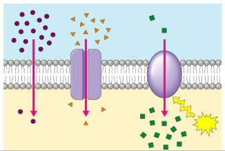
Transport Proteins
Transport proteins allow passage of hydrophilic substances across the membrane
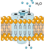
Some transport proteins, called channel proteins, have a hydrophilic channel that certain molecules or ions can use as a tunnel
Channel proteins called aquaporins facilitate the passage of water
Other transport proteins, called carrier proteins, bind to molecules and change shape to shuttle them across the membrane
A transport protein is specific for the substance it moves
Concept 5.3: Passive Transport is Diffusion Requiring NO Energy
Substances diffuse down their concentration gradient, from where it is more concentrated to where it is less concentrated
Substances move down their own concentration gradient, unaffected by concentration gradients of other substances
The diffusion of a substance across a biological membrane is passive transport because no energy is expended by the cell to make it happen
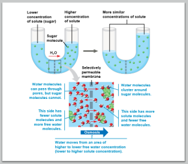
Effects of Osmosis on Water Balance
Osmosis is the diffusion of free water across a selectibely permeable membrane
Water diffuses across a membrane from the region of lower solute concentration to the region of higher solute concentration until the solute concentration is equal on both sides
Water Balance of Cells without Cell Walls
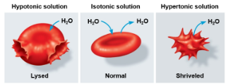
Tonicity is the ability of a surrounding solution to cause a cell to gain or lose water
Isotonic solution: Solute concentration is the same as inside the cell; no net water movement across the plasma membrane
Hypertonic solution: Solute concentration is greater than that inside the cell; cell loses water
Hypotonic solution: Solute concentration is less than that inside the cell; cell gains water
Hypertonic or hypotonic environments create osmotic problems for organisms
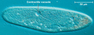
Osmoregulation, the control of solute concentrations and water balance, is a necessary adaptation for life in such environments
The protist Paramecium caudatum, which is hypertonic to its pondwater environment, has a contractile vacuole that can pump excess water out of the cell
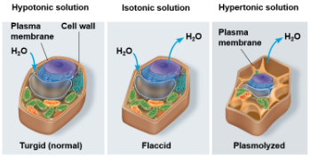
Cell walls help maintain water balance
A plant cell in a hypotonic solution swells until the wall opposes uptake; the cell is now turgid (very firm)
If a plant cell and its surroundings are isotonic, these is no net movement of water into the cell: the cell becomes flaccid (limp), and the plant may wilt
In a hypertonic environment, plant cells lose water; eventually, the membrane pulls away from the wall, a usually lethal effect called plasmolysis
Understanding Water Potential
Water potential predicts which way water diffuses through plant tissues
Abbreviated by the Greek letter psi: Ψ
Water potential is the free energy per mole of water and calculated from two major components
the solute potential Ψs (dependent on solute concentration)
the pressure potential Ψp (results from the exertion of pressure)
Water potential = Pressure Potential + Solute Potential
Ψ=Ψp+Ψs
Water moves from an area of higher water potential to an area of lower water potential
The water potential of pure water in an open beaker is zero (Ψ=0)
An increase in positive pressure raises the pressure potential and the water potential
The addition of solute to the water lowers the solute potential and decreases the water potential
A solution at atmospheric pressure has a negative water potential of the solute
Solute potential (Ψs) = -iCRT
i = ionization constant
C = the molar concentration
R = the pressure constant (R = 0.0831 liter bars/mole-K)
T = the temperature in K (273 + °C)
Facilitated Diffusion: Passive Transport Aided by Proteins
In facilitated diffusion, transport proteins speed the passive movement of specific molecules across the plasma membrane
Channel proteins provide corridors that allow a specific molecule or ion to cross the membrane
Channel proteins include:
Aquaporins, for facilitated diffusion of water
Ion channels that open or close in response to a stimulus (gated channels)
Carrier proteins undergo a subtle change in shape that translocates the solute-binding site across the membrane
The shape change may be triggered by a binding and release of the transported molecule
No net energy input is required
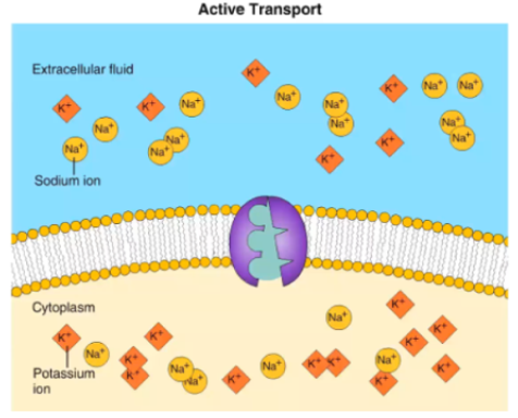
Concept 5.4: Active Transport Uses Energy (ATP) to Move Solutes Against Their Gradients
Active transport moves substances across membranes against their concentration gradients
Active transport allows cells to maintain concentration gradients that differ from their surroundings
Active transport requires energy, usually in the form of ATP
The Need for Energy in Active Transport
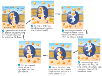
The sodium-potassium pump is one type of active transport system
It exchanges Na+ for K+ across the plasma membrane of animal cells
At top speed, the sodium-potassium pump can transport about 450 Na+ ions and 300 K+ ions per second
Important in nerve cells
How Ion Pumps Maintain Membrane Potential
Membrane potential is the voltage across a membrane
Ranges from -50 millivolts (mV) to -200 mV
Voltage is created by differences in the distribution of anions and cations across a membrane
Two combined forces, collectively called the electrochemical gradient, drive the diffusion of ions across a membrane
A chemical force (the ion’s concentration gradient)
An electrical force (the effect of the membrane potential on the ions movement)
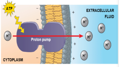
An electrogenic pump is a transport protein that generates voltage across a membrane
The sodium-potassium pump is the major electrogenic pump of animal cells
The main electrogenic pump of plants, fungi, and bacteria is a proton pump
Electrogenic pumps help store energy that can be used for cellular work
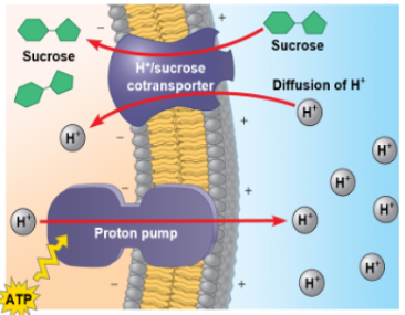
Cotransport: Coupled Transport by a Membrane Protein
Cotransport occurs when a transport protein can couple the “downhill” diffusion of a solute to the “uphill” transport of a second solute against its electrochemical gradient
Plant cells use the gradient of hydrogen ions generated by proton pumps to drive active transport of nutrients into the cell
Concept 5.5: Bulk Transport Across the Plasma Membrane Occurs by Exocytosis and Endocytosis
Water and small solutes enter or leave the cell through the lipid bilayer or by means of transport proteins
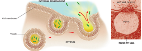
Large molecules, such as polysaccharides and proteins, cross the membrane in bulk by means of vesicles
Bulk transport requires energy
/
Exocytosis
In exocytosis, transport vesicles migrate to the membrane, fuse with it, and release their contents
Many secretory cells use exocytosis to export products
Example: pancreatic cells release insulin into bloodstream
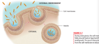
Endocytosis
In endocytosis, the cell takes in molecules ant particulate matter by forming new vesicles in the plasma membrane
Endocytosis is a reversal of exocytosis, involving different proteins
There are three types of endocytosis:
Phagocytosis (cellular eating)
Example: white blood cells
Pinocytosis (cellular drinking)
Example: cells in kidney separate nutrients and fluids from urine
Receptor mediated endocytosis
Example: human cells pull cholesterol from the bloodstream
Human cells use receptor-mediated endocytosis to take in cholesterol for membrane synthesis and the synthesis of other steroids
Cholesterol travels in particles called low-density lipoproteins (LDLs)
In the inherited disease familial hypercholesterolemia, LDL receptor proteins are defective or missing. Thus, cholesterol accumulates in the blood, contributing to atherosclerosis (the buildup of lipids within blood vessel walls, which narrows the space in the vessels and impedes blood flow, potentially resulting in heart damage or stroke)