Dissection 6 - The Face
Dissection Guide
Muscles of Facial Expression
Platysma
F: Tightens skin of the neck and depresses angle of the mouth
I: Cervical branch of CN VII
Frontalis
F: Raise eyebrows
I: Temporal and zygomatic branches of CN VII
Orbicularis oculi
F: Closes eyes
I: Zygomatic branch of CN VII
Zygomaticus major
F: Smiling
I: Upper buccal branches of CN VII
Orbicularis oris
F: Pucker the lips
I: Lower buccal and marginal mandibular branches of CN VII
Buccinator
F: Movement of cheeks
I: Buccal branch of CN VII
Nasalis
F: Flare nostrils
I: Buccal branch of CN VII
Levator labii superioris
F: Elevate upper lip
I: Buccal branch of CN VII
Levator anguli oris
F: Elevate angles of the mouth
I: Buccal branch of CN VII
Depressor labii inferioris
F: Depress lower lip
I: Marginal mandibular branch of CN VII
Depressor anguli oris
F; Depress angles of the lips
I: Marginal mandibular branch of CN VII
Risorius
F: Smile
I: Buccal branch of CN VII
Mentalis
F: Raise lower lip
I: Mandibular branch of CN VII
Face Dissection
Parotid gland and duct
Note that buccal branch of CN VII runs parallel to parotid duct
Parotid duct is deep anterior to masseter m.
Runs through buccal fat pad and pierces buccinator → open adjacent to second maxillary molar
Facial a. passes over corpus of the mandible anterior to masseter
Branch of ECA
Note: The a. is tortuous near the mandible
Facial a. branches into angular a. and inferior/superior labial a.
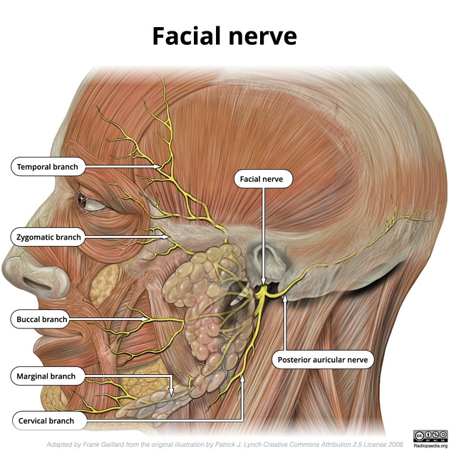
Facial n.
Common facial n. found posterior to the parotid gland and divides within the gland
Bolded Terms
Angular artery
Facial a. once it reaches the side of the nose
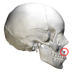
Anterior nasal spine
Pointed, midline bony projection located at the lower margin of the nasal opening, formed by the fusion of the right and left maxillae at the superior end of the intermaxillary suture
Buccal branch of facial n.
Run parallel the parotid duct
Buccal fat pad
Adipose tissue located deep within the cheek
Between the buccinator muscle and more superficial facial muscles such as the masseter and zygomaticus
Buccinator m.
F: Movement of cheeks
I: Buccal branch of CN VII
Cervical branch of facial n.
Found crossing the mandible to reach the platysma
Depressor anguli oris
F: Depresses angle of the the lips
I: Marginal mandibular branch of CN VII
Depressor labii inferioris
F: Depress lower lip
I: Marginal mandibular branch of CN VII
Facial a.
Branch of ECA
Passes over the corpus of the mandible, anterior to the masseter
A. can be compressed here to deduced bleeding from a facial laceration
Tortuous to give the a. more flexibility (lots of movement in the region)
Facial n.
Common facial n. found posterior to parotid gland (exits through the stylomastoid foramen)
Facial v.
Frontalis
F: Raises eyebrows
I: Temporal and zygomatic branches of CN VII
Inferior labial artery
Branch of facial a.
Runs inferior to lips
Infraorbital foramen
Infraorbital VAN exits (CN V2)
Levator anguli oris
F: Elevate angle of the mouth
I: Buccal branch of CN VII
Levator labii superioris
F: Elevate upper lip
I: Buccal branch of CN VII
Marginal mandibular branch of facial n.
Runs along body of mandible
Masseter m.
Mastoid process to angle of mandible
Innervated by CN V3
Mental foramen
Mental VAN exits (CN V3)
Mentalis
F: raise lower lip
I: Mandibular branch of CN VII
Nasalis
F: Flare nostrils
I: Buccal branch of CN VII
Orbicularis oculi
F: Closes eyes
I: Zygomatic branch of CN VII
Orbicularis oris
F: Pucker the lips
I: Lower buccal and marginal mandibular branches of CN VII
Parotid duct
Courses from the anterior border of the parotid gland to exit behind the maxillary 2nd molar
Dives deep anterior to the masseter, through the buccal fat pad, and pierces the buccinator m. before opening into the oral cavity
Parotid gland
Located anterior to the ear
Major salivary duct
CN VII runs inside the gland and parasympathetics from CN IX run to the gland
Platysma
F: Tightens skin of the neck and depresses angle of the mouth
I: Cervical branch of CN VII
Posterior auricular branch of facial
Runs posteriorly
Retromandibular v.
Posterior division joins with posterior auricular v. to drain into the EJV
Anterior division joins facial v. to form the common facial v. and drain into the IJV
Risorius
F: Smiling
I: Buccal branch of CN VII
Superior labial a.
Branch of facial a.
Runs superior to lips
Supraorbital foramen
Supraorbital VAN (CN V1) exits
Temporal branch of facial n.
Found in temporal region
Tragus
Small, cartilaginous projection located on the external ear, positioned immediately anterior to the external auditory (ear) canal
Zygomatic branch of facial n.
Found in zygomatic arch region
Zygomaticus major
F: Smiling
I: Upper buccal branches of CN VII