Chapter 22: The Circulatory System
Introduction to Cardiovascular System
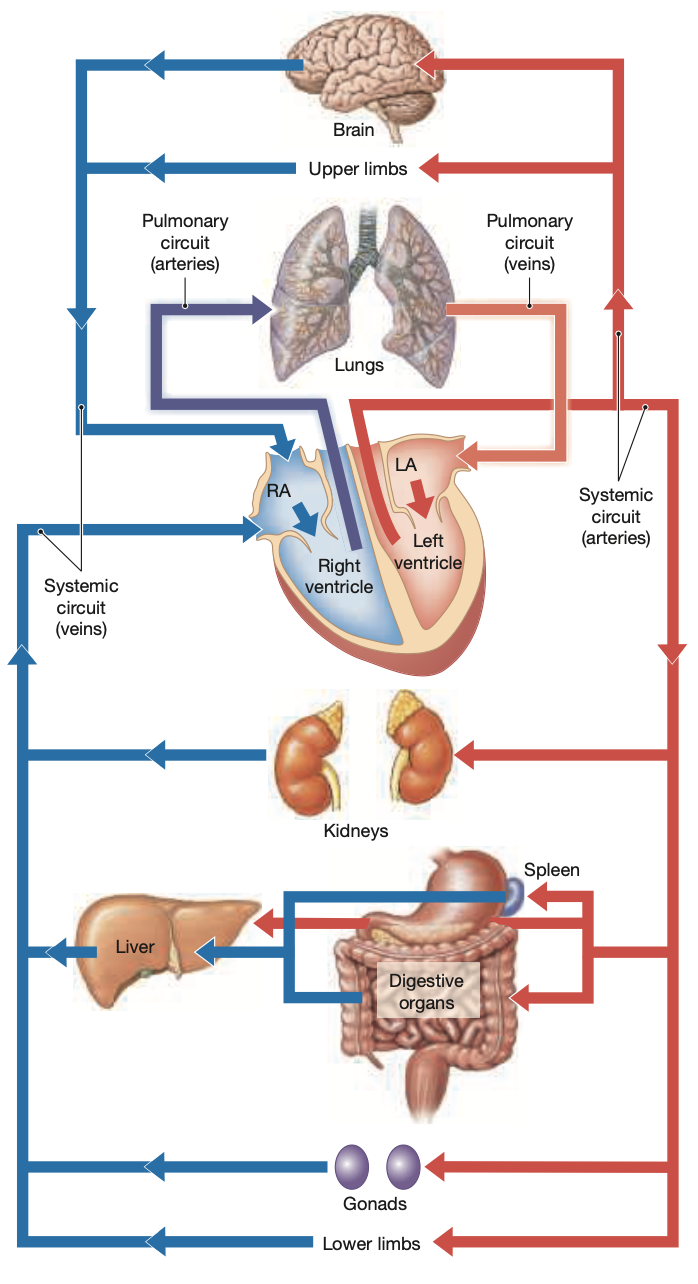
The cardiovascular system is a closed system (begins & ends at the heart) with two main circulatory patterns:
Pulmonary circuit
Begins at the pulmonary valve & ends at the L Atrium
Systemic circuit
Beings at the aoritc valve & ends in the R Atrium
Coronary circuit
Begins at the aorta and ends at the coronary sinus (R Atrium)
Blood circulates through a network of arteries, veins, and capillaries.
Veins carry volumes of blood generally against the force of gravity
Arteries and veins run parallel to each other
The volume of blood pumped by the heart per minute is known as cardiac output, which can vary widely, between 5-30L per minute
Histological Organization of Blood Vessels
Blood vessels consist of three layers:
Intima (tunica intima): Innermost endothelial layer (lining)
Media (tunica media): Middle muscular layer (smooth muscle cells involved in vasoconstriction & dilation)
Adventitia (tunic externa): Outer CT sheath
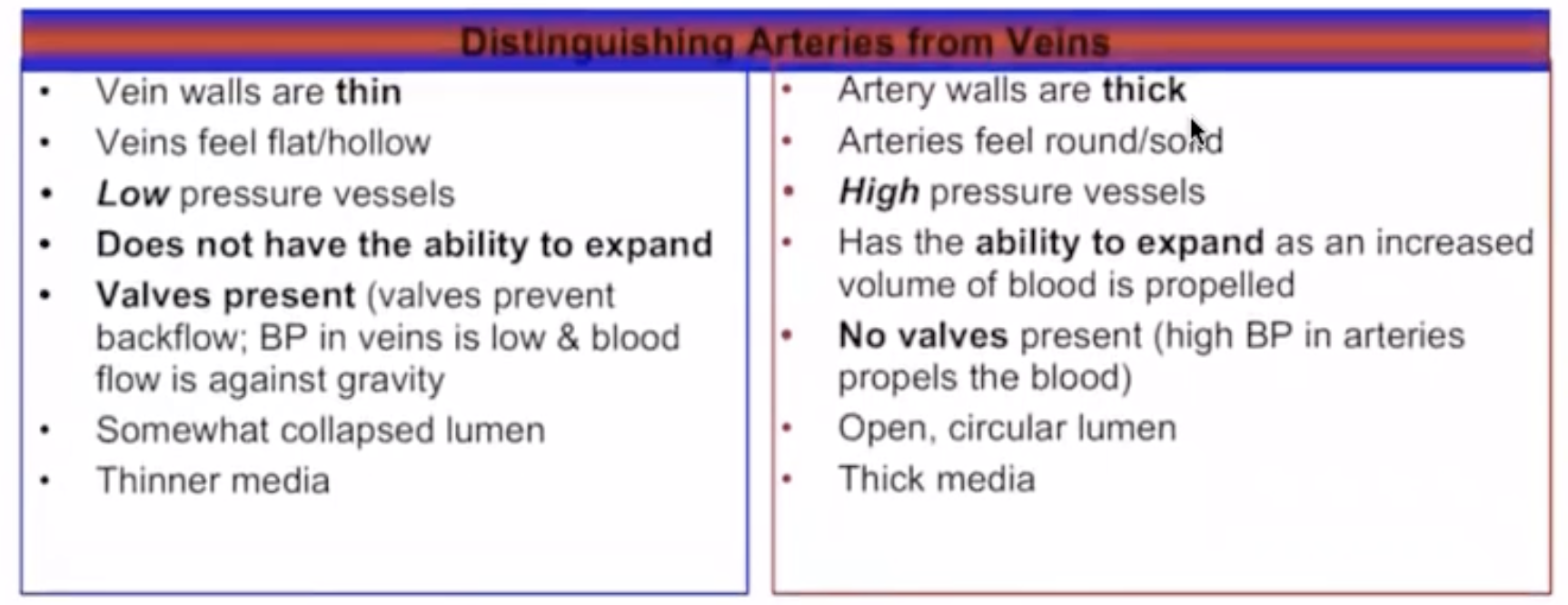
Distinguishing Arteries from Veins
Arteries vs. Veins:
Arterial walls are generally thicker and can withstand greater pressure than veins.
Endothelial lining in arteries appears folded due to lack of contractility.
Arteries can constrict under high blood pressure; veins have little constriction.
Most veins have one-way valves to prevent backflow of blood, arteries do not
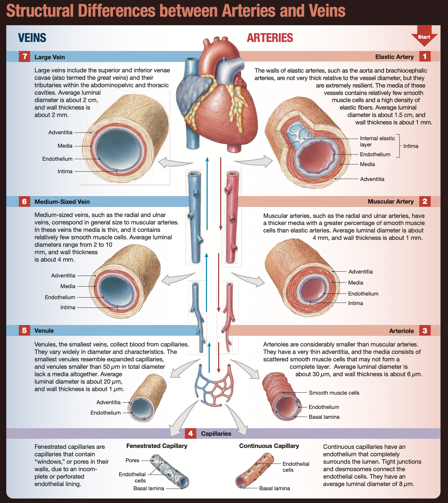
Types of Blood Vessels
Arteries:
Classified into:
Large Elastic Arteries— These arteries are closest to the heart and therefore experience the highest pressures. They contain a high proportion of elastin, allowing them to stretch and recoil to accommodate the pulsatile flow of blood coming from the heart.
ex— Aorta, Brachiocephalic Trunk, Pulmonary Trunk
Muscular (medium-sized) Arteries— Mainly known as distribution arteries characterized by a thick layer of smooth muscle which allows them to regulate blood flow by constricting or dilating.
ex— Radial & Ulnar, External Carotid, Brachial, Femoral, Mesenteric
Smaller Arterioles— These arteries are small and have very thin adventitia which helps them control blood flow between the arteries and capillaries
Capillaries:
Smallest blood vessels, facilitating exchanges between blood and interstitial fluid.
Types of capillaries:
Continuous: Complete endothelial lining, tight junctions or desmosomes
Found in most regions of the body
Fenestrated: Contain small openings/pores ("windows")
Found in brain, edocrine organs, kidneys
Sinusoids: Specialized fenestrated capillaries with large pores, allowing for slow blood flow.
Found in liver, bone marrow, adrenal glands
Capillary Networks
Capillary Beds: The interconnected networks of capillaries that facilitate the exchange of nutrients, gases, and waste products between blood and tissues.
Blood enters the capillary bed from arterioles— oxygen and nutrients leave the blood and enter surrounding tissues as carbon dioxide and metabolic wastes move from the tissues into the blood for removal.
The deoxygenated blood then exits through the venules eventually making its way back to the heart
Metarteriole: Regulates blood flow into capillaries.
Thoroughfare channels: Provide communication between arterioles and venules.
Veins
Veins return oxygen-poor blood from organs to the heart.
Comprised of:
Venules— The smallest veins, collect blood from capillaries
Medium-Sized Veins— Contain one-way valves that prevent backflow of blood, found near muscular arteries
The skeletal muscles of the legs help to propel the blood back to the heart by squeezing veins, the valves close ensuring the blood does not go back down
Large Veins— These vessels are responsible for transporting larger volumes of blood and are often located deep within the body, providing structural support and greater resistance to pressure.
Venous system operates under lower pressure than arteries.
Valves prevent backflow of blood within veins.
Distribution of Blood Volume
The total blood volume is distributed unevenly within the vessels of the body
Approximately 30-35% of blood volume is in the heart, arteries, and capillaries.
The venous system contains about 65-70% of total blood volume.
Circulatory Pathways
Pulmonary Circuit:
Carries oxygen-poor blood to the lungs via pulmonary arteries; returns via pulmonary veins to the left atrium.
Systemic Circuit:
Begins at the aortic valve and ends at the right atrium.
Supplies oxygenated blood to all body tissues excluding the pulmonary circuit.
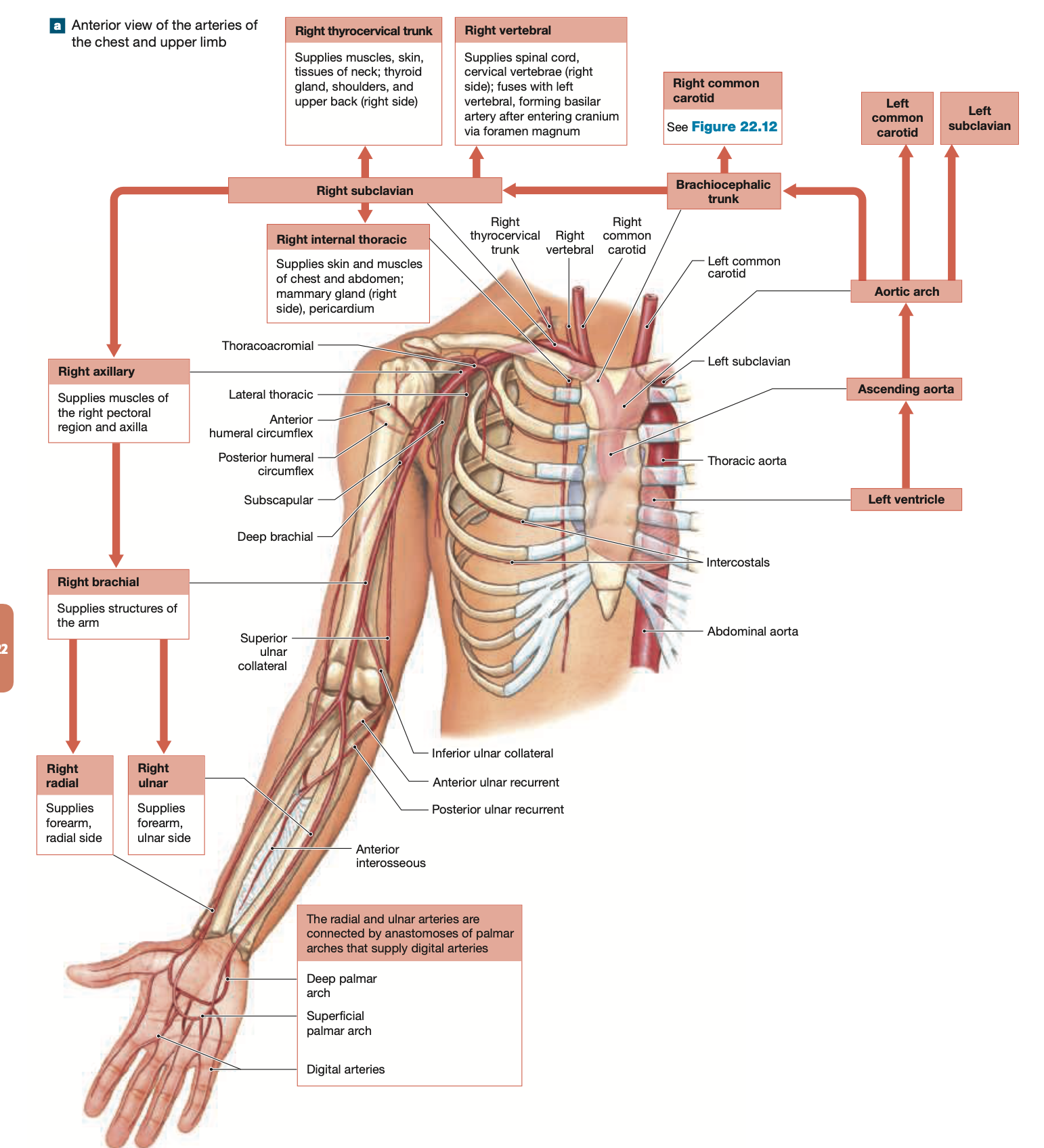
Major Blood Vessels
Aorta:
Ascending aorta gives rise to coronary circulation.
Aortic arch branches into three major arteries supplying the head, neck, shoulders, and upper limbs:
Brachiocephalic Trunk— Supplies blood to the right side of the head, neck, and right upper limb.
Right Subclavian Artery → Supplies blood to the right upper limb and parts of the thorax.
Key Branches:
Vertebral Artery → Supplies blood to the brainstem, cerebellum, and posterior brain.
Right Common Carotid Artery → Supplies the right side of the head and neck.
Key Branches:
Internal Carotid Artery → Supplies blood to the brain and eyes.
External Carotid Artery → Supplies blood to the face, scalp, and neck.
Left Common Carotid Artery— Supplies blood to the left side of the head and neck.
Key Branches:
Internal Carotid Artery → Supplies blood to the brain and eyes.
External Carotid Artery → Supplies blood to the face, scalp, and neck.
(These branches mirror those of the right common carotid artery.)
Left Subclavian Artery—Supplies blood to the left upper limb and parts of the thorax.
Key Branches:
Vertebral Artery → Supplies blood to the brainstem, cerebellum, and posterior brain.
Each common carotid artery divides into external and internal carotids, supplying blood to the neck, face, and brain.
Summary of the Head, Neck, and Upper Limb:
Brachiocephalic Trunk
R & L Axillary
R & L Brachial
R & L Radial & Ulnar
R & L Superfical Palmar Arch
R & L Digitals
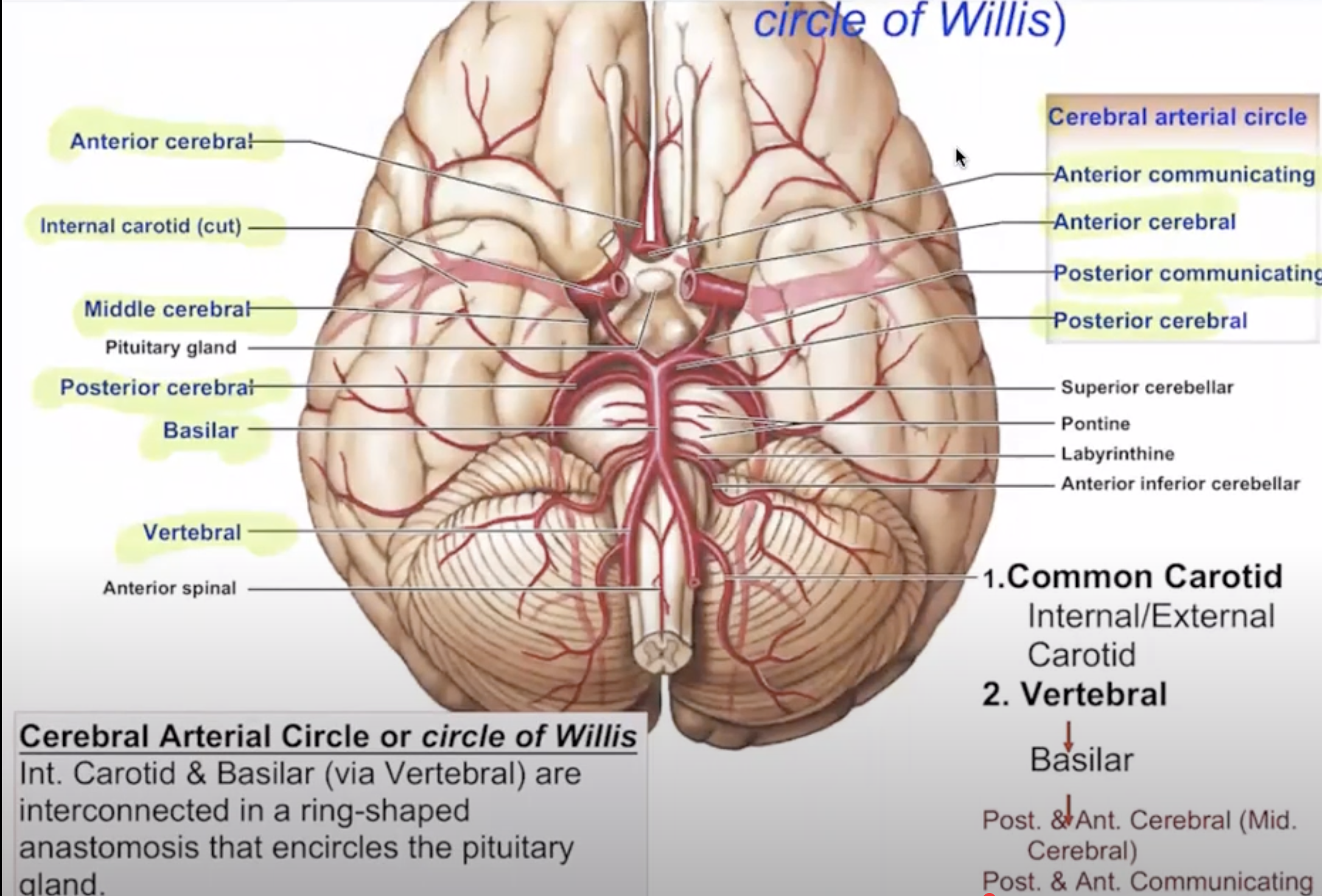
Blood supply to the Brain
The blood in the vertebral arteries reaches the brain
Superior part (above diaphragm): Thoracic aorta
Inferior part (below diaphragm): Abdominal aorta
Supplies blood to the thoracic and abdominal cavities and lower limbs.
The L & R vertebral arteries fuse to form the Basilar Artery, which branches many times in area of the pons
Posterior Cerebral & Posterior Communicating Arteries connect the basilar artery and internal carotid arities
The ring-like anastomosis between the basilar & carotid arteries is called the Cerebral Arterial Circle (Circle of Willis)
The Celiac Trunk
The celiac trunk (also called the celiac artery) is a major artery that branches from the abdominal aorta
The celiac trunk quickly divides into three major arteries:
Left Gastric Artery
Supplies: The stomach (lesser curvature) and the lower esophagus.
Common Hepatic Artery
Supplies: The liver, stomach, duodenum, and pancreas.
Splenic Artery
Supplies: The spleen, pancreas, and stomach.
Venous Systems
Systemic Veins:
Return blood to the right atrium through superior and inferior venae cavae (below diaphragm).
Superior Vena Cava:
Collects blood from the upper regions: head, neck, chest, and arms (above diaphragm).
Inferior Vena Cava:
Collects blood from areas below the diaphragm not drained by the hepatic portal vein.
Unique Portal Systems
Portal vessels connect two capillary beds. The hepatic portal system collects nutrient-rich blood from digestive organs, flowing directly to the liver for processing.
Cardiovascular Changes at Birth
During fetal development, the umbilical arteries/veins connect to the placenta.
After birth, the ductus venosus, foramen ovale, and ductus arteriosus close, becoming fibrous structures (fossa ovalis and ligamentum arteriosum).
Aging and the Cardiovascular System
Age-related changes can lead to increased risk of thrombus and arterial rigidity (arteriosclerosis).
Inelastic arterial walls may cause aneurysms.