Micropara Midterm
Module 1: History and Scope of Microbiology
Microorganisms:
Considered as ubiquitous (it can be found anywhere)
Microbes are here on Earth before man was created
Also known as microbes
Represents the major function of the Earth’s biomass
Plants and animals are engaged in the world of microbes, their evolution and survival are influenced by microbial activities
Microbiology: branch of biology that deals with microorganism and their effects on other living things
Microbial Cells: first appeared between 3.8 to 4.3 billion years ago
First 2 billion years ago: microorganisms are capable to survive without oxygen in the atmosphere
Phototrophic Microorganisms: occurred 1 billion years ago (harvest energy from sunlight)
Purple sulfur bacteria and green sulfur bacteria: were anoxygenic (non-oxygen producing); were the first phototrophs
Cyanobacteria: oxygenic phototrophs; evolved and began the slow process of oxygenating earth’s atmosphere, multicellular life forms eventually evolved
ORIGINS OF MICROBIOLOGY
Microscope is the ultimate tool in studying microbiology
The first known image of microscope and fruiting molds was illustrated by Robert Hooke. Cut from thin slices of cork and observed under the microscope the presence of “tiny little boxes”. He started to formulate the “Cell Theory”.
Antoni van Leeuwenhoek constructed single lens microscope and first to observe bacteria by using pepper-water infusion.
Louis Pasteur experimented the process of fermentation and sterilization. He disproved the “Spontaneous Generation theory”. He developed a vaccine for rabies.
Joseph Lister introduced the aseptic technique in order to kill and prevent microbial infection of surgical patients.
Ignaz Semmelweis introduced the method of hand washing.
Robert Koch discovered the causative agent of diseases like anthrax, cholera and tuberculosis. He used his own Koch’s postulate in identifying these diseases.
Richard Petri developed a transparent double-sided dish known as “Petri dish”, a standard tool for obtaining pure cultures.
DIFFERENT THEORIES
Cell Theory by Robert Hooke: all organisms are composed of cells
Spontaneous Generation Theory: life could arise spontaneously from nonliving matter
Germ Theory or Koch’s Postulate: disease agent-man-disease
KOCH’S POSTULATE
1st Postulate: the suspected pathogen must be present in all cases of the disease and absent in healthy animals
2nd Postulate: the suspected pathogen must be grown in pure culture
3rd Postulate: cells from a pure culture of the suspected pathogen must cause disease in a healthy animal
4th Postulate: the suspected pathogen must be reisolated and shown to be the same as the original
DIVERSITY OF MICROORGANISMS
Estimated 2x 1030 microbial cells on Earth
The total amount of nitrogen and phosphorus (essential nutrients for life) within microbial cells is nearly four times that in all plant and animal cells combined
Microbes also represent a major fraction of the total DNA in the biosphere (about 31%), and their genetic diversity far exceeds that of plants and animals.
Microbes are even abundant in habitats that are too much harsh for other forms of life.
All ecosystems are influenced greatly by microbial activities.
The metabolic activities of microorganisms can change the habitats in which they live, both chemically and physically, and these changes can affect other organisms.
Microbes provide nutritional and other benefits that are essential to human health
IMPACT OF MICROORGANISM
Agent of Disease: microorganisms are the major cause of human death were infectious disease caused by bacteria and viruses
Agriculture: root nodules of plant contain bacteria that fix molecular nitrogen that can be used by plant
Microbes in the rumen of the animal convert cellulose from grass into fatty acids that can be used by animals
Food: cause of foodborne disease and food spoilage; not all microorganisms in foods are harmful
Beneficial microbes are used to improve food safety and to preserve food such as cheese, yogurt; while thru microbial fermentation can produce food like sauerkraut, kimchi, pickles
Colon contains diverse microbial species that assist in the digestion of complex carbohydrates, and that synthesize vitamins and other nutrients essential to host nutrition
Industry: harnessed to produce antibiotics, enzymes, insulin, biofuels, and helps clean up waste
Environment: used in bioremediation, a process that cleans up toxic wastes and pollutants
Decompose or destroy wastes, such as sewage and oil spills
Module 2: Microbial Diversity, Structures, and Function
TYPES OF MICROORGANISM
Cellular Microorganism: organism that contain cells, either unicellular of multicellular
Unicellular: Prokaryotes (Archaea, Bacteria, Cyanobacteria)
Unicellular/Multicellular: Eukaryotes (Algae, Fungi, Protozoa)
Acellular Microorganism: organism that doesn’t contain cell
Examples: virus, viroids, prions
PROKARYOTIC CELL
“No” true nucleus
Ave size: • 0.2 – 2.0 μm (diameter.)
2 – 8 μm (length)
Lack membrane-enclosed organelles
No cytoskeleton
Transverse binary fission
Monomorphic or pleomorphic
EXAMPLES OF PROKARYOTES
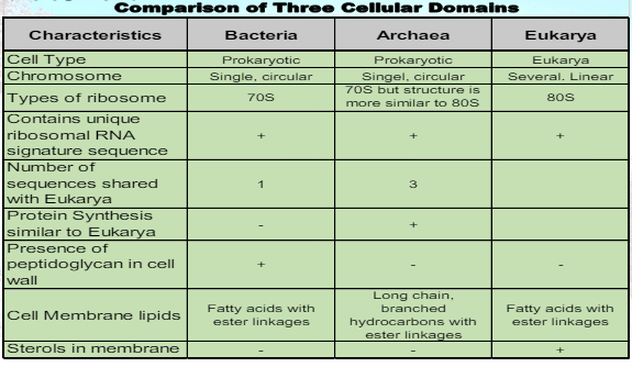
Archaea
Archaea means ancient
Archaea bacteria look identical microscopically to members of the Bacteria domain
Chemical composition of cell wall differs: Archaea do not have peptidoglycan
Have the ability to grow in extreme environments
extreme temperatures: hot or cold
acidic or alkaline conditions
extreme salt concentration
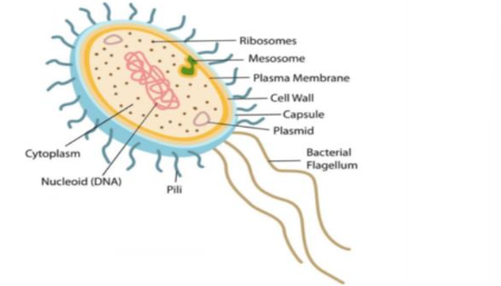
Bacteria
Single-celled prokaryote
prokaryote is a simple cell with a nucleoid region, surrounded by cytoplasm and a cell wall
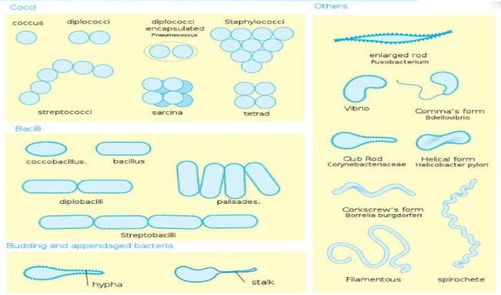
Comprised of specific shapes:
Bacilli: rod shaped
Cocci: spherical shaped
Spirilla: spiral shaped
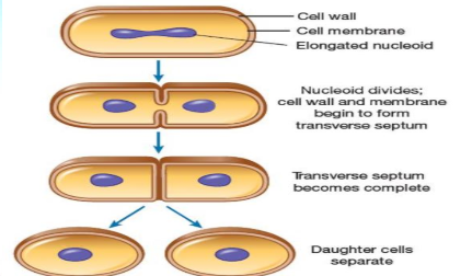
Bacterial cells multiply by binary fission
One cell divides into two cells, identical to original
PROKARYOTIC CELL STRUCTURE
Appendages | Internal to the cell wall |
Glycocalyx | Cell Membrane |
Flagella | Cytoplasm |
Fimbriae | Chromosomes |
Pili | Ribosomes |
Inclusions | |
Endospores (some) |
APPENDAGES
Glycocalyx
The outer layer usually made up of bound polysaccharides on the cell surface and superficial layer of unbound proteoglycans and glycoproteins
Capsule
Anti-phagocytic function
Vaccine target
Attachment
Slime Layer
Glide or slide on surfaces
Formation of biofilm

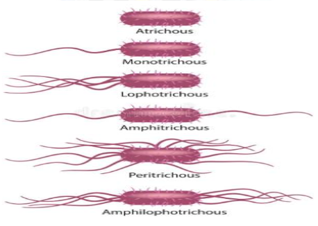
Flagella
Long, slender, threadlike, whip-like extension of certain cells or unicellular organisms used mainly for movements (others for signal transduction).
Arrangement: Atrichous, Monotrichous, Lophotrichous, Amphitrichous, Peritrichous
Fimbriae
Hair-like structures made of “pilin”
Enable bacterial cell to adhere to surfaces

Pili
Short, filamentous projection on a bacterial cell, used not for motility but for adhering to other bacterial cell (especially for mating) or to animal cells.
Joins bacterial cell for DNA transfer during conjugation
“sex pili”
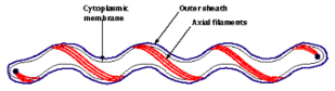
Axial filaments
Flagella-like fibrils arising at the ends
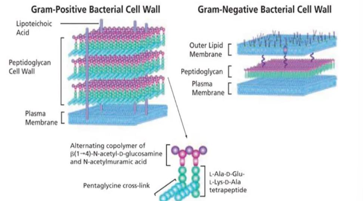
PROKARYOTE: CELL WALL
Composed of peptidoglycan (murein)
N-acetylglucosamine (NAG)
N-acetylmuramic acid (NAM)
Used to characterized bacterial cells
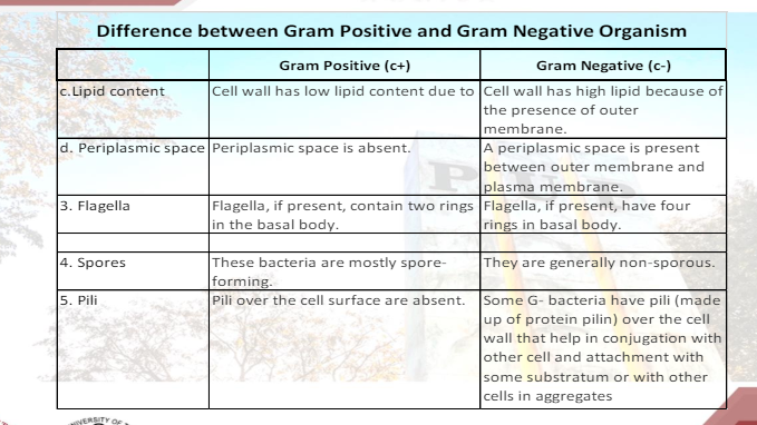
Gram Positive – teichoic acid and lipoteichoic
Gram Negative – lipoproteins, LPS and phospholipid

OTHER STRUCTURES INTERNAL TO THE CELL WALL
Plasmid: Contains extra-chromosomal DNA which carry genes that confer protective trait that may be duplicated and passed on to an offspring
Ribosomes: A minute particle composed of protein and ribonucleic acid (RNA) that serves as the site of protein synthesis.
Inclusions: An abnormal structure in a cell nucleus or cytoplasm having characteristic staining properties
Endospore: Bacteria undergo sporulation during exposure to harsh conditions
EUCARYOTIC CELL (EUKARYOTES)
all members of the living world except the prokaryotes are considered Eucarya
single celled and multi-celled
contain basic organelles: cell membrane, cytoplasm, nucleus
membrane bound “true” nucleus
Ave size: 10 – 30 μm
Mitosis/meiosis
Examples: Algae, Fungi, Protozoa

EXAMPLES OF EUKARYOTES
Algae
photosynthetic eukaryotes with a wide variety of shapes that occur in most habitats, ranging from marine and freshwater to desert sands and from hot boiling springs to snow and ice belonging to Kingdom Protista
small, single-celled forms (filamentous) to most complex multicellular forms.
They are not plants, they are more plantlike than protozoa. Algae lack true roots, stems and leaves.
They important role in balancing the environment.
Examples: Chlamydomonas, Spirogyra, Rhodymenia
Fungi
Fungi can be single celled or very complex multicellular organisms.
Found in just about any habitat but most live on the land, mainly in soil or on plant material rather than in sea or fresh water.
Decomposers grow in the soil or on dead plant matter where they play an important role in the cycling of carbon and other elements
Examples: Yeast and Molds
Protozoa
Protozoa are single celled organisms. They come in many different shapes and sizes ranging from an Amoeba which can change its shape to Paramecium with its fixed shape and complex structure.
They live in a wide variety of moist habitats including fresh water, marine environments and the soil.
Some are parasitic, which means they live in other plants and animals including humans, where they cause disease.
Plasmodium, for example, causes malaria. They are motile and can move by means of flagella, cilia, and amoeboid movement.

ACELLULAR STRUCTURE - without cell parts
Virus
considered acellular, non-living, obligate intracellular parasite
made up of a core containing DNA or RNA surrounded by a protein coat
can reproduce only by using the cellular mechanism of another cell
often considered the parasites of the microbial world
Viroids
very small, circular RNA (may appear linear), and infectious in plants. They do not contain a capsid.
The only human disease known to be caused by a viroid is hepatitis D; in this case the viroid is enclosed in a hepatitis B virus capsule.
Prions
contain only protein
causative agent for some neurodegenerative diseases in humans and animals
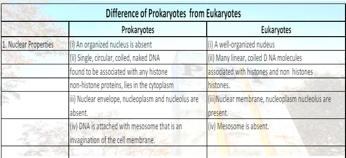
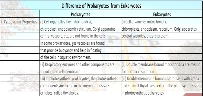
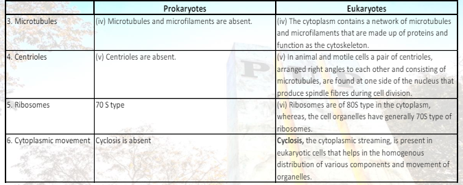
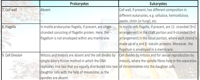
COMPARISON BETWEEN PROKARYOTE AND EUKARYOTE
Eukaryote | Prokaryote |
Typically larger | Typically smaller |
Compartmentalized by membrane-bounded sacs or organelles | Not compartmentalized |
Contain a nucleus with multiple chromosomes | Do not have a nucleus |
Divide by complex process of mitosis | Divide by binary fission |
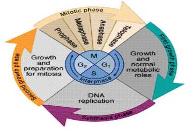
CELL DIVISION
Process by which a parent cell divides into two or more daughter cells
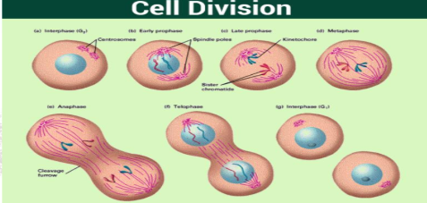
Usually occurs as a part of a larger cell cycle
2 GENETIC STATES
Haploid
One copy of chromosome
In humans, n = 23
Gametes (sperm & ova) are haploid
Diploid
Two copies of chromosome
In humans, n = 46
All body cells are diploid found in skin, blood and muscle cells also known as somatic cells
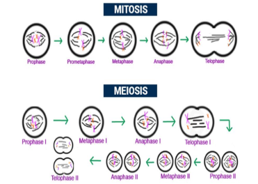
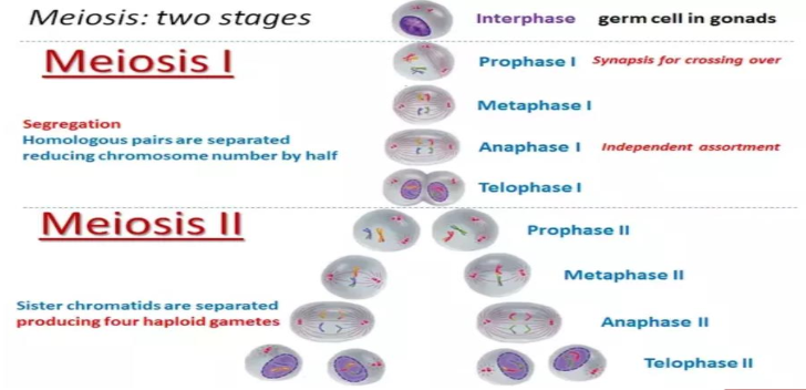
TYPES OF CELL DIVISION
Mitosis
the chromosomes condense, divide, and are separated into two sets, one for each daughter cell
Meiosis I
homologous chromosomes segregate into separate cells, changing the genetic state from diploid to haploid
Meiosis II
the two haploid cells divide to form a total of four haploid cells called gametes
Module 3: Microbial Taxonomy
Taxonomy: way of identifying different organisms, classifying them into categories, and naming them. All organisms, both living and extinct, are classified into distinct groups with other similar organisms and given a scientific name
Hierarchical Classification: one way to help scientists understand, categorize and organize the diversity of life. In this manner, it minimizes confusion and provide reliable means of identifying and naming an organism
ETYMOLOGY OF TAXONOMY (from a Greek word)
Taxis - arrangement or order
Nomos - law
Nemein - means to distribute or govern
DEFINITION
Taxonomy - science of biological classification
Taxa or Taxon - group or level of classification or hierarchy categorized at different levels
Systematics or Phylogeny - study of diversity of organisms and their evolutionary relationship
MAJOR TAXONOMIC CHARACTERISTICS
Morphological Characters: general external and internal morphology, special structures, embryology, karyology and other cytological factors
Physiological Characters: metabolic factors, body secretions, genic sterility factors
Geographic and Behavioral Characters
Molecular Characters: immunological distance, electrophoretic differences, DNA hybridization, DNA-RNA sequence
Ecological Characters: habitats, food, seasonal variation, parasite and host
COMPONENTS OF TAXONOMY
Classification: taxa are classified based on the similarities in phenotypic (phenetic) characteristics which are expressed in an organism and can be examined visually or can be tested by other means
Nomenclature: branch of taxonomy concerned with the assignment of names to taxonomic groups in agreement with published rules
Identification: the process of determining a particular (organism) belongs to a recognized taxon
Classification
TYPES OF CLASSIFICATION
Artificial System: share the same characteristics but they are not closely related to one another genetically
Natural System: with many of the same characteristics and highly predictive
Phylogenetic (Phyletic) System: classifying organism on the basis of descent from a common ancestor
METHODS OF CLASSIFICATION
Phenotypic (Phenetic) Classification System
Groups do not necessarily reflect genetic similarity or evolutionary relatedness.
Instead, groups are based on convenient, observable characteristics.
e.g. morphology, motility, metabolism, cell chemistry, physiologic, biochemical, pathogenicity, antibiotic sensitivity, serological.
Genotypic Classification System
considers characteristics of the genome
e.g. 16S rRNA, DNA base content (G-C ratio), DNA–DNA hybridization, DNA fingerprinting,MLST, RFLP, REP-PCR, Ribotyping, genome analyses
CLASSIFICATION
FAMILY
encompasses a group of organisms that may contain multiple genera and consists of organisms with a common attribute.
GENUS
Grouping similar genera into common families and similar families into common orders is used for classification of plants and animals, higher taxa designations are not useful for classifying bacteria. (i.e., division, class, and order)
SPECIES
population or groups of populations that can potentially interbreed freely within and among themselves.
collection of bacterial strains that share common physiologic and genetic features and differ notably from other microbial species
SUBSPECIES
Taxonomic subgroups within a species
Biotype: group of organisms having the same or nearly the same genotype
Serotype: a group of organisms within a species that have the same type and number of surface antigen
Genotype: may be given to groups below the subspecies level that share specific but relatively minor characteristics
CLONE
is a population of cells derived from a single parent cell and identical.
STRAIN
Came from pure cultures of the same species are not identical in all ways
Serovar: a strain differentiated by serological means. Strains vary in their antigenic properties
Biovar (biotype): strains that are differentiated by biochemical or other non-serological means.
Morphovar (morphotype): a strain which is differentiated on the basis of morphological distinctions
Isolate: a pure culture derived from a heterogeneous, wild population of microorganisms. The term isolate is also applicable to eucaryotic microorganisms as well as to viruses.
Nomenclature
rules governing microbial nomenclature is limited to two taxa known as binomial nomenclature.
Carolus Linnaeus introduced a formal system of classification dividing living organisms into two kingdoms— Plantae and Animalia.
Every organism is assigned a genus and a species of Latin or Greek derivation by the addition of the appropriate suffix
naming of microorganisms according to established rules and guidelines set forth in the International Code of Nomenclature of Bacteria (ICNB) or the Bacteriological Code (BC)
The taxonomic classification scheme for prokaryotes is found in Bergey’s Manual of Systematic Bacteriology
NOMENCLATURE (SYSTEMIC NAME)
Each organism assigned to two names, genus and species are known as binomial nomenclature.
Suffixes for order and family are written as -ales and –aceae
e.g. Streptococcaceae family type genus is Streptococcus
The genus and specific epithet (species), both names are printed underlined or italicized.
e.g. Streptococcus pyogenes
The genus name is always capitalized in first letter and is always a noun. The species name is lowercase in first letter and is usually an adjective.
e.g. Klebsiella pneumoniae
The name may be abbreviated by using the uppercase form of the first letter of the genus designation followed by a period (.) and the full species name, which is never abbreviated
e.g. S. aureus
Identification
the process by which a microorganism’s key features are delineated
IDENTIFICATION METHOD
Genotypic Characteristics
relate to an organism’s genetic makeup, including the nature of the organism’s genes and constituent nucleic acids.
E.g. hair color, height
Phenotypic Characteristics
are based on features beyond the genetic level, including both readily observable characteristics and features that may require extensive analytic procedures to be detected
E.g. skin color
Module 4: Microbial Genetics
Microbial Genetics is the study of the mechanisms of heritable information in microorganisms (bacteria, archaea, viruses and some protozoa and fungi)
This also involves the study of the genotype of microbial species and also the expression system in the form of phenotypes
The Central Dogma of Molecular Biology mainly involves the conversion of DNA encoded information into RNA, that is then essential to form proteins
The Central Dogma of Molecular Biology is therefore divided into three major events: DNA replication, mRNA Transcription, and protein Translation
Mutation is any heritable alteration in the base sequence of the genetic material
Gene transfer mechanisms in prokaryotes can either be via vertical gene transfer (movement of genetic material by descent) or horizontal gene transfer (movement of genes between cells that are not direct descendants of one another)
Nucleic Acids - structural units are called the nucleotides
3 parts of nucleotide:
Nitrogen-containing base
Purines - Adenine, Guanine
Pyrimidines - Cytosine, Thymine, and Uracil
Pentose (five-carbon sugar)
DNA - deoxyribose
RNA - ribose
Phosphate group (phosphoric acid)
Structure of DNA:
A molecule of DNA consists of two strands that form a double helix structure
Each DNA strand is composed of nucleotides
The sequences of nitrogenous bases on the two strands of a DNA molecule are complementary
The nitrogenous base pairs are joined by hydrogen bonds
The two strands of DNA are antiparallel
Structure of RNA:
RNA is made up of nucleotide
Uses ribose instead of deoxyribose
Generally single-stranded
Contains uracil in place of thymine
3 types of RNA:
Messenger RNA (mRNA) - generated from transcribing DNA; Carries information for the translation of a particular protein.
Ribosomal RNA (rRNA) - structural component of ribosomes
Transfer RNA (tRNA) - carries amino acids to the ribosome during translation to help build an amino acid chain

Central Dogma of Molecular Biology (Replication, Transcription, Translation):
DNA contains the complete genetic information that defines the structure and function of an organism
Proteins are formed using the genetic code of the DNA.
Conversion of DNA encoded information to RNA is essential to form proteins
Within most cells, the genetic information flows from DNA to RNA to Protein
Genotype - the organism’s genetic makeup - all its DNA—the information that codes for all the particular characteristics of the organism
Phenotype - refers to actual, expressed properties (proteins)
Example: Chromobacterium violaceum has the vioC gene, expresses the vioC gene, leading to the production of violacein pigment
DNA Replication
Semi-conservative mode - resulting daughter molecules each have one parental (old) strand and one newly synthesized strand
Watson and Crick base pairing maintained
DNA is synthesized in the 5’ to 3’ direction
A primer is needed for initiation - stretch of DNA or RNA nucleotides that provide 3’ OH end
A complex process involving several enzymes and proteins
Stages in DNA Replication
Initiation
Origin of Replication - sequence of DNA at which replication is initiated on a chromosome, plasmid or virus
In prokaryotes and viruses: begins at a defined chromosomal locus (usually unique) ~300 nuc
Eukaryotes: begins at various replication origins; faster replication
Example: Drosophila embryos - DNA replicates in three minutes
100x more DNA but 6x faster than E.coli
DNA gyrase and topoisomerases relaxes supercoiling ahead of the replication fork
Replication fork - The point at which replication actively occurs
The two strands of parental DNA are unwound by helicase
Primers signal the starting point of DNA replication
Synthesized by primase
Elongation
Both parental strands serve as template for the DNA replication.
DNA polymerase synthesizes only at the 5’ to 3’ direction
DNA replication is biredirectional
Leading strand - continuous
One primer
DNA polymerase; 5’ to 3’
Lagging strand – synthesized opposite to the fork movement; discontinuous
Several primers
DNA polymerase; 5’ to 3’
Okazaki fragments
Termination - occurs when two replication forks meet on the same stretch of DNA, during which the following events occur, although not necessarily in this order
Forks converge until all intervening DNA is unwound
Any remaining gaps are filled and ligated (DNA ligase)
Replication proteins are unloaded
Transcription - The synthesis of complementary strand of RNA from a DNA template
Recall the Central Dogma:
DNA gene (genotype)
Transcription to RNA
Translation to protein (biological function; phenotype)
Transcription - Strand of mRNA is synthesized using a specific portion of the cell’s DNA as a template; steps:
RNA polymerase binds to the DNA at a site called the promoter
Only one of the two DNA strands serves as the template for RNA synthesis for a given gene.
Coding strand and template strand
RNA polymerase synthesize mRNA in the 5’ – 3’ direction
RNA synthesis continues until RNA polymerase reaches a site on the DNA called the terminator
Translation - also known as protein synthesis and form mRNA to protein
The language of mRNA is the form of codons
Groups of 3 nucleotides, such as AUG, GGC, or AAA
The sequence of codons on an mRNA molecule determines the sequence of amino acids that will be in the protein being synthesized
Each codon codes for a particular amino acid
The Genetic Code - there are 61 possible codons but only 20 amino acids, meaning most amino acids are signaled by several alternative codons
Codons for same amino acid usually differ in 3rd position only
Third base degeneracy or Wobble Hypothesis
Translation - usually a number of ribosomes are attached to a single mRNA, all at various stages of protein synthesis
In prokaryotic cells, the translation of mRNA into protein can begin even before transcription is complete
Co-transcription translation
Steps in Translation:
The ribosome binds to mRNA at a specific area.
The ribosome starts matching tRNA anticodon sequences to the mRNA codon sequence.
Each time a new tRNA comes into the ribosome, the amino acid that it was carrying gets added to the elongating polypeptide chain.
The ribosome continues until it hits a stop sequence, then it releases the polypeptide and the mRNA.
The polypeptide forms into its native shape and starts acting as a functional protein in the cell.
Mutation - any heritable alteration in the base sequence of the genetic material
appear suddenly without any transitional stage between the initial and final states of the organisms
when established, mutation may be permanently present whether or not the conditions of development of the mutated organism allow their detection
can either be spontaneous or induced
Spontaneous mutation - occur without external intervention, and most result from occasional errors in the pairing of bases by DNA polymerase during DNA replication
Induced mutation - caused by agents in the environment and include mutations made deliberately by humans; result from exposure to natural radiation that alters the structure of bases in the DNA, or from a variety of chemicals that chemically modify DNA
Types of Mutation:
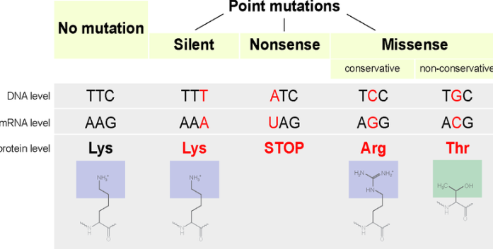
Base Substitution or Point Mutation
a single base at one point in the DNA sequence is replaced with a different base during replication
Can either be:
Transition: purine to purine or pyrimidine to pyrimidine
Transversion: purine to pyrimidine or vice versa
Consequences of Base Pair changes:
Missense mutation: changes a codon for one amino acid to a codon for another amino acid; results in an amino acid substitution in the protein product
Nonsense mutation: changes a codon for an amino acid with a codon for chain termination (UAG, UAA, UGA)
Silent mutation: a change in codon composition that has no effect on the resulting polypeptide
Frameshift Mutation - adds or deletes one or two bases (or any non-multiple of 3) from a coding sequence in a DNA, so that the genetic code is read out-of-phase
Consequence: incorrect amino acid or premature termination; severe phenotypic effects
Deletion - mutation in which a region of the DNA has been eliminated
Insertion - occurs when new bases are added to the DNA
Mutagens - physical or chemical agents that changes the genetic material
Physical Mutagens
High energy radiations
Penetrate living cells
Types: Electromagnetic radiations and Particulate radiations
Electromagnetic Radiations:
Gamma rays, X-rays, and ultraviolet rays
Penetration power is inversely proportional to their wavelength
Result (Gamma Rays and X-rays; ionizing radiation):
Direct effect: single or double stranded breaks in the DNA molecules
Indirect effect: free radicals created
Form compounds, hydrogen peroxide - initiate harmful chemical reactions within the cells
Can lead to cell death
Result (Ultraviolet rays; non-ionizing radiation):
Formation of pyrimidine dimers
Most are immediately repaired, but some escape repair and inhibit replication and transcription
Particulate Radiations:
In the form of sub-atomic particles emitted from the atoms with high energy
Alpha particles, beta particles, and neutrons
Penetrating power: beta particles > alpha particles because of its smaller size
Neutrons: extremely penetrant, can cause severe damage to the living tissues as well as genetic material
Result: single strand or double-strand breaks in the DNA
Chemical Mutagens - classified into 4 major groups, on the basis of their specific reaction within DNA
Deaminating Agents - cause the loss of the amino group (e.g. nitric oxide, nitrous acid, or N-nitrosoindoles)
Base Analogs - structurally resemble purines and pyrimidines and may be incorporated into DNA in place of the normal bases during DNA replication. They induce mutations because they often have different base-pairing rules than the bases they replace
Alkylating Agents - chemicals that donate alkyl groups (methyl, ethyl) to other molecules. The alkylated base may then degrade to yield a baseless site, which is mutagenic and recombinogenic, or mispair to result in mutations upon DNA replication
e.g. methylmethane sulphonate (MMS), ethyl methane sulfonate (EMS),N-methyl-N’-nitro-N-nitrosoguanidine (MNNG)
Intercalating Agents - molecules that may insert between bases in DNA, causing frameshift mutation during replication
E.g. acridine derivatives (acridine orange, proflavine, acriflavine), nitrogen mustards ethidium bromide
Gene Transfer Mechanisms in Prokaryotes:
Vertical Gene Transfer
movement of genetic material by descent where information travels through the generations as the cell divides
e.g. antibiotic resistance due to spontaneous mutation transferred directly to all the bacteria's progeny during DNA replication
Horizontal Gene Transfer
movement of genes between cells that are not direct descendants of one another.
3 mechanisms: Transformation, Conjugation, and Transduction
“10,000 unique genes flowing via HGT among 2,235 bacterial genomes," providing the bacteria with genetic information they didn't inherit from their parent cells.
multiple drug resistance
Recombination
the physical exchange of DNA between genetic elements
Horizontally-transferred DNA from a donor should undergo recombination with the recipient’s DNA in order to be retained in the recipient
Not necessarily for plasmids
Can replicate itself
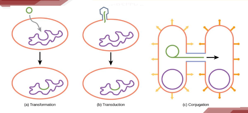
Transformation
the uptake of exogenous DNA from the environment.
involves recombination and integration in the recipient’s genome.
sources of exogenous DNA:
native bacterial chromosome fragments
Plasmid
bacteriophage DNA
Cell-cell contact is not required
Conjugation
plasmid-encoded mechanism that can mediate DNA transfer between bacterial cells
requires cell-to-cell contact (mating)
Occurs via a conjugal/membrane pore
DNA transfer occurs in one direction - from donor (contains F plasmid) to recipient not vice versa.
F plasmid (“F” stands for Fertility)
Found in donor cells which are designated as F+ cells.
Encode genes that allow conjugative transfer of genes.
F-encoded (sex) pilus
Allow specific pairing of donor (F+) and recipient cells (F-)
Transduction
the transfer of genetic information between cells through the mediation of a virus (phage) particle
does not require cell to cell contact.
common tool used by molecular biologists to stably introduce a foreign gene into a host cell’s genome on both bacterial and mammalian cells
Two ways:
Generalized transduction
DNA derived from virtually any portion of the host genome is packaged inside the mature virion in place of the virus genome
the bacterial donor genes cannot replicate independently and are not part of a viral genome
Specialized transduction
DNA from a specific region of the host chromosome is integrated directly into the virus genome— usually replacing some of the virus genes
Module 5: Microbial Metabolism
Metabolism - the series of biochemical reactions by which the cell breaks down or biosynthesizes various metabolites
Microbial Nutrients - Cell are primarily composed of elements C, H, O, N, P, and S. These chemical elements are predominant in the cell:
C is needed in the largest amount (50% of a cell’s dry weight)
O and H are next (combined, 25% of dry weight)
N follows (13%).
P, S, K, Mg, and Se combine for less than 5% of a cell’s dry weight
All microbes require a core set of nutrients
Macronutrients: required in large amounts
Micronutrients: required in minute amounts
E.g trace elements as co-factor of certain enzymes, vitamins as growth factors, and Iron (Fe) plays a major role in cellular respiration
Transporting Nutrients into the Cell
The active transport of nutrients into the cell is an energy-requiring process driven by ATP (or some other energy-rich compound) or by the proton motive force.
Three classes of transport systems: simple, group translocation, and ABC systems. Each functions to accumulate solutes against the concentration gradient
Simple Transport - reactions that are driven by the energy inherent in the proton motive force
Major Transport:
Symport reactions - solute and proton are cotransported in one direction
Antiport reactions - solute and a proton are transported in opposite directions
Group Translocation
Differs from simple transport in two important ways:
The transported substance is chemically modified during the transport process
An energy-rich organic compound (rather than the proton motive force) drives the transport event
ABC Systems - ‘ABC’ standing for ATP-binding cassette
ATP-binding cassette - a structural feature of proteins that bind ATP
Transport systems that employ a periplasmic binding protein along with transmembrane and ATP-hydrolyzing components
Energy Classes of Microorganism:
All microorganisms conserve energy from either the oxidation of chemicals or from light
Catabolism - energy-yielding reactions
Chemotrophs - organisms that conserve energy from chemicals
Chemoorganotrophs - use organic chemicals as their electron donors
Chemolithotrophs - use inorganic chemicals as their electron donors
Phototrophic organisms - convert light energy into chemical energy (ATP) and include both oxygenic and anoxygenic species
Heterotroph - cell carbon is obtained from one or another organic compound
Autotroph - uses carbon dioxide (CO2) as its carbon source; primary producers as they synthesize new organic matter from inorganic carbon (CO2)
Chemolithotrophs & Photothrops - most are autotrophs
Calvin Cycle - major biochemical pathway by which phototrophic organisms incorporate CO2 into cell material
Enzymes - protein catalysts that increase the rate of biochemical reactions by activating the substrates that bind to their active site; highly specific in the reactions they catalyze, and this specificity resides in the three-dimensional structures of the polypeptide(s) that make up the protein(s)
Redox - chemical reactions in the cell are accompanied by changes in energy, expressed in kilojoules; reactions either release or consume free energy
∆G0 is a measure of the energy released or consumed in a reaction under standard conditions and reveals which reactions can be used by an organism to conserve energy
Oxidation-reduction - reactions require electron donors and electron acceptors. The tendency of a compound to accept or release electrons is expressed by its reduction potential (E0’)
The substance oxidized (H2) as the electron donor, and the substance reduced (O2) as the electron acceptor.
Redox reactions in a cell often employ redox coenzymes such as NAD+/NADH as electron shuttles.
The energy released in redox reactions is conserved in compounds that contain energy-rich phosphate or sulfur bonds
ATP - prime energy carrier in the cell; consists of the ribonucleoside adenosine to which three phosphate molecules are bonded in series
Catabolism: Fermentation and Respiration - universal pathway for the catabolism of glucose is the Embden– Meyerhof–Parnas pathway, better known as glycolysis
Glycolytic pathway - used to break down glucose to pyruvate and is a widespread mechanism for energy conservation by fermentative anaerobes that employ substrate-level phosphorylation
The pathway releases a small amount of ATP (2–3/glucose) and large amounts of fermentation products. Besides glucose, the fermentation of other sugars, amino acids, nucleotides and polymeric compounds is possible
Respiration - offers an energy yield much greater than that of fermentation
Citric acid cycle - generates CO2 and electrons for the electron transport chain. The pathway by which pyruvate is oxidized to CO2
Glyoxylate cycle - necessary for the catabolism of two-carbon electron donors, such as acetate
Electron transport chains - composed of membrane associated redox proteins that are arranged in order of their increasing E0’ values
functions in a concerted fashion to carry electrons from the primary electron donor to the terminal electron acceptor, which is O2 in aerobic respiration
Biosyntheses - Polysaccharides are important structural components of cells and are biosynthesized from activated forms of their monomers
Gluconeogenesis - production of glucose from nonsugar precursors
Amino acids - are formed from carbon skeletons to which ammonia is added from glutamate, glutamine, or a few other amino acids
Nucleotides - biosynthesized using carbon skeletons from several different sources
Fatty acids - synthesized from the three-carbon precursor malonyl-ACP, and fully formed fatty acids are attached to glycerol to form lipids.
Only the lipids of Bacteria and Eukarya contain fatty acids
Module 6: Microbial Growth
The requirements for microbial growth can be divided into two main categories:
Physical Requirements
Temperature - Most microorganisms grow well at the temperatures that humans favor. Classified into three primary groups:
Psychrophiles - cold-loving microbes at 0⁰C
Mesophiles (moderate-temperature– loving microbes) - 25–40°C, are the most common type of microbe.
Thermophiles (heat-loving microbes) – 50-60⁰C.
Hyperthermophiles - have an optimum growth temperature of 80°C or higher.
Extreme thermophiles – 121⁰C and above.
pH - refer to acidity or alkalinity of a solution
Most bacteria grow best in narrow pH range near neutrality, between 6.5 and 7.5
Very few bacteria grow at an acidic pH below about pH 4. This is why a number of foods, such as sauerkraut, pickles, and many cheeses, are preserved from spoilage by acids produced by bacterial fermentation
Acidophiles are bacteria that loves acids environment
Osmotic Pressure - require water for growth, and their composition is 80–90% water
High osmotic pressures have the effect of removing necessary water from a cell
Hypertonic - whose concentration of solutes is higher than in the cell
Plasmolysis - shrinkage of cell cytoplasm
Types of Halophiles:
Extreme Halophiles - require high salt concentration
Obligate Halophiles - require 30% of salt for growth
Facultative Halophiles - require 15% of salt for growth
Chemical Requirements
Carbon
Sulfur, Nitrogen, and Phosphorus
Trace elements such as iron, copper, molybdenum, and zinc
Organic growth factors
Oxygen
Aerobes - microbes that use molecular oxygen
Anaerobes - microbes that do not use oxygen
Types of Oxygen Requirement
Obligate aerobe - organisms that require oxygen to live
Facultative anaerobe - ability to continue growing in the absence of oxygen; example: E. coli and yeast
Obligate anaerobe - bacteria that are unable to use molecular oxygen for energy-yielding reactions; example: Clostridium
Aerotolerant anaerobe - cannot use oxygen for growth, but they tolerate it fairly well; example: Lactobacilli
Microaerophile - they are aerobic; they do require oxygen. They grow only in oxygen concentrations lower than those in air
Cell Division and Population Growth
Binary Fission - forms a totally new daughter cell, with the mother cell retaining its original identity
Budding Division - forms a totally new daughter cell, with the mother cell retaining its original identity
Generation time - When one cell eventually separates to form two cells, we say that one generation has occurred
During one generation, all cellular constituents increase proportionally.
Each daughter cell receives a copy of the chromosome(s) and sufficient copies of ribosomes and all other macromolecular complexes, monomers, and inorganic ions to begin life as an independent entity.
Biofilms - attached polysaccharide matrix containing embedded bacterial cells
Quantification of Microbial Growth
Growth - an increase in the number of cells
Exponential growth - repetitive pattern where the number of cells doubles in a constant time interval
The increase in cell number in an exponentially growing bacterial culture can be expressed mathematically as a geometric progression of the number 2
As one cell divides to become two cells, we express this as 2^0 -> 2^1 . As two cells become four, we express this as 2^1 -> 2^2 and so on
Batch culture - enclosed vessel wherein bacteria grow quickly, and cell numbers increase dramatically in a short period of time
Growth curve - measures the rate of cell population increase over time, depicts growth
Microbial Growth Cycle
Lag phase - growth begins only after a period of time
Exponential or Log phase - cell population doubles at regular intervals
Stationary phase - cells in the population grow while others die
Death phase - growth ceases
Continuous culture - a type of open system that must be done in order to control both specific growth rate and cell density independently
Most common type is the chemostat
In the continuous culture growth vessel, a known volume of sterile medium is added at a constant rate while an equal volume of spent culture medium (which also contains cells) is removed at the same rate.
Culture media - laboratory cultures of microorganisms are grown in
Defined Media - prepared by adding precise amounts of pure inorganic or organic chemicals to distilled water. Exact composition is known
Complex Media - made from digests of microbial, animal, or plant products
Total Cell Count
Microscopic Counting - quick and easy way of estimating microbial cell numbers; can be performed either on samples dried on slides or on liquid samples
Stained samples - to increase contrast between cells and their background
Liquid samples - counting chambers consisting of a grid with squares of known area etched on the surface of a glass slide are used.
Viable cell - one that is able to divide and form offspring, and in most cell-counting situations
Methods of Viable Plate Count
Spread plate method - a volume (usually 0.1 ml or less) of an appropriately diluted culture is spread over the surface of an agar plate using a sterile glass spreader
Pour plate method - known volume (usually 0.1-1.0 ml) of culture is pipetted into a sterile Petri plate
Direct Measurement of Microbial Growth
Plate count - most frequently used method of measuring bacterial populations. Often reported as colony-forming units (CFU)
Serial dilution - to ensure that some colony counts will be within this range, the original inoculum is diluted several times in a process
Pour plates and spread plates
Pour Plate - colonies will grow within the nutrient agar (from cells suspended in the nutrient medium as the agar solidifies) as well as on the surface of the agar plate
Spread Plate - positions all the colonies on the surface and avoids contact between the cells and melted agar
Filtration - applied frequently to detection and enumeration of coliform bacteria, which are indicators of fecal contamination of food or water
Most Probable Number (MPN) method - the greater the number of bacteria in a sample, the more dilution is needed to reduce the density to the point at which no bacteria are left to grow in the tubes in a dilution series
microbes being counted will not grow on solid media (such as the chemoautotrophic nitrifying bacteria).
useful when the growth of bacteria in a liquid differential medium is used to identify the microbes (such as coliform bacteria, which selectively ferment lactose to acid, in water testing)
Direct Microscopic Count - a measured volume of a bacterial suspension is placed within a defined area on a microscope slide
Petroff-Hausser cell counter - used for direct microscopic counting
Coulter counter - used for electronic cell counters
Estimating Bacterial Numbers by Indirect Methods
Turbidity - As bacteria multiply in a liquid medium, the medium becomes turbid, or cloudy with cells
Spectrophotometer or Colorimeter - instrument used to measure turbidity
Metabolic Activity - assumes that the amount of a certain metabolic product, such as acid or CO2, is in direct proportion to the number of bacteria present
Dry Weight - the fungus is removed from the growth medium, filtered to remove extraneous material, and dried in a desiccator. It is then weighed
Module 7: Microbial Control
Sterilization - the removal or destruction of all living microorganisms
Heating - most common method used for killing microbes, including the most resistant forms, such as endospores
Filtration - method used to sterilize liquids or gases
Sterilization - the killing or removal of all microorganisms (including viruses), absolutely essential.
Decontamination - the treatment of an object or surface to make it safe to handle. As a result, it needs wiping off to remove fragments before using.
Disinfection - process that directly targets pathogens although it may not eliminate all microorganisms. It requires agents called disinfectants that actually kill microorganisms or severely inhibit their growth
2 Method in Microbial Control:
Physical Method - used extensively in industry, medicine, and home
Chemical Method - routinely used to control microbial growth; it may be a solid, liquid, or gases
3 Classes of Physical Control
Heat - the most widely used method of physically treating an object or substance to render it sterile; higher temperature and shorter time required to kill pathogens; 2 factors to heat:
Temperature
Time
How to quantify and characterize the Heat Sensitivity of Organism
Decimal Reduction Time - quantified by the time required for a 10-fold reduction in the viability of a microbial population at a given temperature
Heat killing proceeds more rapidly as the temperature rises.
The relationship between time (D) and temperature is exponential, as the logarithm of D plotted against temperature yields a straight line.
Interpretation: time and temperature are directly proportional with each other
Thermal Death Time - the time it takes to kill all cells at a given temperature
Greatly affected by population size
A longer time is required to kill all cells in a large population than in a small one.
Type of Heat
Moist Heat - has more penetrating power and inhibits growth or kills cells more quickly than does dry heat (e.g. autoclave and pasteurization)
Autoclave - sealed heating device that uses steam under pressure to kill microorganisms
Steam under a pressure of 1.1 kg/cm2 (15 lb/in2), which yields a temperature of 121°C for 15 mins.
It is not the pressure inside the autoclave that kills the microorganisms but the high temperatures that are achieved when steam is placed under pressure
Pasteurization - uses heat to significantly reduce rather than totally eliminate the microorganisms found in liquids, such as milk
Flash Pasteurization - 71°C for 15 seconds (or even higher temperatures for shorter time periods, after which it is rapidly cooled
Ultrahigh-temperature (UHT) pasteurization - of milk requires heat treatment at 135°C for 1–2 sec and actually sterilizes the milk such that it can be stored at room temperature for long periods without spoilage
Dry Heat - effective sterilization of metals, glasswares, some powders, oils and waxes (e.g. incineration and flaming)
Incineration (burning) - effective means of destroying contaminated disposable materials
Intense heat ignites and reduces microbes to ashes and gas
Limited to metals and heat-resistant glass materials
Flaming - accomplished by holding the end of the loop or forceps in the yellow portion of the gas flame.
flaming the surface metal forcep and wire bacteriologic loops is an effective way to kill microorganisms
Radiation
UV Radiation - useful for disinfecting surfaces and air.
widely used to decontaminate and disinfect the work surface of laboratory laminar flow hoods equipped with a “germicidal” UV light, air circulating in hospital and food preparation rooms
It has very poor penetrating power, limiting its use to the disinfection of exposed surfaces or air rather than bulk objects such as canned foods or surgical clothing
Ionizing Radiation - electromagnetic radiation of sufficient energy to produce ions and other reactive molecular species from molecules with which the radiation particles collide
Filtration - used to separate cells, larger viruses, bacteria, certain microbes from liquids or gases in which they are suspended.
Cotton plug in test tube, flask, pipette is a good filter for preventing the entry of microbe
Dry gauze and paper mask prevent the outward passage of microbes from the mouth and nose, protects from inhaling airborne pathogens and foreign particles that could damage the lungs
Type of Filters:
Depth Filter - important in biosafety applications such as in a biological safety cabinet
E.g. HEPA filter (does not ensure sterilization)
Membrane Filter - most common filters used for liquid sterilization
Nucleopore Filter - commonly used to isolate specimens for scanning electron microscopy
Chemical Method
Antimicrobial agent - natural or synthetic chemical that kills or inhibits the growth of microorganisms
-cidal agents - Agents that actually kill microbes. The prefix indicating the type of microorganism killed. (bactericidal, fungicidal, viricidal)
-static agents - -Agents that do not kill but only inhibit growth (bacteriotatic, fungistatic, viristatic)
Chemical Antimicrobial Agent
Sterilants - e.g. ethylene oxide or aldehydes such as formaldehyde or glutaraldehyde
Disinfectants - e.g. phenol and cationic detergents Sanitizers, by contrast, are less harsh than disinfectants and reduce microbial numbers but do not sterilize.
Antiseptics or germicides - chemicals that kill or inhibit the growth of microorganisms but are sufficiently nontoxic to animals to be applied to living tissues. Most germicides are used for hand washing or for treating surface wounds
Module 8: Microbial Pathogenesis
Pathogenesis - a manner in which the disease develops
Pathology - scientific study of disease. Concerned with the structural and functional changes brought about by disease and with their final effects on the body
Infection - is the invasion of pathogenic microorganisms in the body
Disease - abnormal state in which part or all of the body is incapable of performing normal functions
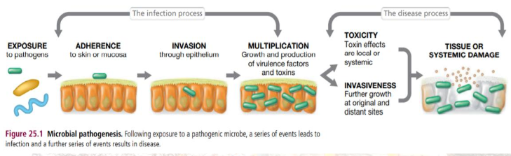
Adherence - enhanced ability of a microorganism to attach to a cell or a surface; structures:
Capsules - important for protecting pathogenic bacteria from host defenses besides adherence; capsule surface contains specific receptors that facilitate adherence to host tissues, but the inherently sticky nature of the capsule itself also assists in the overall attachment process
Pili - typically longer and fewer in number than fimbriae, and in addition to attachment, some pili function in the bacterial genetic transfer process of conjugation
Fimbriae (and Pili) - bacterial cell surface protein structures that function in attachment; function by specifically binding to host cell surface glycoproteins, thereby initiating adherence
Flagella - may also facilitate adherence of bacterial cells to host cells although their role is thought to be less important than that of fimbriae and pili
Portal of entry - mucous membranes, the skin surface, or under mucous membranes or the skin during penetration of these sites from puncture wounds, insect bites, cuts, or other abrasions; Pathogens gain access to host tissues by way of a portal of entry of one sort or another
Colonization - growth of a microorganism after it has gained access to host tissues, begins at birth as a newborn is naturally exposed to a suite of harmless (and in many cases necessary) bacteria and viruses that will be the infant’s initial normal microbiota
typically begins at sites in the mucous membranes.
Our human body is rich in organic nutrients and provides conditions of controlled pH, osmotic pressure, and temperature that are favorable for the growth of microorganisms
Mucus - a thick liquid secretion that contains water-soluble proteins and glycoproteins secreted by mucous membranes of epithelial cells; retains moisture and naturally inhibits microbial attachment because most microbes are swept away by physical processes like swallowing or sneezing
Nevertheless, some microbes—both pathogens and nonpathogens—adhere to the epithelial surface and colonize. If these attached microbes are pathogens, it sets the stage for infection, invasion, and disease
Invasion - e ability of a pathogen to enter into host cells or tissues, spread, and cause disease
Bacteremia - the mere presence of bacteria in the blood; this condition is typically self-limiting as the bacterial cells do not grow in the bloodstream and thus the immune system quickly removes them. The symptoms of bacteremia may be mild or none
Septicemia - bacteria multiply in the bloodstream and the organism spreads systemically from an initial focus and produces toxins or other poisonous substances
Viremia - term used to describe viruses present in the bloodstream
Normal flora or Normal Microbiota - microorganisms that colonize and reside in local part of the body that do not cause infection
Factors in the distribution and composition of normal microbiota:
Nutrients
Physical and chemical factors
Defences of the host
Mechanical factors
Microbial antagonism or competitive exclusion - normal microbiota protect the host against colonization by potentially pathogenic microbes by competing for nutrients, producing substances harmful to the invading microbes, and affecting conditions such as pH and available oxygen
Opportunistic pathogen - ordinarily do not cause disease in their normal habitat in a healthy person but may do so in a different environment
Physical and Chemical factors that can affect the growth of microorganism: temperature, pH, sunlight, available oxygen and carbon dioxide, salinity
Factors that also affect the normal microbiota: age, diet, disability, climate, lifestyle, personal hygiene, living conditions, occupation, emotional stress, nutritional status, health status, stress, geography, hospitalization
Pathogenicity - overall ability to cause disease
Virulence - ability of a pathogen to cause disease
Virulence factors - toxic or destructive substances produced by the pathogen that directly or indirectly enhance invasiveness and host damage by facilitating and promoting infection
Attenuation - decrease or loss of virulence of a pathogen
Bacterial Pathogens - damage host tissues (or the entire host) in two major ways:
By secreting tissue-destructing enzymes
By secreting or shedding toxins that target specific host tissues or the entire host
Viral Pathogens - damage host tissues by lysing cells directly, although some viruses are non-lytic and instead introduce genes into host cells that may eventually harm the host
Enzymes as Virulence Factors - Some pathogenic bacteria produce enzymes that function to either destroy host tissues or disarm host defenses. The activity of these enzymes releases nutrients to support growth of the pathogen and facilitate further invasiveness
Adherence, colonization, and infection by a pathogen, invasiveness requires the breakdown of host tissues and access to nutrients released from host cells. This is accomplished through the activity of enzymes that attack and destroy cells in one type of tissue or another.
Tissue destroying enzymes: hyaluronidase, collagenase, streptokinase, protease
Enzyme Activities on Mucosal Surfaces - Host mucosal surfaces are bathed in immune substances including enzymes such as lysozyme, an enzyme that cleaves the peptidoglycan of bacterial cells and promotes their osmotic lysis
Antibodies are also present on mucosal surfaces, in particular a class of antibody called IgA (example source: breastmilk). These “secretory antibodies,” as they are called, help prevent pathogen adherence to host tissues
Pathogens can produce enzymes both as offensive weapons
Objective - The invasiveness of the pathogen is increased and this allows it to ultimately extract more resources from its host
To destroy host tissue as defensive weapons
To destroy or inactivate offensive weapons of the host
AB - Type Exotoxins
Toxicity - ability of an organism to cause disease by means of a toxin that inhibits host cell function or kills host cells (or the host itself)
Exotoxins - toxic proteins and major virulence factors. Each exotoxin affects a specific host cell function
Enterotoxins - exotoxins that affect the small intestine
Three categories of Exotoxins:
AB toxins
Cytolytic toxins
Superantigen toxins
AB Toxins - two subunits, A and B
The B component binds to a host cell surface molecule, facilitating the transfer of the A subunit across the cytoplasmic membrane, where it damages the cell
Some of the best-known exotoxins are AB toxins, including those expressed in the diseases diphtheria, tetanus, botulism, and cholera
Diphtheria toxin - inhibits protein synthesis in eukaryotic cells
Neurological Exotoxins - Botulinum and Tetanus Toxins
Clostridium botulinum and Clostridium tetani
are endospore-forming bacteria commonly found in soil
both diseases are caused by the secretion of highly poisonous AB exotoxins that function as neurotoxins
Botulinum toxin and tetanus toxin both block the release of neurotransmitters that control muscle activities, but the mode of action and disease symptoms
flaccid paralysis in a botulism victim
botulinum toxin, which prevents muscle contraction,
tetanus toxin prevents muscle relaxation
Cholera Enterotoxin
Cholera - AB-type exotoxin produced by Vibrio cholerae, the causative agent of the waterborne disease cholera
characterized by massive fluid loss from the intestines, resulting in severe diarrhea, life-threatening dehydration, and electrolyte depletion.
The organism travels to the small intestine, where it colonizes and secretes cholera toxin
Cytotoxins and Superantigen Exotoxins
Cytotoxins and superantigens - toxic proteins that lyse host cells and trigger a massive immune response against host tissues
Cytotoxins (cytolytic exotoxins) - are soluble proteins secreted by a variety of pathogens. Cytotoxins damage the host cytoplasmic membrane, causing cell lysis and death
Hemolysis - on a blood agar plate is a classic cytotoxic effect, whereas toxic shock syndrome, a potentially fatal condition, is one result of superantigen activity
Endotoxin poisoning - symptoms include fever and intestinal distress
Toxic Shock Syndrome (TSS) - classic example of the systemic effects of a toxic superantigen and results from exposure to any of a series of exotoxins secreted during infection by certain strains of S. aureus or S. pyogenes
S. pyogenes TSS - typically the result of a systemic infection where bacteremia or septicemia is present and tissue damage including extensive tissue necrosis occurs.
Endotoxins - toxic lipopolysaccharides found in the cell walls of most gram-negative Bacteria
Cause a variety of physiological effects
Stimulates host cells to release cytokines
Lipopolysaccharide (LPS) - major component of the gram-negative cell outer membrane
LPS of three covalently linked subunits:
Membrane-distal O-specific polysaccharide
Membrane-proximal core polysaccharide
Lipid A - a phosphoglycolipid and the membrane anchoring portion of LPS
Fever - almost universal result of endotoxin exposure because endotoxin stimulates host cells to release cytokines, soluble proteins secreted by certain cells of the immune system that function as endogenous pyrogens, proteins that affect the temperature-controlling center of the brain, causing fever