Unit 1: Biological Bases of Behaviour
1.1 Interaction of Heredity & Environment
Nature vs Nurture influences:
personality characteristics & persona; preference
intelligence & psychological health
language acquisition
Nature vs Nurture
genetic influences on behaviour and traits
environmental factors shaping individual experiences
the interplay between heredity and upbringing in development
Noam Chomsky (nature side): believed that stuff like language acquisition is in our nature to learn and that’s why babies pick it up so easily.
John B Watson (nurture side): said that he could train a dozen healthy infants to be anything he wanted to, because he believed that a person’s traits are influenced by their environment & condition
This debate highlights the ongoing discussion in psychology regarding the extent to which genetics versus environment influences human behavior and development.
Epigenetics
helps us understand how genetics & nurture influence us
Rat licking studies: research that showed how environment can alter genetics
Twin Studies
Identical Twins: same DNA, same sex, monozygotic (one egg).
Fraternal Twins: different DNA, no more genetically similar than non-twin siblings, might be same/opposite sex, dizygotic (two eggs).
Example - Minnesota Twin Study: separated identical twins at birth for 35 years; when brought back together, they had insane similarities — wives had the same name, had dogs of the same name, named their sons almost the same thing etc.
Heritability
the amount of variation among individuals that we can attribute to genetics
provides insight into nature vs nurture
studied in twin studies, adoption studies, family studies, DNA studies.
1.2 The Nervous System
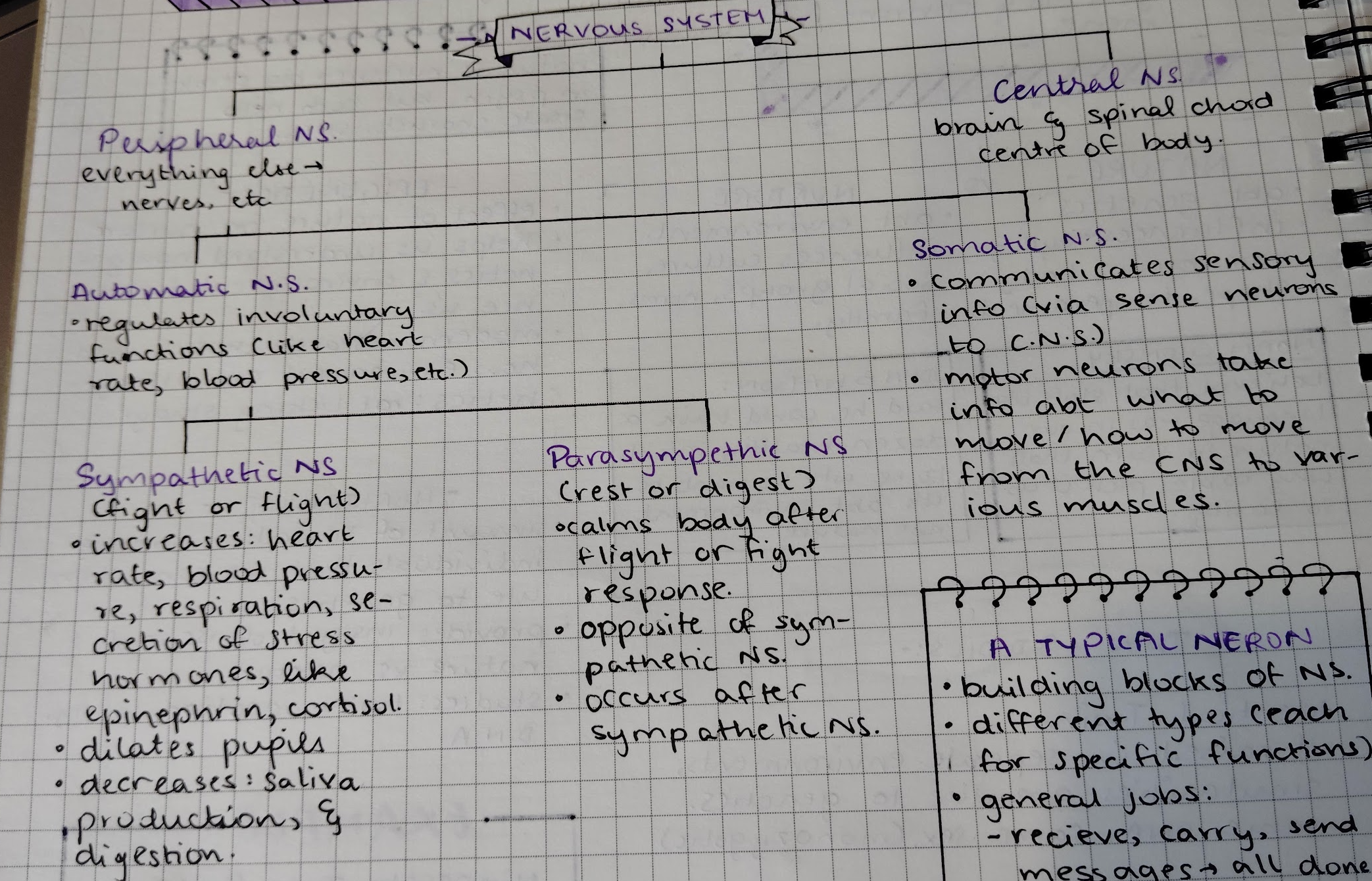
Neurons
building blocks of nervous system
general jobs: receive, carry, send message (all done within milliseconds, speed approx 30 mph).
neuron cells fire in a particular patter for thoughts to occur.
Types of a Neuron
Sensory Neurons: measures sensation.
respond to non-chemical stimulation.
receives raw materials from the body’s sense organs like free nerve endings in the epidermis.
carry messages from the senses to the brain (ARRIVE at the brain).
Motor Neurons: measures reflexes.
are connected to muscle fibres and can make the muscles contract.
can react to voluntary and involuntary signals.
carry messages from the brain to the senses (EXIT the brain).
Interneurons: connect neurons within the brain and spinal cord.
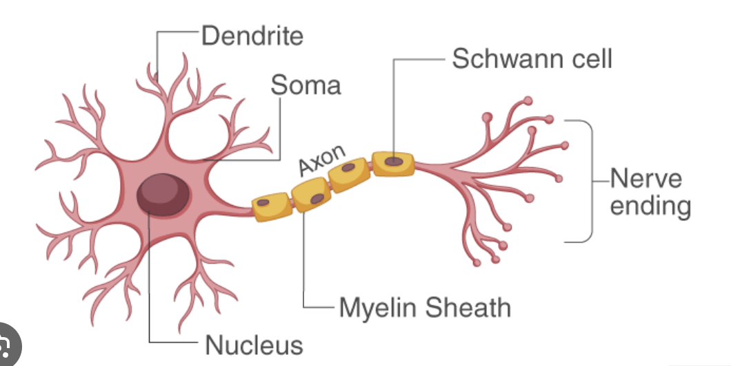
Structure of a Neuron
Dendrites: Receive signals from other neurons.
Cell Body (Soma): Contains the nucleus and processes signals.
Axon: Transmits signals away from the cell body to other neurons.
Myelin Sheath: Insulates the axon to speed up signal transmission.
Axon Terminals: Release neurotransmitters to communicate with other neurons.
Synapse: The junction where communication between neurons occurs via neurotransmitters.
Neural Firing
Basics of Neural Firing
Resting Potential: Polarised (-70 millivolts).
Firing Threshold: Reached if enough volts are there.
All or None Law: Once at threshold, the neuron WILL fire, and it will fire at the same intensity every time.
Action Potential: An electric impulse that travels down the axon. This change in electricity creates a positive electric charge (+30 millivolts) inside the neuron - this is called depolarisation.
Refractory Period: A brief period when the neuron can’t fire again.
Reuptake: The sending neuron recollects neurotransmitters.
Process of Neural Firing
Neurons fire when there is a shift in electric energy - creating an action potential.
When the action potential reaches the terminal buttons, neurotransmitters are released into the synapse.
Neurotransmitters then lock into the next neuron.
Summary of Neural Firing
Neuron fires → Synaptic vesicles release neurotransmitters from axon terminals → Neurotransmitters travel across the synapse & fit into the post-synaptic receptors.
1.3 Neurotransmitters
chemical messengers of the nervous system.
excitatory - pushes neurons "accelerator" - encites response
inhibitory - pushes neurons "brake" - blocks response
antagonist - binds to dendrites & prevents or blocks a response.
agonist - bind to receptor sites & mimic the affects of a specific neurotransmitter.
Types of Neurotransmitters
Neurotransmitter | Function | Malfunction |
Glutamate (major exhibitory NT) | enhances learning & memory by strengthening synaptic connections. | oversupply → migraines & seizures. |
GABA (major inhibitory NT) | associated with anxiety-related disorders. | undersupply → depression, anxiety, moodiness. |
Acetycholine (ACh) | involved in movement, learning, memory, etc. | undersupply → alzheimer’s. |
Dopamine | influences movement, learning, attention, emotion. | undersupply → parkinson’s, oversupply → schizophrenia. |
Endorphins | involved in pain reduction & reward. | undersupply → depression. |
Epinephrin | both a NT & hormone - boosts energy. primary chemical in “fight or flight” response. | undersupply → low blood pressure, depression, anxiety, etc. |
Norepinephrin | arousal, alertness, sleep cycle. | undersupply → depression. |
Serotonin | mood, apetite, sleep & dreams. | undersupply → depression. |
Agonists
Agonists enhance the actions of neurotransmitters.
Direct Agonists
Direct Agonists mimic the neurotransmitters and bind with the receptors of the next neurons.
Examples:
Heroin
agonist for endorphins.
mimic → receptor sites can’t distinguish between an endorphin and the chemical structure of heroin.
Nicotine
agonist for acetycholine.
stimulates skeletal muscles & causes increased heart rate.
Black Widow Venom (a toxin, not a drug)
agonist for acetycholine.
causes ACh to be continuously released at neuromuscular junctions.
increase in heart rate, spasms → leads to death.
Indirect Agonists
Indirect Agonists block the reuptake of a neurotransmitter; thus are also known as reuptake inhibitors. (these are introduced at the synapse).
Examples:
Prozac
inhibits the reuptake of serotonin → floods the synapse with serotonin.
decreases symptoms of depression & anxiety for some people.
Cocaine
inhibits reuptake of dopamine.
also decreases symptoms of depression & anxiety, can slow down parkinson’s effects.
Antagonists
inhibit the actions of neurotransmitters in many ways.
often bind to a receptor but do not stimulate it.
block a neurotransmitter from being released by the terminal or from binding to the receptor site.
this inhibits the normal functions of an neurotransmitter.
Examples:
Botox (a toxin)
an antagonist for ACh.
blocks ACh from reaching receptors.
effected muscles can’t move → freeze muscle from firing to prevent wrinkles.
Thorazine
an antagonist for dopamine.
blocks dopamine receptors and starves the synapse of this neurotransmitter.
early drug for schizophrenia (as oversupply of dopamine is linked to schizophrenia and this slows down the supply of it).
Psychoactive drugs
What do all psychoactive drugs have in common?
they alter mental states.
activate dopamine-producing neurons in the brain’s reward system.
this increase in dopamine is associated with greater reward, which can lead to a stronger desire to take the drug again.
many drugs can create tolerance - needing increasing amounts of the drugs to create the original high/desired effect.
many drugs lead to physical dependance - with repeated use, a person may need to administer the drug to prevent withdrawal symptoms (here, drug acts as a negative reinforcement to take these symptoms away).
the effect of a drug is primarily dependant on which neurotransmitter is affected.
Blood-Brain Barrier: barrier that allows some chemicals to pass from the blood into the brain but prevents other chemical structures from entering.
Categories of Psychoactive drugs: Depressants, Opiates, Stimulants, Hallucinogens / Psychedelics.
Depressants
show or inhibit CNS functions.
create drowsiness, sedation or sleep → relieve anxiety or lower inhibitions.
combining depressants can be deadly.
Examples:
Alcohol
2nd most widely used depressant in the U.S.
agonist for GABA (gamma-aminobutyric acid).
lessens inhibitions by depressing brain centres for judgement and self-control.
Opiates/Opiods
agonist for endorphins.
incredibly addictive & creates powerful withdrawal symptoms.
Examples: heroin, oxytocin, fentanyl.
Stimulants
active sympathetic nervous system.
increase brain activity, arouse behaviour, & increase mental alertness.
Examples:
Caffiene
most widely used drug in the U.S.
promotes wakefulness, mental alertness, & faster thought processes by stimulating release of dopamine.
antagonist for adenosine → blocks sleep-inducing effects.
is physically addictive and creates withdrawal symptoms.
Cocaine
dopamine antagonist (reuptake inhibitor).
also elevates serotonin & norepinephrin.
creates intense euphoria, alertness, & heightened self-confidence.
highly addictive & causes a crash after the high dissipates.
Hallucinogens / Psychedelics
create sensory & perceptual disorders, alter mood, and affect thinking.
there is lots of current research on psychedelics in therapeutic settings (anxiety, depression & more) but it’s still in the experimental phase.
Examples:
Tetrahydrocannabinol (THC or cannabis)
very mind hallucinogen.
produces sense of well-being, mild euphoria, dreamy state of relaxation.
interferes with muscle coordination, learning, memory, and overall cognitive function.
has various therapeutic uses.
1.4 The Brain
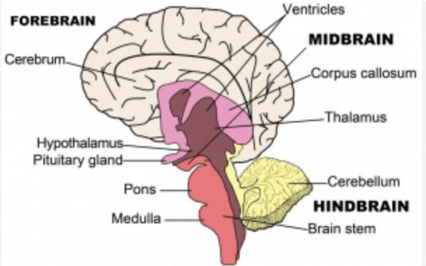
Structure of a Brain
Structure of the brain divided into three main regions:
Hindbrain: Bottom of the brain.
Midbrain: Center region, connects upper and lower brain parts.
Forebrain: Top region containing central structures and outer layer.
Hindbrain
Located at the base of the brain, near the brain stem.
Brain Stem
Function: Responsible for essential life functions.
Medulla Oblongata
Location: At the bottom of the brain stem, above the spinal cord.
Function: Automatic functions (heart rate, breathing, blood pressure), reflex coordination (swallowing, coughing, sneezing).
Description: Looks like a small ice cream cone shape.
Pons
Note: Not an essential concept according to College Board.
Description: Resembles an Adam's apple/curved letter P.
Function: Unconscious processes (sleep/wake cycles).
Reticular Activating System (RAS)
Description: A bundle of nerve fibers running through the brain stem.
Function: Influences consciousness and alertness.
Example: Electrical stimulation in a cat caused a coma, illustrating its role in consciousness.
Cerebellum
Location: Underneath the backside of the brain, behind the brain stem.
Description: About the size of a fist, referred to as the "little brain."
Function: Coordinates voluntary movements, balance, posture, and fine-tuning motor activities.
Role in learning motor skills and refining movements.
Midbrain
nerve system connecting higher and lower portions of the brain.
relays info between the brain & the eyes & the ears.
Forebrain
Thalamus
Location: Egg-shaped structures deep within the brain, above the brain stem.
Function: Relay system for sensory information directing it to appropriate brain areas for interpretation.
Role in regulating alertness and consciousness.
Limbic System
Location: Encircles the top of the brain stem, includes the hippocampus, amygdala, and hypothalamus.
Function: Emotional and behavioral responses.
Hippocampus
Location: The green arm-like structures in the limbic system.
Function: Memory formation, especially in long-term memory consolidation.
Amygdala
Location: Blue almond-shaped structures at the ends of the hippocampus.
Function: Processes emotions (fear, aggression, pleasure), recognizes emotional experiences, influences emotional memory formation.
Hypothalamus
Location: Center of the limbic system, maroon triangular shape.
Function: Regulates basic drives (hunger, thirst, body temperature), connects nervous system to endocrine system via pituitary gland.
Cerebrum
Location: Largest part covering the limbic system, thalamus, and brain stem.
Function: Higher brain functions (thinking, reasoning, planning, problem-solving) and sensory processing.
Cerebral Cortex: Outer layer responsible for complex functions.
Corpus Callosum
Description: Thick band of nerve fibers connecting right and left hemispheres.
Function: Facilitates communication between hemispheres for coordinated complex functions (movement, language, problem-solving).
Pituitary Gland
Location: Small, pea-sized structure below hypothalamus.
Function: "Master gland" of the endocrine system, regulates growth, metabolism, stress responses through hormone production; works closely with the hypothalamus.
Cerebral cortex structure
The cerebral cortex is the most evolutionarily advanced part of the brain, responsible for our most sophisticated mental capabilities. It consists of two hemispheres connected by the corpus callosum, with each hemisphere specialized for different types of processing.
The cortex is organized into distinct regions:
Right hemisphere:
Processes spatial info
Handles nonverbal communication
Takes a big-picture approach Left hemisphere:
Manages language processing
Handles logical operations
Processes analytical tasks Four main lobes plus the limbic system:
Frontal, parietal, temporal, and occipital lobes
Limbic system includes structures crucial for emotion and memory
Occipital lobe
The occipital lobe is dedicated to processing visual information. Located at the back of the brain, it contains multiple specialized areas for different aspects of vision. 👀 🖼
Main functions:
Processes visual information from the eyes
Interprets color, shape, depth, and motion
Damage can cause visual deficits (blindness, agnosia, hallucinations)
Temporal lobe
The temporal lobe processes auditory information and plays a crucial role in memory and emotion. Located on the sides of the brain, it contains several important structures including the hippocampus and amygdala. 😭💬
This region is responsible for:
Processing auditory information and language comprehension via Wernicke's area
Memory formation and retrieval via the hippocampus
Emotion, social perception, and memory via the amygdala
Damage can affect hearing, language, or personality
Parietal lobe
The parietal lobe integrates sensory information and helps us understand our position in space. It serves as a hub for processing various types of sensory input and coordinating responses.
Critical functions include:
Processing sensory information (touch, pressure, temperature, pain)
Integrating sensory input with motor output
Spatial processing and navigation
Body awareness and attention
Frontal lobe
The frontal lobe is the seat of our highest cognitive abilities and personality. It's involved in planning, decision-making, and emotional regulation.
This region handles:
Higher-order cognitive processes (planning, decision-making, problem-solving)
Voluntary movements and fine motor skills via motor cortex
Emotional responses and social behavior
Language production via Broca's area
Split brain research
Split-brain studies have provided remarkable insights into how the two hemispheres of the brain function independently and together. This research began as a treatment for severe epilepsy but revealed fundamental principles about brain organization.
Key findings include:
Demonstrates specialized functions of left and right hemispheres
Shows how the brain processes information in isolation
Reveals the brain's adaptive capabilities
Language areas in brain
Language processing involves multiple specialized regions working together. The two main areas are Broca's and Wernicke's areas, each serving distinct functions in language use. Most language functions are lateralized to the left hemisphere.
Broca's area (left frontal lobe)
Controls speech production
Damage leads to trouble producing speech Wernicke's area (left temporal lobe)
Manages language comprehension
Damage results in comprehension difficulties
Cortex specialization testing
Research on cortical specialization helps us understand how different brain regions process information. This is particularly evident in split-brain patients, where researchers can study each hemisphere in isolation.
Testing methods include:
Presenting stimuli to specific visual fields
Studying hemispheric differences in perception
Examining contralateral organization of pathways These tests reveal:
Hemispheric differences in perception
Specialized cognitive processing
Motor pathway organization
Brain plasticity concept
Brain plasticity refers to the brain's remarkable ability to change and adapt throughout life. This property allows for learning, memory formation, and recovery from injury.
Key aspects of plasticity:
Formation of new neural connections
Strengthening or weakening of existing connections
Most pronounced during development but continues throughout life
Enables compensation for damage
Brain research methods
Modern neuroscience employs various techniques to study brain structure and function. Each method provides unique insights into brain activity and organization.
Current research tools include:
EEG for measuring electrical activity
fMRI for tracking blood flow changes
PET scans for visualizing metabolism and neurotransmitter activity
Case studies of brain lesions
Transcranial magnetic stimulation (TMS) for temporary disruption studies
Optogenetics for precise neural control
1.5 Sleep
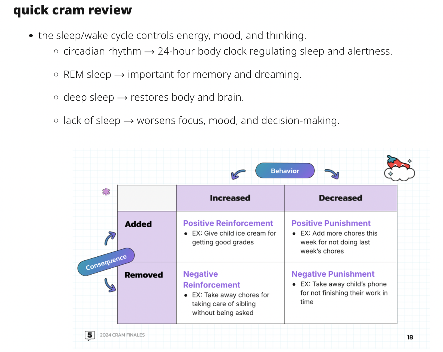
*ultradian rhythm: every 90 mins.
Sleep/wake cycle effects on behavior
Levels of consciousness
Consciousness = how aware we are of stuff going on inside and outside our bodies.
😳 During wakefulness, we experience:
High levels of awareness and responsiveness
Active engagement with our environment
Clear perception of stimuli
😴 In contrast, sleep is characterized by:
Reduced awareness of surroundings
Decreased responsiveness to external stimuli
Altered states of consciousness
Circadian rhythm and disruptions
Our body's internal clock runs on about a 24-hour cycle, controlled by the suprachiasmatic nucleus (SCN) in the brain. This biological timekeeper responds to environmental cues, especially light and dark.
Common disruptions include:
Jet lag from crossing time zones
Shift work schedules, especially if they keep changing
Irregular sleep schedules
Exposure to artificial light at night (aka doom scrolling)
These disruptions can lead to:
Trouble falling asleep, staying asleep, or staying awake
Feeling foggy-headed
Changes in appetite (hangry) or mood
Feel physically off (tired, queasy)
Stages of sleep
Sleep progresses through multiple stages throughout the night, each serving distinct biological purposes.
NREM sleep consists of three stages:
Stage 1: Light sleep transition
Brief duration
Easily awakened
Hypnagogic sensations
Stage 2: Intermediate sleep with some specific brain wave patterns
Sleep spindles appear
K-complexes present on EEG
Decreased body temperature
Stage 3: Deep sleep with slow brain waves
Slow-wave activity
Difficult to wake
Critical for physical restoration
These stages change throughout the night, with the deepest sleep happening earlier on.
REM sleep characteristics
REM sleep is a unique state that combines aspects of both sleep and wakefulness. During this phase, the brain becomes highly active while the body remains largely paralyzed.
Key features include:
Rapid eye movements (even though they are closed)
Muscle atonia (so you don't act out your dreams)
Vivid dreaming
Increased brain activity
The timing and duration of REM periods follow a predictable pattern:
First REM period occurs about 90 minutes after falling asleep
Later REM periods become progressively longer, with longest happening in the morning
REM rebound occurs after REM deprivation
Theories of dream function
Dreams serve multiple potential functions in cognitive processing and emotional regulation. The two main scientific theories explain dreams through different mechanisms.
Activation-synthesis theory proposes that:
Dreams result from the brain interpreting random neural firing
The brain creates narratives to make sense of this activity
Dream content reflects this synthesis process
Memory consolidation theory suggests that:
Dreams help process daily experiences
Emotional memories are integrated during dreaming
Dream content often incorporates recent events
🚫 Exclusion Note: The psychoanalytic theory of dreams is not assessed on the AP Psych exam.
Sleep's role in memory
Sleep plays a vital role in learning and memory formation. During sleep, the brain processes and stabilizes new information while clearing away unnecessary data. aka you need to sleep before big tests!
Memory consolidation occurs through:
Neural replay of daytime experiences
Strengthening of important synaptic connections
Transfer of information to long-term storage
The restoration process involves:
Clearing metabolic waste from the brain
Replenishing neurotransmitters
Optimizing neural networks for new learning
Sleep disorders and effects
Sleep disorders can significantly impact daily functioning and overall health. Understanding these conditions is crucial for proper diagnosis and treatment.
Common sleep disorders include:
Insomnia (difficulty falling or staying asleep)
Narcolepsy (excessive daytime sleepiness or suddenly falling asleep)
Sleep apnea (interrupted breathing during sleep)
REM sleep behavior disorder (physically acting out dreams)
Somnambulism (sleepwalking)
🚫 Exclusion Note: The AP Psych exam will only cover the sleep disorders listed above.
These conditions can lead to various effects:
Cognitive impacts:
Reduced attention span
Impaired decision-making
Memory problems
Physical effects:
Increased accident risk
Weakened immune system
Weight gain
Cardiovascular issues
Emotional consequences:
Mood swings
Irritability
Increased anxiety
Depression risk
1.6 Sensation
Sensation is how we detect and process environmental stimuli. Our sensory systems work together to pick up information from light, sound, taste, and touch.
Our brains take this sensory input and turn it into meaningful perceptions. This includes getting used to constant stimuli, noticing changes, and sometimes experiencing weird sensory combos like synesthesia. Understanding sensation helps explain how we interact with the world around us.
Sensation and behavior
Detection of sensory information
Sensation begins when environmental stimuli reach our sensory organs and are converted into neural signals. This process requires stimuli to meet certain thresholds before being detected and processed by the brain.
Key detection concepts:
Absolute threshold: minimum stimulus intensity detected 50% of the time
Just-noticeable difference: smallest detectable change in stimulus intensity
Sensory adaptation: decreased sensitivity to constant stimulation Our senses rarely work alone. Instead, they team up through:
Cross-modal processing combining multiple senses
Sensory interaction enhancing overall perception
Unique phenomena like synesthesia where senses overlap
Change detection and adaptation
Our sensory systems are particularly attuned to detecting changes in stimulation rather than constant levels of stimulation. This helps us notice important environmental changes while conserving energy by reducing responses to ongoing stimulation.
Weber's law governs how we detect differences:
The just-noticeable difference (JND) is proportional to stimulus intensity
Larger changes needed to detect differences in stronger stimuli
Applies across different sensory modalities Adaptation helps us:
Tune out constant background noise
Stay sensitive to new or changing stimuli
Adjust to different environments
Optimize sensory processing for what's happening now
Sensory interaction and synesthesia
The brain integrates information from multiple senses to create coherent experiences. This integration happens automatically and continuously, enhancing our ability to understand and navigate our environment.
Common sensory interactions include:
Taste enhanced by smell and visual cues
Speech comprehension improved by watching lip movements
Balance maintained through vision and inner ear input Synesthesia represents an unusual form of sensory interaction where:
One sensory experience automatically triggers another
Associations are consistent and automatic
Experiences can involve any combination of senses and can boost memory and creativity
Visual system and behavior
Retina and image processing
The retina serves as the primary visual receptor, converting light into neural signals. This complex tissue contains multiple cell layers that begin processing visual information before it reaches the brain.
Initial processing includes:
Detecting light intensity
Basic edge and motion detection
Color processing in cone-rich areas The brain makes up for retinal limitations by:
Filling in the blind spot
Maintaining perceptual stability
Integrating information from both eyes
Lens accommodation and vision
The lens adjusts to focus images clearly on the retina. This process of accommodation involves:
Lens shape changes for near and far vision
Pupil size adjustments for light intensity
Eye muscle coordination for binocular vision Vision problems can occur when:
Myopia: images focus in front of the retina (nearsightedness)
Hyperopia: images focus behind the retina (farsightedness)
Astigmatism: irregular cornea shape causes distortion
Rod cells and light adaptation
Rod cells give us vision in low light and are crucial for detecting movement in our peripheral vision. These cells adapt significantly as lighting conditions change.
Light adaptation happens fast when entering bright areas:
Rod sensitivity decreases
Cone cells become more active
Pupil constricts to reduce light entry Dark adaptation is slower and involves:
Increased rod sensitivity
Reduced cone activity
Pupil dilation
Rhodopsin regeneration
Theories of color vision
Color vision relies on multiple mechanisms working together. Two main theories explain how we perceive color:
Trichromatic Theory explains initial color processing:
Three types of cone cells – 💙 short-wavelength (blue), 💚 medium-wavelength (green), and ❤ long-wavelength (red)
Each responds to different wavelengths
Combining signals creates color perception Opponent-Process Theory describes how the brain processes color information:
Opposing pairs of colors (red-green, blue-yellow)
Black-white opposition for brightness
Explains afterimages and color contrast effects
Brain damage and vision disorders
Damage to visual processing areas can create unique disorders that reveal how the visual system works. The complexity of vision becomes apparent through various conditions:
Common disorders include:
Prosopagnosia (face blindness)
Blindsight
Visual agnosia Impact varies based on:
Location of damage
Extent of injury
Timing of damage during development
Auditory system and behavior
Sound perception and processing
Sound travels through air as pressure waves at various frequencies and amplitudes. Our auditory system converts these waves into neural signals that we interpret as meaningful sounds.
Key sound properties include:
Pitch determined by wave frequency (measured in Hz)
Loudness determined by wave amplitude (measured in dB)
Timbre determined by sound wave complexity The ear processes sound through:
Outer ear collecting and channeling sound waves
Middle ear amplifying vibrations
Inner ear converting mechanical energy to neural signals
Theories of pitch perception
Multiple theories work together to explain how we perceive pitch across different frequency ranges. Each theory addresses specific aspects of auditory processing.
Place theory explains high-frequency perception:
Different frequencies stimulate different areas of the basilar membrane
Higher frequencies activate the base of the cochlea
Lower frequencies activate the apex of the cochlea Frequency theory works for lower pitches:
Neurons fire at the same rate as sound wave frequency
Works best below 1000 Hz
Neural firing patterns match sound wave patterns Volley theory handles mid-range frequencies:
Groups of neurons fire in alternating patterns
Multiple neurons together can represent frequencies up to 4000 Hz
Combines aspects of both place and frequency theories
Sound localization mechanisms
Our ability to locate sound sources in space relies on comparing input between our ears and integrating this with other sensory information.
Localization depends on:
Interaural time differences (sound reaches one ear before the other)
Interaural intensity differences (sound is louder in one ear)
Head-related transfer functions (how the ear shape filters sound) The brain processes these cues in the:
Superior olive (initial binaural processing)
Inferior colliculus (integration of spatial information)
Auditory cortex (conscious perception of sound location)
Hearing loss and disorders
Hearing impairment can result from various factors and affect different parts of the auditory system. Understanding these conditions reveals how our hearing system functions.
Conduction deafness involves:
Problems in the outer or middle ear
Difficulty transmitting sound waves to the cochlea
Often temporary and treatable
Caused by earwax buildup, ear infections, or ossicle damage Sensorineural deafness involves:
Damage to the cochlea or auditory nerve
Usually permanent hearing loss
Common causes include aging (presbycusis), loud noise exposure, and certain medications
Often treated with hearing aids or cochlear implants Other hearing conditions include:
Tinnitus (phantom ringing or buzzing sounds)
Auditory processing disorders (brain struggles to process sound)
Hyperacusis (increased sensitivity to everyday sounds)
Chemical sensory systems and behavior
Olfactory (smell)
The olfactory system detects airborne chemicals and converts them into meaningful smell perceptions. It's the only sense not processed first in the thalamus.
Olfactory processing involves:
Odorant molecules binding to receptors in nasal epithelium
Signals traveling directly to the olfactory bulb
Information bypassing the thalamus (unique among senses)
Direct connections to the limbic system for emotional processing Pheromones represent specialized chemical signals that:
Communicate between members of the same species
May influence mood, attraction, and physiological states
Are processed by the vomeronasal organ in many mammals
May play subtle roles in human behavior
Basic taste qualities and perception
Gustation allows us to evaluate what we're about to consume. This chemical sense helps us identify nutritious foods and avoid potential toxins.
The primary taste qualities include:
Sweet (sugars and some proteins)
Sour (acids)
Salty (sodium and essential minerals)
Bitter (potentially toxic compounds)
Umami (savory, protein-rich foods)
Oleogustus (fatty acids) Taste receptor distribution creates:
Supertasters with abundant taste buds and heightened sensitivity
Medium tasters with average taste perception
Nontasters with fewer taste buds and reduced sensitivity
Gustatory structures and taste sensitivity
Taste information follows specific neural pathways from the tongue to conscious perception. This processing helps us make rapid decisions about food consumption.
The taste system includes:
Taste buds containing specialized receptor cells
Cranial nerves carrying taste information
Brainstem nuclei for initial processing
Thalamic relay to the gustatory cortex
Integration in the orbitofrontal cortex Taste preferences develop through:
Innate preferences for sweet and umami
Innate aversions to bitter
Cultural and personal experience
Conditioning and learning
Interaction between taste and smell
Flavor perception results from the integration of multiple sensory inputs. This multisensory experience enhances our ability to identify and remember foods.
Taste and smell interact through:
Retronasal olfaction during chewing and swallowing
Shared neural pathways in the orbitofrontal cortex
Complementary information processing Without smell, taste perception is:
Limited to basic taste qualities
Significantly reduced in intensity
Missing the complexity we call "flavor"
Often described as "bland" or "flat" Other factors influencing flavor include:
Texture (somatosensory input)
Temperature
Visual appearance
Sound (crunchiness)
Prior expectations
Touch sensory system and behavior
Somatosensory receptors and processing
The tactile system provides crucial information about objects we contact and our position in space. Various receptor types in the skin detect different aspects of touch.
Specialized mechanoreceptors include:
Merkel cells for pressure and texture
Meissner corpuscles for light touch and vibration
Pacinian corpuscles for deep pressure and rapid vibration
Ruffini endings for skin stretch and joint position Neural pathways for touch include:
Sensory neurons carrying signals to the spinal cord
Ascending pathways to the thalamus
Projections to the somatosensory cortex
Secondary processing in association areas
Temperature perception mechanisms
Temperature sensation helps us maintain homeostasis and avoid tissue damage. Our perception of hot and cold relies on specialized thermoreceptors.
Temperature processing involves:
TRPM8 receptors activated by cold
TRPV1 receptors activated by heat
Paradoxical activation creating mixed sensations The sensation of "hot" results from:
Simultaneous activation of warm and cold receptors
Integration of these signals in the central nervous system
Contextual interpretation based on baseline temperature
Cross-activation of pain receptors at extreme temperatures
Vestibular and kinesthetic systems and behavior
Vestibular structures and balance
The vestibular system provides constant information about head position and movement. This system is essential for maintaining balance and coordinating movements.
Vestibular processing involves:
Semicircular canals detecting rotational movements
Otolith organs (utricle and saccule) sensing linear acceleration
Hair cells converting mechanical movement to neural signals
Vestibular nuclei in the brainstem integrating signals Balance maintenance relies on:
Vestibular input about head position
Visual information about the environment
Proprioceptive feedback from joints and muscles
Cerebellar integration of these sensory inputs
Kinesthetic sensing and movement
Kinesthesis gives us awareness of body position and movement without visual input. This proprioceptive sense allows for smooth, coordinated actions.
Key kinesthetic structures include:
Muscle spindles detecting muscle stretch
Golgi tendon organs monitoring tension
Joint receptors sensing position
Somatosensory cortex integrating body position information Kinesthesis enables:
Coordinated movements without visual monitoring
Automatic postural adjustments
Spatial awareness of limb positions
Skilled motor learning through body awareness
Unit Overview
What's This Unit All About?
Explores the biological underpinnings of behavior and mental processes
Focuses on the structure and function of the brain and nervous system
Examines how neurons communicate with each other to transmit information
Investigates the role of neurotransmitters in regulating behavior and mood
Discusses the influence of hormones on behavior and psychological processes
Covers the central and peripheral nervous systems and their respective functions
Delves into various brain disorders and their effects on behavior and cognition
Highlights real-life applications of understanding the biological bases of behavior (treating mental illnesses, developing medications, improving learning and memory)
Key Brain Parts and What They Do
Cerebral cortex: responsible for higher-order cognitive functions (thinking, reasoning, decision-making)
Divided into four lobes: frontal, parietal, temporal, and occipital
Each lobe specializes in processing specific types of information (visual, auditory, sensory, motor)
Limbic system: involved in emotional processing, memory formation, and motivation
Includes the amygdala, which plays a crucial role in fear and emotional responses
Hippocampus is essential for forming new memories and spatial navigation
Hypothalamus: regulates basic biological needs and drives (hunger, thirst, sleep, sexual behavior)
Controls the release of hormones from the pituitary gland
Brainstem: connects the brain to the spinal cord and regulates vital functions (breathing, heart rate, blood pressure)
Cerebellum: coordinates fine motor movements, balance, and posture
Basal ganglia: involved in motor control, learning, and decision-making
Thalamus: relays sensory and motor information between the brain and body
Neurons: The Brain's Building Blocks
Neurons are specialized cells that transmit electrical and chemical signals throughout the nervous system
Consist of three main parts: dendrites, cell body (soma), and axon
Dendrites receive incoming signals from other neurons
Cell body contains the nucleus and other organelles necessary for cellular functions
Axon is a long, thin fiber that carries electrical signals away from the cell body
Neurons are surrounded by a fatty substance called myelin, which insulates the axon and speeds up signal transmission
There are three main types of neurons: sensory neurons, motor neurons, and interneurons
Sensory neurons detect stimuli from the environment and send signals to the central nervous system
Motor neurons carry signals from the central nervous system to muscles and glands
Interneurons connect sensory and motor neurons and process information within the central nervous system
Neurons do not divide or regenerate once they are damaged or lost, making brain injuries and neurodegenerative diseases particularly devastating
How Neurons Talk to Each Other
Neurons communicate with each other through electrical and chemical signals
Electrical signals, called action potentials, travel along the axon of a neuron
Action potentials are generated when the neuron's membrane potential reaches a certain threshold
The action potential is an all-or-none response, meaning it either occurs at full strength or not at all
Chemical signals, called neurotransmitters, are released from the axon terminals of one neuron and bind to receptors on the dendrites of another neuron
Neurotransmitters can have either excitatory or inhibitory effects on the receiving neuron
Common neurotransmitters include dopamine, serotonin, norepinephrine, and GABA
The junction between two neurons where neurotransmitters are released is called a synapse
Synaptic transmission can be modulated by various factors (neurotransmitter availability, receptor sensitivity, reuptake mechanisms)
Long-term potentiation (LTP) and long-term depression (LTD) are processes that strengthen or weaken synaptic connections, respectively, and are thought to underlie learning and memory
The Nervous System: Central vs. Peripheral
The nervous system is divided into two main parts: the central nervous system (CNS) and the peripheral nervous system (PNS)
The CNS consists of the brain and spinal cord and is responsible for processing information and generating responses
The brain is the control center of the nervous system and is involved in higher-order cognitive functions, emotion, and behavior
The spinal cord relays sensory and motor information between the brain and the rest of the body
The PNS consists of all the nerves and ganglia outside the brain and spinal cord
Divided into the somatic nervous system and the autonomic nervous system
The somatic nervous system controls voluntary movements and receives sensory input from the environment
The autonomic nervous system regulates involuntary functions (heart rate, digestion, respiration) and is further divided into the sympathetic and parasympathetic nervous systems
The sympathetic nervous system activates the "fight or flight" response during stress or emergencies
The parasympathetic nervous system promotes "rest and digest" functions and helps maintain homeostasis
Hormones and Behavior
Hormones are chemical messengers released by endocrine glands that travel through the bloodstream and affect target cells throughout the body
Hormones play a crucial role in regulating behavior, mood, and various physiological processes
Examples include cortisol (stress response), testosterone (aggression, sexual behavior), and oxytocin (social bonding, maternal behavior)
The hypothalamus-pituitary-adrenal (HPA) axis is a key system that regulates the body's response to stress
The hypothalamus releases corticotropin-releasing hormone (CRH), which stimulates the pituitary gland to secrete adrenocorticotropic hormone (ACTH)
ACTH then stimulates the adrenal glands to release cortisol, which helps the body cope with stress
Hormonal imbalances can lead to various behavioral and psychological disorders (depression, anxiety, mood swings)
Sex hormones, such as estrogen and testosterone, play a significant role in sexual differentiation and the development of gender-specific behaviors
Hormones interact with neurotransmitters and other signaling molecules to modulate brain function and behavior
When Things Go Wrong: Brain Disorders
Brain disorders can arise from a variety of factors, including genetic predisposition, environmental influences, and injury or illness
Neurodegenerative diseases, such as Alzheimer's and Parkinson's, involve the progressive loss of neurons in specific brain regions
Alzheimer's disease is characterized by memory loss, cognitive decline, and changes in behavior and personality
Parkinson's disease affects motor function, causing tremors, rigidity, and difficulty with movement
Mental illnesses, such as depression, anxiety disorders, and schizophrenia, are associated with imbalances in neurotransmitter systems and abnormalities in brain structure and function
Depression is linked to reduced levels of serotonin and norepinephrine in the brain
Schizophrenia involves disruptions in dopamine signaling and abnormal brain development
Substance use disorders, such as addiction to drugs or alcohol, involve changes in the brain's reward system and can lead to compulsive drug-seeking behavior
Traumatic brain injuries (TBIs) can result in a range of cognitive, emotional, and behavioral deficits, depending on the location and severity of the injury
Understanding the biological basis of brain disorders is crucial for developing effective treatments and interventions
Real-Life Applications
Knowledge of the biological bases of behavior has led to the development of various pharmacological treatments for mental illnesses (antidepressants, antipsychotics, anxiolytics)
These medications work by modulating neurotransmitter systems in the brain to alleviate symptoms and improve functioning
Cognitive-behavioral therapy (CBT) is a psychotherapeutic approach that aims to modify maladaptive thoughts and behaviors by targeting the underlying neural circuits involved in emotional processing and decision-making
Neurofeedback is a technique that uses real-time monitoring of brain activity to help individuals learn to regulate their own brain function and improve symptoms of various disorders (ADHD, anxiety, depression)
Understanding the role of the brain in learning and memory has informed educational practices and the development of strategies to enhance academic performance (spaced repetition, retrieval practice, elaborative encoding)
Research on the biological basis of addiction has led to the development of medications and behavioral interventions to help individuals overcome substance use disorders (methadone, buprenorphine, contingency management)
Advances in neuroimaging techniques (fMRI, PET, EEG) have allowed researchers to study the living brain and gain insights into the neural basis of behavior, cognition, and mental disorders
Knowledge of the biological factors underlying aggression and violence has informed the development of prevention and intervention strategies to reduce antisocial behavior and promote prosocial behavior