Chapter 17 Sense Organs
EYES
Vocabulary
Definitions of Key Terms Related to the Eye:
Accommodation: The normal adjustment of the eye to focus on objects from far to near. The ciliary body adjusts the lens by rounding it, and the pupil constricts when focusing on close objects. When focusing from near to far, the ciliary body flattens the lens and the pupil dilates.
Anterior Chamber: The space behind the cornea and in front of the lens and iris, containing aqueous humor.
Aqueous Humor: A fluid produced by the ciliary body, found in the anterior chamber of the eye. It helps maintain intraocular pressure and provides nutrients to the eye.
Biconvex: Describing a shape with two rounded, elevated, and evenly curved surfaces, resembling part of a sphere. The lens of the eye is biconvex in shape.
Choroid: The middle layer of the eye, containing blood vessels, located between the retina and the sclera. It provides nourishment to the retina.
Ciliary Body: The structure surrounding the lens that connects the iris to the choroid. It contains ciliary muscles, which control the shape of the lens, and also secretes aqueous humor.
Cone: A type of photoreceptor cell in the retina that converts light energy into nerve impulses. Cones are primarily responsible for color vision and central vision.
Conjunctiva: The thin, transparent membrane lining the inside of the eyelids and covering the white part of the eyeball up to the cornea.
Cornea: A transparent, fibrous layer of tissue that covers the front of the eye. It plays a major role in focusing light onto the retina.
Fovea Centralis: A small depression in the retina, located within the macula, that is responsible for sharp central vision.
Fundus of the Eye: The posterior, inner part of the eye, visible through an ophthalmoscope. It includes the retina, optic disc, and macula.
Iris: The pigmented, colored part of the eye that controls the size of the pupil, regulating the amount of light that enters the eye.
Lens: A transparent, biconvex structure located behind the pupil that helps focus light rays onto the retina.
Macula: The area near the optic disc that contains the fovea centralis, providing the sharpest vision.
Optic Chiasm: The point in the brain where the optic nerve fibers from both eyes cross, allowing visual information from both eyes to be processed by both sides of the brain.
Optic Disc: The region at the back of the eye where the optic nerve meets the retina. It is the blind spot of the eye because it lacks photoreceptor cells (rods and cones).
Optic Nerve: The cranial nerve that transmits visual information from the retina to the brain for processing.
Pupil: The central opening in the iris that allows light to pass into the eye, appearing dark due to the absorption of light.
Refraction: The bending of light rays as they pass through the cornea, lens, and other fluids in the eye, helping focus the rays on the retina.
Retina: The light-sensitive layer at the back of the eye containing photoreceptor cells (rods and cones), which convert light into nerve impulses.
Rod: A photoreceptor cell in the retina that is essential for vision in low light conditions and for peripheral vision.
Sclera: The tough, white outer layer of the eyeball, providing structure and protection.
Thalamus: The relay center in the brain through which optic nerve fibers pass on their way to the cerebral cortex for visual processing.
Vitreous Humor: A jelly-like substance located behind the lens in the vitreous chamber. It helps maintain the shape of the eyeball and supports the retina.
Terminology
aque/o: Water
blephar/o: Eyelid (see palpebr/o)
conjunctiv/o: Conjunctiva
cor/o: Pupil (see also pupill/o)
corne/o: Cornea (see also kerat/o)
cycl/o: Ciliary body or muscle of the eye
dacry/o: Tears, tear duct (see also lacrim/o)
ir/o, irid/o: Iris (colored portion of the eye around the pupil)
kerat/o: Cornea
lacrim/o: Tears
ocul/o: Eye
ophthalm/o: Eye
opt/o, optic/o: Eye, vision
palpebr/o: Eyelid
papill/o: Optic disc; nipple-like
phac/o, phak/o: Lens of the eye
pupill/o: Pupil
retin/o: Retina
scler/o: Sclera (white of the eye); hard
uve/o: Uvea; vascular layer of the eye (iris, ciliary body, and choroid)
vitre/o: Glassy
ambly/o: Dull, dim
dipl/o: Double
glauc/o: Gray
mi/o: Smaller, less
mydr/o: Widen, enlarge
nyct/o: Night
phot/o: Light
presby/o: Old age
scot/o: Darkness
xer/o: Dry
-opia: Vision
-opsia: Vision
-tropia: To turn
Errors of refraction
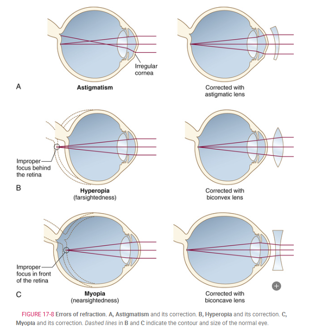
Astigmatism: Defective curvature of the cornea or lens of the eye, causing uneven light focus on the retina and resulting in a distorted image.
Hyperopia (Hypermetropia): Farsightedness, where the eyeball is too short or the lens has insufficient refractive power. Light rays focus behind the retina, causing a blurred image. A convex lens corrects this by bending rays inward.
Myopia: Nearsightedness, where the eyeball is too long or the lens has excessive refractive power. Light rays focus in front of the retina, resulting in a blurred image. Concave glasses correct this by spreading rays outward before they reach the cornea.
Presbyopia: Impairment of vision due to aging. The loss of elasticity in the ciliary body reduces its ability to adjust the lens for near vision, causing light rays to focus behind the retina. A convex lens is needed to correct this condition.
Pathology
Cataract: Clouding of the eye’s lens, causing blurry vision. Treated with surgery to remove the lens and replace it with an artificial one.
Chalazion: A small, hard bump on the eyelid caused by a blocked gland. Treatment may include draining the cyst.
Diabetic Retinopathy: Damage to the retina from diabetes, leading to blurred vision. Treated with laser therapy or injections to reduce swelling.
Glaucoma: Increased pressure in the eye that can damage the optic nerve and cause vision loss. Treated with medication or surgery to lower eye pressure.
Macular Degeneration: A condition that causes loss of central vision while peripheral vision stays normal. It can be treated with supplements or injections, depending on the type.
Nystagmus: Rhythmic, uncontrollable eye movements, often caused by brain or inner ear issues.
Retinal Detachment: When the retina separates from the eye, causing flashes of light or floaters. Treated with surgery to reattach the retina.
Strabismus: When the eyes don’t align properly, causing one eye to turn in, out, up, or down. Treated with glasses, exercises, or surgery. It can lead to lazy eye or double vision.
Clinical Procedure
Fluorescein Angiography: A test where a dye is injected into the bloodstream to take pictures of the retina, checking for blood flow and problems like diabetic retinopathy.
Ophthalmoscopy: A test where a doctor looks inside the eye to examine the retina, using a special light and tool.
Optical Coherence Tomography (OCT): A non-invasive scan that uses light to create detailed images of the retina and optic nerve, helping to detect glaucoma and retinal diseases.
Slit Lamp Microscopy: A test that uses a microscope to examine the front structures of the eye, like the cornea, iris, and lens, and measures eye pressure to check for glaucoma.
Visual Acuity Test: A test that checks how well you can see by reading letters on a chart, with results shown as a ratio (like 20/20 vision).
Visual Field Test: A test to measure the entire range of your vision, including peripheral vision.
Enucleation: Surgery to remove the entire eyeball, often due to tumors or painful, blind eyes.
Keratoplasty (Corneal Transplant): Surgery to replace a damaged or cloudy cornea with a donor cornea to restore vision.
Laser Photocoagulation: Using a laser to treat retinal issues, such as tears, leaking blood vessels, or diabetic retinopathy, by sealing them.
LASIK: A laser surgery to correct vision problems like nearsightedness, farsightedness, and astigmatism by reshaping the cornea.
Phacoemulsification: A common cataract surgery where ultrasound vibrations break up the cloudy lens, and pieces are removed, followed by inserting an artificial lens.
Scleral Buckle: Surgery to treat retinal detachment, where a silicone band is placed around the eye to push the retina back into place.
Vitrectomy: Surgery to remove the vitreous (the gel inside the eye) when it’s clouded with blood or scar tissue, often after retinal problems.
Abbreviations
ALT (Argon Laser Trabeculoplasty): A laser treatment used to lower eye pressure in glaucoma.
AMD (Age-Related Macular Degeneration): A condition that causes vision loss in the center of the visual field due to aging.
PERRLA (Pupils Equal, Round, Reactive to Light and Accommodation): A term used to describe normal pupil responses during an eye exam.
HEENT (Head, Eyes, Ears, Nose, and Throat): A common term referring to the routine examination of these areas.
POAG (Primary Open-Angle Glaucoma): A type of glaucoma where the eye's drainage system is blocked, leading to increased pressure in the eye.
IOL (Intraocular Lens): A lens implanted in the eye, often after cataract surgery, to restore vision.
PRK (Photorefractive Keratectomy): A laser surgery to correct nearsightedness by reshaping the surface of the cornea.
IOP (Intraocular Pressure): The pressure inside the eye, important for detecting glaucoma.
SLT (Selective Laser Trabeculoplasty): A type of laser treatment for glaucoma that helps improve drainage of eye fluid.
LASIK (Laser In Situ Keratomileusis): A laser surgery to correct vision by reshaping the cornea.
VA (Visual Acuity): The sharpness or clarity of vision.
OCT (Optical Coherence Tomography): A non-invasive scan to take detailed images of the retina and optic nerve.
VF (Visual Field): The area visible to the eyes when looking straight ahead.
OS (Left Eye): Abbreviation for "Oculus Sinister" (Latin for left eye).
EAR
Vocabulary
Auditory Canal: The channel from the outer ear (pinna) to the eardrum.
Auditory Meatus: Another term for the auditory canal.
Auditory Nerve Fibers: Nerve fibers that transmit sound information from the inner ear to the brain.
Auditory Tube (Eustachian Tube): A passage connecting the middle ear to the throat, helping to equalize pressure.
Auricle: The outer, visible part of the ear.
Cerumen: Earwax, a waxy substance secreted in the ear.
Cochlea: A spiral-shaped structure in the inner ear responsible for hearing.
Endolymph: Fluid inside the inner ear, within the labyrinth.
Eustachian Tube: Same as the auditory tube.
Incus: The anvil-shaped bone in the middle ear, part of the ossicle chain.
Labyrinth: A complex system of canals in the inner ear, including the cochlea and semicircular canals.
Malleus: The hammer-shaped bone in the middle ear, part of the ossicle chain.
Organ of Corti: The part of the cochlea that contains sensory cells for hearing.
Ossicle: The tiny bones in the middle ear, which include the malleus, incus, and stapes.
Oval Window: The membrane separating the middle ear from the inner ear.
Perilymph: Fluid surrounding the inner ear's labyrinth.
Pinna: The outer part of the ear, also called the auricle.
Semicircular Canals: Structures in the inner ear responsible for balance and equilibrium.
Stapes: The stirrup-shaped bone in the middle ear, part of the ossicle chain.
Tympanic Membrane (Eardrum): The membrane that separates the outer ear from the middle ear.
Vestibule: The central part of the inner ear that connects to the semicircular canals and cochlea, helping with balance.
Terminology
Prefixes:
acous/o: Hearing.
audi/o: Hearing or the sense of hearing.
audit/o: Hearing.
aur/o, auricul/o: Ear.
cochle/o: Cochlea.
mastoid/o: Mastoid process.
myring/o: Eardrum, tympanic membrane.
ossicul/o: Ossicle (small bone of the ear).
ot/o: Ear.
salping/o: Eustachian tube.
staped/o: Stapes (third bone of the middle ear).
tympan/o: Eardrum, tympanic membrane.
vestibul/o: Vestibule (part of the inner ear).
Suffixes:
-acusis or -cusis: Hearing.
-meter: Instrument to measure.
-otia: Ear condition.
Medical Professionals:
Audiologist: A healthcare professional who specializes in hearing loss evaluation and rehabilitation.
Otolaryngologist: A doctor specializing in the ear, nose, and throat.
Procedures and Conditions:
Audiogram: A chart produced from testing a person's hearing.
Myringotomy: A procedure where a small incision is made in the eardrum to drain fluid.
Stapedectomy: Surgery where the stapes bone is removed and replaced with a prosthetic device.
Tympanoplasty: Surgical reconstruction of the middle ear bones, reconnecting the eardrum to the oval window.
Pathology
Acoustic neuroma: A benign tumor on the vestibulocochlear nerve (eighth cranial nerve) that causes tinnitus, vertigo, and hearing loss. It may be treated with surgery or radiosurgery.
Cholesteatoma: A growth of skin cells and cholesterol in the middle ear that often results from chronic ear infections and causes a foul-smelling discharge.
Deafness: Loss of the ability to hear. It can be nerve (sensorineural) or conductive deafness, depending on where the problem occurs in the ear.
Meniere disease: A disorder of the inner ear causing elevated fluid pressure, leading to symptoms like tinnitus, hearing loss, vertigo, nausea, and headaches. It can be treated with medication and rest.
Otitis media: Inflammation or infection of the middle ear. Acute otitis media is an infection often caused by a cold, while serous otitis media involves fluid buildup without infection. Chronic cases may require a surgical procedure like myringotomy.
Otosclerosis: Hardening of the middle ear bones, particularly the stapes, leading to hearing loss. A stapedectomy can restore hearing by replacing the stapes with a prosthesis.
Tinnitus: A sensation of ringing or other sounds in the ears without an external source, often associated with hearing conditions like Meniere disease or otosclerosis.
Vertigo: A sensation of spinning or dizziness, often caused by problems in the inner ear or the nerves responsible for balance.
Clinical Procedures
Audiometry: A test to measure hearing ability using an audiometer, which delivers sounds at different frequencies and volumes. Results are displayed on an audiogram.
Cochlear implant procedure: A surgery to insert a device that helps people with sensorineural hearing loss by sending electrical signals directly to the auditory nerve through electrodes in the cochlea.
Ear thermometry: A quick method to measure body temperature by detecting infrared radiation from the eardrum using a device inserted into the ear canal.
Otoscopy: A visual examination of the ear canal using an otoscope.
Tuning fork test: A test for ear conduction where a vibrating tuning fork is placed on the bone behind the ear (for bone conduction) and in front of the ear canal (for air conduction). It helps check hearing ability in both air and bone conduction.
Abbreviations
AD: Right ear (Latin, auris dextra)
ENT: Ears, nose, and throat
AOM: Acute otitis media (middle ear infection)
ETD: Eustachian tube dysfunction (blocked or not working properly)
AS: Left ear (Latin, auris sinistra)
HEENT: Head, eyes, ears, nose, and throat
BPPV: Benign paroxysmal positional vertigo (a condition causing brief, sudden episodes of dizziness due to head movement)
PE tube: Pressure-equalizing tube (a tube placed in the eardrum to help treat ear infections)
EENT: Eyes, ears, nose, and throat
SOM: Serous otitis media (fluid in the middle ear without infection)
ENG: Electronystagmography (test to evaluate balance by assessing eye movement